Dear Editor,
NGENUITY 3D visualization system (Alcon Laboratories, Fort Worth, TX, USA) is equipped with an adaptable high-dynamic range (HDR) surgical camera, a high-speed image processor, and an upgraded organic light-emitting diode (OLED) surgical display for real-time visualization. It has been traditionally employed in vitreoretinal surgeries, but in the recent years; its efficacy in anterior segment procedures such as phacoemulsification and minimally invasive glaucoma surgery (MIGS) has been thoroughly investigated and proven.[1,2,3] During these surgeries by employing this 3D system, we discovered several advantages and downsides.
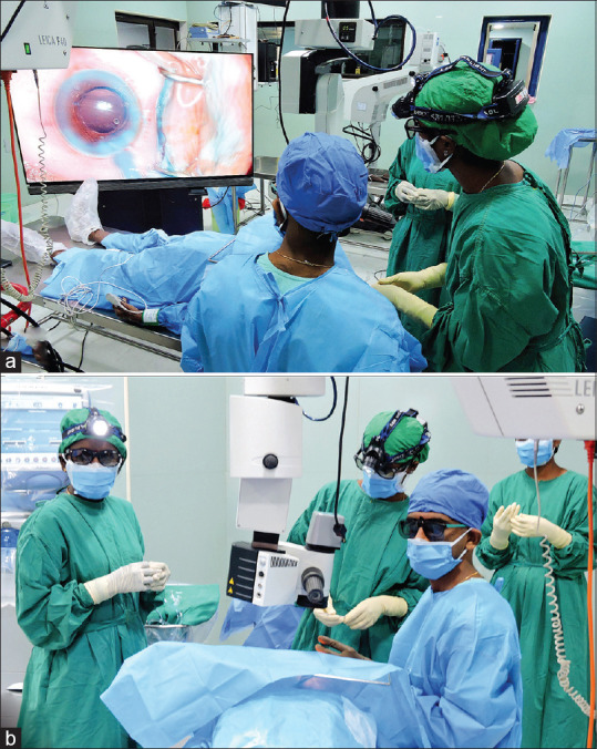
One of the most significant gains for the surgeon was the increased depth of the surgical field, which allowed him to see everything in stereoscopic detail [Fig. 1a]. According to the Alcon studies, when compared to standard microscopes, the NGENUITY 3D system provides a 42% increase in depth resolution and a 48% increase in magnification of details.[4] This increased clarity was also appreciated by us. Additionally, it was discovered that, there was no need to alter the focus regularly while operating. Another feature of the system was the level of ergonomic comfort, it provided. A study by Gauba et al. had revealed that by the age of 55, over 70% of ophthalmic surgeons have musculoskeletal issues.[5] The “heads-up” nature of 3D surgery reduced the neck strain which can occur after lengthened neck flexion linked with traditional microscopes [Fig. 1b].
Figure 1.

(a) Photograph showing the surgeon operating on the patient while sitting erect to face the screen where the video is relayed. (b) Image showing the surgeon with his operating team wearing the appropriate gear constituting the 3D glasses and the headlight for the assisting team, using the NGENUITY 3D microscope
Residents can benefit from the improved depth perception provided by the 3D technology to follow the surgical maneuvers from a training standpoint better. They can start their hands-on training with ocular surface procedures, such as pterygium excision, to improve their understanding of the surgical field and confidence in their abilities. Once they are confident with the hand–eye coordination, we can slowly shift them from extraocular to intraocular surgeries.
The system also delivers an excellent learning experience for the operating surgeon and his/her assistants by allowing them to view things in real-time or “as if they were there.” This immersive environment also allows the operating room assistants to anticipate the next maneuver that the surgeon would do. It is possible to record these films and use them in telementoring trainees all over the world.
From our point of view, there were a few things that made us uncomfortable. We had the impression that the assistants had trouble in adapting and recovering instruments due to poor ambient lighting, despite having headsets provided with headlights to guide in illumination [Fig. 1b]. In one instance, the haptic of the intraocular lens (IOL) was damaged during ejection, due to improper loading into the cartridge under poor ambient lighting. As a result, we recommend using the microscope always to load the IOL correctly. Using only one’s own eyes to handle equipment is made more difficult by the weak illumination in the operating theater. The learning curve for surgeons using the NGENUITY system may be significantly longer than the traditional microscopes. Also, due to the larger volume occupied by the 3D system, it might not be amenable for the small operating theaters. Lastly, the high procuring costs of the machine might not suit the high-volume Indian scenario, thereby restricting its widespread use.
Despite the aforementioned, surgeons should not deter from experimenting with the future avatar of phacoemulsification. More accessibility to 3D technologies and sufficient training in their use may aid us in better understanding and expanding the scope of anterior segment procedures such as MIGS.
Declaration of patient consent
The authors certify that they have obtained all appropriate patient consent forms. In the form the patient(s) has/have given his/her/their consent for his/her/their images and other clinical information to be reported in the journal. The patients understand that their names and initials will not be published and due efforts will be made to conceal their identity, but anonymity cannot be guaranteed.
Financial support and sponsorship
Nil.
Conflicts of interest
There are no conflicts of interest.
References
- 1.Ohno H. Utility of three-dimensional heads-up surgery in cataract and minimally invasive glaucoma surgeries. Clin Ophthalmol. 2019;13:2071–3. doi: 10.2147/OPTH.S227318. [DOI] [PMC free article] [PubMed] [Google Scholar]
- 2.Qian Z, Wang H, Fan H, Lin D, Li W. Three-dimensional digital visualization of phacoemulsification and intraocular lens implantation. Indian J Ophthalmol. 2019;67:341–3. doi: 10.4103/ijo.IJO_1012_18. [DOI] [PMC free article] [PubMed] [Google Scholar]
- 3.Weinstock RJ, Diakonis VF, Schwartz AJ, Weinstock AJ. Heads-up cataract surgery:Complication rates, surgical duration, and comparison with traditional microscopes. J Refract Surg. 2019;35:318–22. doi: 10.3928/1081597X-20190410-02. [DOI] [PubMed] [Google Scholar]
- 4.NGENUITY®3D Visualization System |MyAlcon Professional [Internet] [[cited 2022 Jan 1]]. Available from: https://professional.myalcon.com/vitreoretinal-surgery/visualization/ngenuity-3d-system/
- 5.Gauba V, Tsangaris P, Tossounis C, Mitra A, McLean C, Saleh GM. Human reliability analysis of cataract surgery. Arch Ophthalmol. 2008;126:173–7. doi: 10.1001/archophthalmol.2007.47. [DOI] [PubMed] [Google Scholar]


