Abstract
Methicillin-resistant Staphylococcus aureus (MRSA) regulates resistance to β-lactams via preferential production of an alternative penicillin-binding protein (PBP), PBP2a. PBP2a binds many β-lactam antibiotics with less affinity than PBPs which are predominant in methicillin-susceptible (MSSA) strains. A novel, rather frequent in vitro phenotype was recently identified among clinical MRSA bloodstream isolates, termed “NaHCO3-responsiveness”. This phenotype features β-lactam susceptibility of certain MRSA strains only in the presence of NaHCO3. Two distinct PBP2a variants, 246G and 246E, have been linked to the NaHCO3-responsive and NaHCO3-non-responsive MRSA phenotypes, respectively. To determine the mechanistic impact of PBP2a variants on β-lactam susceptibility, binding profiles of a fluorescent penicillin probe (Bocillin-FL) to each purified PBP2a variant were assessed and compared to whole-cell binding profiles characterized by flow cytometry in the presence vs. absence of NaHCO3. These investigations revealed that NaHCO3 differentially influenced the binding of the fluorescent penicillin, Bocillin-FL, to the PBP2a variants, with binding intensity and rate of binding significantly enhanced in the 246G compared to the 246E variant. Of note, the NaHCO3-β-lactam (oxacillin)-responsive JE2 strain, which natively harbors the 246G variant, had enhanced Bocillin-FL whole-cell binding following exposure to NaHCO3. This NaHCO3-mediated increase in whole-cell Bocillin-FL binding was not observed in the NaHCO3-non-responsive parental strain, COL, which contains the 246E PBP2a variant. Surprisingly, genetic swaps of the mecA coding sites between JE2 and COL did not alter the NaHCO3-enhanced binding seen in JE2 vs. COL. These data suggest that the non-coding regions of mecA may be involved in NaHCO3-responsiveness. This investigation also provides strong evidence that the NaHCO3-responsive phenotype in MRSA may involve NaHCO3-mediated increases in both initial cell surface β-lactam binding, as well as ultimate PBP2a binding of β-lactams.
Keywords: penicillin-binding proteins, sodium bicarbonate, β-lactams, methicillin-resistant Staphylococcus aureus
1. Introduction
The methicillin-resistant Staphylococcus aureus (MRSA) phenotype is a major obstacle to the effective treatment of severe S. aureus clinical infections, including bacteremia and infective endocarditis [1]. MRSA strains are typically resistant to nearly all β-lactams (first-line treatment of choice for methicillin-susceptible S. aureus (MSSA)), on standard in vitro testing, linked to their preferential production of an alternative penicillin-binding protein (PBP), PBP2a [2,3]. Most β-lactam antibiotics bind poorly to PBP2a when tested in standard microbiologic growth media, allowing cell wall synthesis and microbial proliferation to occur in their presence [4,5].
Recently, a novel phenotype was identified amongst clinical MRSA strains, whereby certain isolates exhibit susceptibility to early generation β-lactams (e.g., cefazolin and/or oxacillin) in the presence of NaHCO3 supplementation of standard media in vitro [6,7,8,9,10]. Physiological relevance of this phenomenon in terms of β-lactam re-sensitization has been verified in both ex vivo and in vivo models [6,11]. This β-lactam hypersusceptibility appears to be linked, at least in part, to NaHCO3-mediated repression of the mecA/blaZ/PBP2a axis, as well as alteration in several accessory genes required for proper PBP2a functionality and maturation, ultimately impacting cell wall synthesis [6,9,12]. However, the precise effects of NaHCO3 on specific properties of the PBP2a protein itself are currently unknown.
Of interest, another novel β-lactam hypersusceptibility phenotype was recently identified in MRSA, wherein strains with particular mecA genotypes were rendered susceptible to the combination of β-lactams and β-lactamase inhibitors [13]. Importantly, two particular PBP2a protein variants, 246E (wild-type PBP2a [13]) and 246G, were observed to have differing binding affinities for penicillin in the presence of clavulanic acid [13]. This difference was hypothesized to contribute to altered susceptibilities to penicillin + clavulanic acid in strains harboring these variants.
In the current study, we queried whether such specific PBP2a variants could also influence the NaHCO3-responsive/non-responsive phenotypes in a parallel manner. Furthermore, we aimed to determine whether NaHCO3 affected PBP2a-β-lactam binding affinities and temporal binding profiles, in a genotype-specific process (similar to penicillin + clavulanate).
Herein, purified PBP2a variants 246G and 246E, prototypic of the NaHCO3-responsive and -non-responsive phenotypes, respectively [10], were compared for their binding affinities for the fluorescently labeled penicillin derivative, Bocillin-FL, in the presence and absence of NaHCO3. Furthermore, responsive and non-responsive strain sets harboring either their native or genetically swapped PBP2a variants (246G and 246E) were also assessed for ability to bind Bocillin-FL in the presence vs. absence of NaHCO3 by whole-cell flow cytometry.
2. Results
2.1. Bocillin-FL Binding to Purified PBP2a
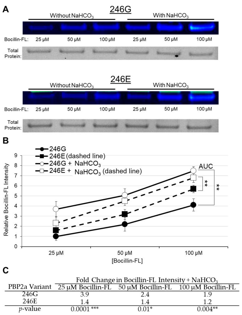
To study penicillin binding in vitro, mecA genes of S. aureus COL and USA300 JE2, encoding for PBP2 variants 246E and 246G, respectively, were cloned into expression vectors for heterologous overexpression and Ni-NTA purification. Purified PBP2a proteins of each variant type were utilized in a Bocillin-FL binding assay to determine whether alteration at the 246th amino acid position impacts direct binding of Bocillin-FL to either purified variant. In the absence of NaHCO3 in the binding reaction, both variants bound similar amounts of Bocillin-FL (Figure 1A,B). Corroborating this result, in a β-lactam competition assay with either “cold” penicillin or “cold” oxacillin, both variants had similar IC50 values for these two antibiotics (Supplementary Materials Figure S1A,B). These findings correspond to those observed by Harrison et al. [13], wherein all PBP2a variants studied had similar affinities for β-lactams under standard assay conditions.
Figure 1.
Bocillin-FL binding to purified PBP2a variants 246G and 246E. (A) Representative gel images for PBP2a-Bocillin binding and total protein loading; 50 µg/mL PBP2a was incubated with 25, 50, or 100 µM Bocillin-FL in each gel. (B) Bocillin-FL binding with and without 44 mM NaHCO3; 50 µg/mL PBP2a was used in each Bocillin binding reaction. Relative Bocillin intensity was determined by normalizing absolute intensity values for each sample to the average intensity of 25 µM Bocillin bound to 50 µg/mL 246G PBP2a in the absence of NaHCO3 (with this value being set equal to 1.0). Additionally, an intra-gel normalization control was run on each gel consisting of 25 µM Bocillin-FL incubated with 50 µg/mL 246G PBP2a to which all bands were initially normalized. The data presented are the composite of four separate gel runs. Asterisks represent the significant difference in the area under the curve (AUC) for each variant incubated with and without NaHCO3 (Student’s t-test, ** p < 0.01). (C) Fold changes in Bocillin-FL binding to each PBP2a variant in the presence and absence of 44 mM NaHCO3 (calculated from data in part (B)). Fold change is calculated as [Bocillin Intensity]NaHCO3/[Bocillin Intensity]No NaHCO3. Values greater than 1 indicate increased Bocillin-FL binding to PBP2a in the presence of NaHCO3. Statistical significance is calculated using the average fold-change in Bocillin-FL binding for each PBP2a variant in the presence vs. absence of NaHCO3 by Student’s t-test (* p < 0.05, ** p < 0.01, *** p < 0.001).
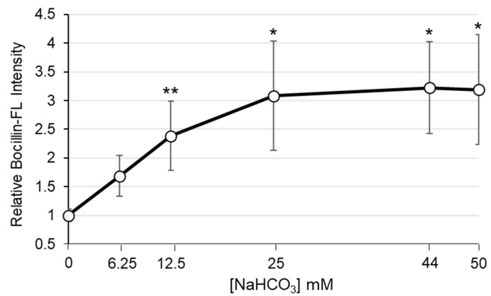
Next, in parallel studies, NaHCO3 (44 mM) was added to each of the PBP2a-Bocillin-FL binding reactions. This concentration of NaHCO3 is the standard used in prior in vitro and ex vivo assays to disclose β-lactam susceptibility and, thus, identify NaHCO3-responsive strains [6,7,8,10,11]. When NaHCO3 was added to the binding reactions, Bocillin-FL binding to both PBP2a variants was substantially enhanced (Figure 1A,B). However, the binding enhancement to the 246G variant was significantly greater than that observed with the 246E variant (Figure 1C). The impact of NaHCO3 on Bocillin-FL binding to the 246G variant was most evident at 25 µM Bocillin-FL, in which NaHCO3 stimulated a nearly 4-fold increase in Bocillin-FL intensity, compared to only a 1.4-fold change in intensity in the 246E variant (Figure 1C). Furthermore, to examine the NaHCO3 concentration-dependent impact on binding to purified PBP2a, we exposed 50 µg/mL of the 246G variant to 50 µM Bocillin with increasing concentrations of NaHCO3 ranging from 0 to 50 mM. There was a significant increase in Bocillin binding at the lower NaHCO3 concentrations (** p = 0.007 [Student’s t-test] comparing 0 mM vs. 12.5 mM), with a more gradual increase in binding between 12.5 and 50 mM (Figure 2).
Figure 2.
NaHCO3 dose response impact on Bocillin-FL binding to purified PBP2a variant 246G; 50 µg/mL PBP2a was incubated with 50 µM Bocillin-FL and either 0, 6.25, 12.5, 25, 44, or 50 mM NaHCO3. Relative Bocillin intensity was normalized to a control lane consisting of 25 µM Bocillin-FL incubated with 50 µg/mL 246G PBP2a run on each gel. Final data was normalized to the intensity of 50 µg/mL PBP2a incubated with 50 µM Bocillin-FL and no NaHCO3 (with this value set equal to 1.0). Asterisks represent significant differences in Bocillin-FL intensity at the indicated NaHCO3 concentration as compared to no NaHCO3 (Student’s t-test, * p < 0.05, ** p < 0.01).
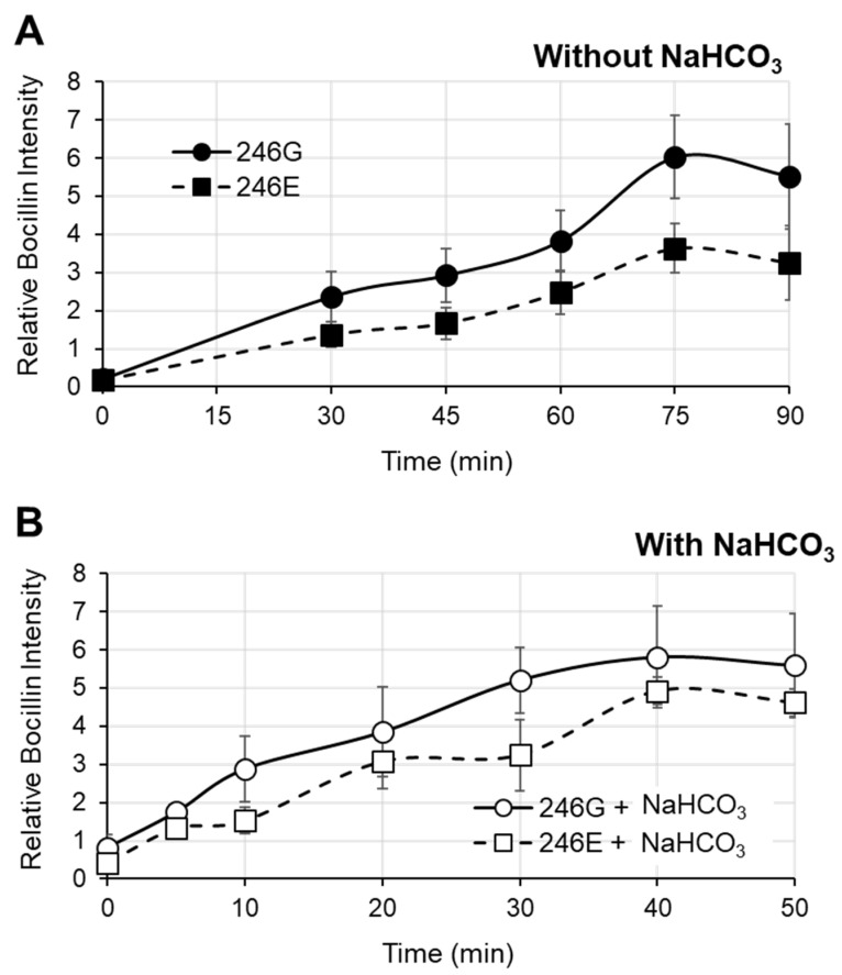
To investigate the differential impact of NaHCO3 on the kinetics of Bocillin-FL binding to each PBP2a variant, 246G PBP2a and 246E PBP2a were incubated with or without NaHCO3 and assessed for binding at various time points (Figure 3A,B). These data query time points were established as optimal for this experimentation based on extensive pilot studies. Based on the binding curves, the time to reach 50% maximal binding was calculated for each variant in the presence vs. absence of NaHCO3. In the absence of NaHCO3, both variants exhibited similar times to reach 50% maximal binding (Figure 3A; 246G 50% binding time = 44.2 ± 4.6 min, 246E 50% binding time = 40.3 ± 8.8 min, Student’s t-test p = NS). However, in the presence of NaHCO3, the 246G variant reached 50% maximal binding significantly faster than the 246E variant (Figure 3B; 246G—50% binding time = 7.6 ± 1.1 min, 246E—50% binding time = 13.2 ± 4.8 min, Student’s t-test * p = 0.04). For both variants, NaHCO3 significantly decreased the time until 50% maximal binding vs. in the absence of NaHCO3 (246G without vs. with ** p = 0.006; 246E without vs. with * p = 0.03).
Figure 3.
Kinetic binding of Bocillin-FL to purified PBP2a variants 246G and 246E. (A) Kinetic Bocillin-FL binding without NaHCO3. (B) Kinetic Bocillin-FL binding with NaHCO3. A total of 50 µg/mL PBP2a was used in each Bocillin binding reaction. Relative Bocillin intensity was determined by normalizing absolute intensity values for each sample to the intensity of 25 µM Bocillin bound to 50 µg/mL 246G PBP2a in the absence of NaHCO3 incubated for 20 min run on the same gel.
2.2. Bocillin-FL Binding to Whole JE2 and COL Cells by Flow Cytometry
To determine the impact of NaHCO3 on binding of Bocillin-FL to whole cells, we compared two well-characterized MRSA strains: JE2 (NaHCO3-responsive (Supplementary Materials Table S1); 246G PBP2a variant); vs. COL (-non-responsive (Table S1); 246E PBP2a variant). Neither strain produces β-lactamase [14]. Cells were incubated with 100 µM Bocillin-FL, and then assessed for overall binding by flow cytometry. Prior to staining, cells were grown to mid-log phase (OD600 nm = 0.5) in CA-MHB Tris media plus half the minimum inhibitory concentration (MIC) of oxacillin (Table S1), with or without 44 mM NaHCO3.
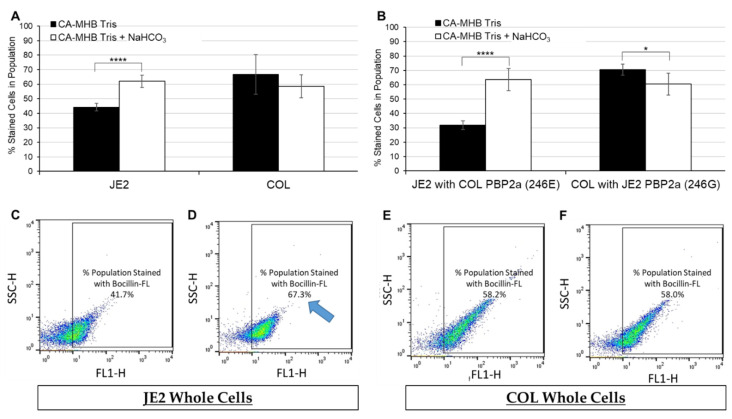
Upon analysis, JE2 cells grown in the presence of NaHCO3 displayed a significantly greater number of Bocillin-FL-stained cells within the population vs. cells grown in the absence of NaHCO3 (Figure 4A,C,D). This NaHCO3-induced enhancement of Bocillin-FL binding to JE2 cells corresponds to the NaHCO3-stimulated β-lactam sensitization observed in this strain by MIC testing (Table S1). Growth in the presence of NaHCO3 had no impact on Bocillin-FL binding to the -non-responsive strain, COL (Figure 4A,E,F), corresponding to the lack of impact of NaHCO3 on β-lactam susceptibility by MIC for this strain (Table S1).
Figure 4.
Percentage of JE2 and COL cells out of total population (10,000 cells) stained with Bocillin-FL following growth in cation-adjusted Mueller Hinton broth (CA-MHB Tris) ± 44 mM NaHCO3. (A) Summary of average Bocillin-FL binding for JE2 and COL. (B) Summary of average Bocillin-FL binding for JE2 and COL strains with “swapped” mecA coding regions. (C–F) Representative dot-plots for (C) JE2 grown in CA-MHB Tris (D) + NaHCO3, vs. COL grown in (E) CA-MHB Tris (F) + NaHCO3. Gates on dot-plots (box surrounding cells) depict percentage of cells in total population of 10,000 cells that have taken up the Bocillin-FL dye. Note enhanced proportion of JE2 cells stained with Bocillin-FL in the presence vs. absence of NaHCO3 (blue arrow). Statistics were calculated by a Student’s t-test, * p < 0.05, **** p < 0.0001.
To further investigate the specific impact of NaHCO3 on Bocillin-FL whole-cell binding in the presence of each PBP2a variant within a given strain background, “swap” mutants were constructed in JE2 and COL so that each strain possessed the mecA coding region of the other strain (JE2 with mecA 246E, COL with mecA 246G). As expected, swap of only the mecA coding regions did not alter the NaHCO3 responsiveness of these variants to oxacillin by MIC testing (Table S1). This relates to the requirement of both the mecA coding regions and ribosomal-binding sites (RBS) for full expression of this phenotype (Ersoy et al.; manuscript submitted—in review Antimicrobial Agents and Chemotherapy).
Interestingly, upon flow analysis of these mutant constructs in the presence vs. absence of NaHCO3, each “swap” strain derivative displayed the same phenotype as its parental strain (Figure 4B and Figure S2). This indicates that the NaHCO3 enhancement of Bocillin-FL binding to JE2 whole cells is not due solely to increased affinity of this particular PBP2a variant for Bocillin-FL in the presence of NaHCO3. This outcome may implicate a likely role of differences in NaHCO3 impacts on selected cell surface properties specific to each strain background.
In parallel, the mean fluorescence intensity (MFI) of the cell population was assessed in the presence or absence of NaHCO3 exposures. Despite the significant enhancement of the proportion of JE2 (vs. COL) cells binding Bocillin-FL in the presence of NaHCO3, the amount of fluorophore taken up per cell population (10,000 cells) did not differ between cells grown in the presence vs. absence of NaHCO3 for JE2 vs. COL parental strains, nor COL possessing JE2 PBP2a (Figure S3).
3. Discussion
Investigations with purified PBP2a variants, as well as whole cells harboring differing PBP2a variants, revealed that NaHCO3 substantially and differentially influenced the ability of a β-lactam probe, Bocillin-FL, to bind each PBP2a variant. The purified 246G PBP2a variant (previously associated with NaHCO3-responsiveness [10]) displayed enhanced binding of Bocillin-FL when NaHCO3 was incorporated into the reaction mixture (vs. the 246E variant) in a NaHCO3 dose-dependent manner. Prior in vitro data demonstrated that in NaHCO3-responsive MRSA harboring the 246G PBP2a variant, β-lactam MICs also decreased in a NaHCO3 concentration-dependent-manner. For example, oxacillin and cefazolin MICs for NaHCO3-responsive strain MRSA 11/11 were each 4 µg/mL at 25 mM of NaHCO3, decreasing 8-fold to 0.5 µg/mL at 44 mM NaHCO3 [6].
In parallel to the binding studies with purified PBP2a, whole-cell binding assays demonstrated that exposure to NaHCO3 during the early to mid-log growth phases resulted in enhanced Bocillin-FL binding to the NaHCO3-responsive strain, JE2, harboring the 246G variant vs. COL, harboring the 246E variant. These NaHCO3-stimulated increases in Bocillin-FL whole-cell binding again correspond to NaHCO3 impacts on β-lactam susceptibility by MIC testing in vitro for each strain harboring these PBP2a variants (as detailed above). Interestingly, further in-parallel studies utilizing JE2 and COL mutant strains with “swapped” PBP2a variants (JE2 now with 246E vs. COL now with 246G) displayed similar whole-cell binding affinities in the presence vs. absence of NaHCO3 as their parental counterparts. These data suggest the possibility that, in addition to NaHCO3-mediated enhancements of Bocillin-FL binding to the 246G PBP2a variant, NaHCO3 may also be differentially affecting specific surface factors which influence overall Bocillin-FL binding to whole cells. In particular, we have previously shown that NaHCO3 can specifically repress transcription of the prsA gene and PrsA protein production in NaHCO3-responsive but not NaHCO3-non-responsive MRSA [9]. The prsA gene encodes the chaperone PrsA; this protein is important for the optimal translocation, folding, and maturation of PBP2a [15,16]. In addition to its chaperone function, PrsA is known to regulate the expression of a multitude of surface proteins and exoproteins. Moreover, alteration of PrsA expression can result in a variety of other phenotypic alterations to the cell surface, including perturbations of lipid composition and localization, which can also impact surface protein distribution and function [17,18]. Thus, NaHCO3-mediated impacts on prsA could plausibly impact net whole-cell binding of Bocillin-FL. In addition to NaHCO3 impacts on PrsA, NaHCO3 is known to differentially affect the expression of a variety of surface-associated genes/proteins, including the cap operon, pbp2, and fmtA, among others, in NaHCO3-responsive vs. non-responsive strain backgrounds [12].
As reported previously [13], both the 246G and 246E PBP2a variants had similar intrinsic binding affinities for Bocillin-FL, as well as to the β-lactams, penicillin, and oxacillin, in the absence of NaHCO3. This indicates that factors in the extracellular environment during NaHCO3 exposure likely play a crucial role in dictating binding affinities of such fluorophores, as would generally be expected for biochemical interactions. It should be noted that the precise surface factors (e.g., cell wall proteins or membrane lipids) that Bocillin-FL initially binds to in whole cells are not known.
The profound genetic regulatory effects of NaHCO3 on PBP2a expression and function have been recently detailed [9]. However, the precise and direct physio-chemical impacts of NaHCO3 on the PBP2a molecule itself following whole-cell binding and intracellular transport are unknown [19]. Since NaHCO3 is a weak base, it is likely that pH effects may impact certain aspects of tertiary folding of the PBP2a protein, such as hydrogen bonding and the formation of salt bridges [20,21]. Furthermore, alterations to environmental pH and temperature are known to have catastrophic effects on PBP2a protein folding and functionality [22]. It is unclear whether alterations in Bocillin-FL-PBP2a binding stimulated by NaHCO3 are specific to the bicarbonate molecule or are a more generalized weak acid–base effect. Further studies with other weak acids and bases are needed to verify if these impacts are NaHCO3-specific or related to more generalized impacts on PBP2a protein structure. In this regard, previous studies by us and others suggest that the “NaHCO3 effect” microbiologically is not seen with other weak acids (e.g., salicylic acid; boric acid [6,23]).
Regulation of PBP2a protein activity occurs via allosteric binding interactions, wherein peptidoglycan precursors bind to the allosteric site, causing a conformational change that reveals the active site and allows peptidoglycan synthesis to progress [20,24,25]. Traditional β-lactams (with the exception of ceftaroline) are unable to efficiently occupy this allosteric binding site, allowing the active site to remain closed unless peptidoglycan precursor substrates are present [24,25,26,27]. Although it is difficult to determine the specific impact of NaHCO3 on PBP2a protein structure, it is possible that this weak base may alter the allosteric site in a manner that allows better β-lactam binding to both the allosteric and subsequently to the active sites (e.g., as seen in the 246G variant). Indeed, the position of 246th amino acid residue is centrally located within the PBP2a allosteric site [20]. As the 246E variant is considered the “wild type” form of PBP2a [13], it is plausible that the glutamic acid at this residue aids in stabilization of the allosteric binding site compared to variants possessing a glycine at this residue. Thus, it could be hypothesized that the weak acid nature of glutamic acid buffers NaHCO3 better than glycine, allowing more stable protein interactions.
Overall, these data support the notion that NaHCO3 differentially enhances the binding affinities of selected β-lactams to both the whole cell surface, as well as to the 246G PBP2a variant. This event may, in turn, contribute to the NaHCO3-responsive phenotype observed in strains possessing these particular protein variants. Our current studies with mutant strains harboring each of these mecA alleles yielded additional mechanistic insights concerning specific PBP2a protein variants and the NaHCO3-responsive/-non-responsive phenotypes. Furthermore, since the mecA variants we have studied also differ in their ribosomal-binding site (RBS) sequences [10,13], the specific impacts of these latter differences on NaHCO3-responsiveness remain to be determined. We are currently examining this concept by transgenic (“swapping”) methods similar to the present investigation, targeting both the mecA coding and/or RBS regions between the same NaHCO3-responsive/-non-responsive strains used currently. Moreover, we recognize that the data presented here are only comparing β-lactam binding metrics in a single strain set, and need to be validated in additional MRSA strains featuring these two mecA variants. Finally, the impact of these two PBP2a variants on binding profiles to other prominent PBPs (e.g., PBP2) needs to be adjudicated. Such studies are in progress.
4. Materials and Methods
4.1. Bacterial Strains and Growth Conditions
The NaHCO3-responsive strain, JE2 (PBP2a 246G variant), and -non-responsive strain, COL (PBP2a 246E variant), utilized in these studies are both β-lactamase-negative. This latter phenotype prevented testing these strains for differential penicillin-clavulanate vs. penicillin-alone MICs [13]. NaHCO3-responsive phenotypes were determined following MIC testing in cation-adjusted Mueller Hinton Broth (CA-MHB, Difco) containing 100 mM Tris (added to maintain a stable pH 7.3 ± 0.1 in the presence and absence of NaHCO3 as in our prior studies [6,7,9,10]); 44 mM NaHCO3 was employed as previously described, and represents tissue-level NaHCO3 concentrations [6,28,29]. MIC values for each strain in media with and without NaHCO3 are listed in Table S1. The specific PBP2a genotypes for each strain were determined by sequencing the PCR-amplified mecA product (GeneWiz LLC) [10]. Primers used for mecA amplification and sequencing are listed in Table S2.
4.2. Isolation of Purified PBP2a: Plasmid Construction
Plasmids pET21b-mecA246E, pET21b-mecA246G, pET28b-mecA246E, and pET28b-mecA246G were constructed using the In-Fusion Cloning kit (Takara Bio). mecA genes were amplified by PCR from genomic DNA of S. aureus strain COL and strain USA300 JE2 using the primer pairs A/B (for cloning into pET21b) or C/D (for cloning into pET28b). Plasmid backbones were generated by inverse PCR of pET21b and pET28b (Novagen) using primer pairs E/F and E/G, respectively. The resulting constructs were verified by Sanger DNA sequencing. Oligonucleotide primers (Table S2) were synthesized by Eurofin Genomics.
4.3. Isolation of Purified PBP2a: Overexpression and Purification of S. aureus PBP2a246E and PBP2a246G
Expression cultures of Escherichia coli C43(DE3) transformed with the recombinant plasmids (pET21b containing mecA with a C-terminal His6-tag or pET28b containing mecA with an N-terminal His6-tag, respectively) were grown in 2 L of LB containing 100 µg/mL ampicillin for pET21b or 50 µg/mL kanamycin for pET28b at 37 °C with shaking. At an optical density (OD600) of 0.5, isopropyl β-D-thiogalactopyranoside (IPTG) was added to a concentration of 0.5 mM to induce expression of recombinant PBP2a. Expression cultures were cooled down to 18 °C and incubation was continued for 16 h. Cells were harvested by centrifugation (8000× g, 15 min, 4 °C) and resuspended in 20 mL of ice-cold lysis buffer (50 mM Tris-HCl, pH 7.5, 500 mM NaCl, 1% v/v Triton X-100). All subsequent steps were performed at 4 °C. Resuspended cells were incubated with 5 µg/mL RNase, 20 µg/mL DNase, and 0.5% v/v N-lauroylsarcosine for 1 h and disrupted by sonication (amplitude: 60%, pulse cycle 0.5). Cell debris was pelleted by centrifugation (48,000× g, 30 min, 4 °C) and the supernatant was added to 1 mL Ni-NTA-agarose (Macherey-Nagel). After gentle stirring for 1 h, the suspension was loaded to a column support. To remove weakly bound material, the column was washed with 10 mL wash buffer (50 mM Tris-HCl pH 7.5, 500 mM NaCl) supplemented with 10 mM imidazole and then with 10 mL wash buffer supplemented with 20 mM imidazole. His-tagged proteins were eluted 5 times with 500 μL wash buffer supplemented with 200 mM of imidazole and stored in 40% v/v glycerol at −20 °C. The purity of elution fractions was analyzed by SDS-PAGE (Figure S4) and concentrations were measured using Pierce™ BCA protein assay with BSA as standard.
4.4. Construction of JE2 and COL mecA 246-Residue Point Mutation Strains
To determine the contribution of the amino acid position at 246-residue (Glu/Gly) of the mecA gene in NaHCO3-responsive or non-responsive S. aureus strains, we constructed chromosomal point mutation of the mecA region in S. aureus strains JE2 (RBS: AGGAGT and 246G) and COL(tetR)(RBS:AGGAGG and 246E) using routine procedures as described [30]. To construct point mutation constructs, 3.2 kb DNA fragments were amplified that contained the intact mecA and mecR genes by PCR using primers flanked with BamHI sites at both ends and chromosomal DNA as the template of both JE2 and COL (Table S2). The DNA fragment was cloned into a temperature-sensitive shuttle vector pMAD (β-gal, ermR) [31], and then selected in E. coli IM08B [32] for the correct construct by digesting with BamHI of the isolated plasmid. To construct point mutation at the 246-residue position, site-specific mutagenesis was performed with pMAD constructs as the template and various mutagenized primers using a PCR-based method with pfu Taq-polymerase (Phusion, Thermo Scientific, Waltham, MA, USA). PCR products were treated with a KLD enzymes mix (NE Biolabs Inc., Ipswich, MA, USA) and transformed to E. coli IM08B for the selection on LB agar plates containing ampicillin (100 μg/)mL-X-Gal (40 μg/)mL. Final constructs were verified by restriction digestion and DNA sequencing. Authenticated desired respective constructs were introduced into JE2 and COL strains by electroporation and selected on erythromycin and X-Gal-containing plates (40 μg/)mL for blue colonies at 30 °C. Plasmid DNA was isolated and digested with BamHI for the authentication of the presence of DNA fragments in the respective constructs in the strains. The construction of chromosomal mutations in the respective strain by recombination or two-point cross-over was performed by routine procedure as described previously [30]. Briefly, two-point cross-over of the mecA-mecR region was performed by temperature shift by growing strains at 43 °C with erythromycin followed by 30 °C sub-culturing without any antibiotics. Cells were plated with and without erythromycin in the presence of X-Gal for selection and incubated at 37 °C. White/non-blue colonies were cross-streaked to select ermS colonies for the potential two-point cross-over clones or mutants. The mutants were verified by chromosomal PCR and DNA sequencing of the PCR product for both RBS and 246-residue positions. Final clones were designated as ALC9165 (JE2, RBS: AGGAGT and 246E) and ALC9167 (COL, RBS:AGGAGG and 246G).
4.5. Bocillin-FL Binding to Purified PBP2a and β-Lactam Competition Studies
Bocillin-FL is a fluorescently labeled derivative of penicillin V [33], and was purchased from Thermo Fisher Scientific (Waltham, MA, USA). Based on in vitro MIC data performed with strain JE2, Bocillin-FL has similar antibacterial activity to “cold” penicillin G (Table S3). Based on numerous pilot studies, 50 µg/mL of N-terminal His6-tagged PBP2a246E or PBP2a246G were deemed as ideal, and utilized in all purified PBP2a binding studies. Purified proteins were incubated with 25 µM, 50 µM, or 100 µM Bocillin-FL for 20 min at 37 °C in a 20 µL final reaction volume and then incubated for 5 min at 95 °C with LDS loading dye. The reaction mixtures were then run on a NuPAGETM 4–12% Bis-Tris protein gel and imaged with an Azure c400 imager (Azure Biosystems). Fluorescence intensities of the gel images were analyzed and quantified by ImageJ software (version 1.52a). For competition studies, “cold” penicillin G or oxacillin (purchased from Sigma-Aldrich, St. Louis, MO, USA) was incubated with 50 µg/mL PBP2a at various concentrations (0, 0.5, 1, 5, 10, 50, 100, 500 µg/mL) for 10 min at 37 °C. Then, 50 µM Bocillin-FL was added to each reaction mixture, and incubated for an additional 20 min at 37 °C. A control comprising 50 µg/mL PBP2a246G and 25 µM Bocillin-FL was run on each gel, to which the intensity of all other reactions on that gel were normalized. Following protein gel electrophoresis, imaging, and ImageJ analysis, the IC50 for each PBP2a variant and antibiotic was calculated by linear regression analysis in Excel.
Fold changes in Bocillin-FL fluorescence in the presence vs. absence of NaHCO3 were calculated by measuring the Bocillin-FL intensity in paired gels, with Bocillin-FL-PBP2a binding reactions performed with and without NaHCO3. The intensity observed in the presence of NaHCO3 was divided by the intensity observed in the absence of NaHCO3 for each Bocillin-FL concentration (25, 50, 100 µM) to determine the fold change in intensity for a given pair of gels at each Bocillin-FL concentration. The fold change in intensity for three gel pairs was used to determine the average fold change and calculate statistical significance (p-value) by a Student’s t-test for each Bocillin-FL concentration indicated for the two PBP2a protein variants.
For kinetics studies, 50 µg/mL PBP2a246G and PBP2a246E were incubated at 37 °C with 50 µM Bocillin-FL, with or without 44 mM NaHCO3. At each indicated time point, the reaction sample was removed, incubated for 5 min at 95 °C with LDS loading dye, and run on a NuPAGETM 4–12% Bis-Tris protein gel, as indicated previously. A control sample of 50 µg/mL PBP2a246G and 25 µM Bocillin-FL, incubated for 20 min, was run with each gel as a normalization control. The time to reach “maximal binding” was assessed by identifying the first point at which Bocillin-FL intensity no longer increased or began to decrease for one or more subsequent time points, following extensive pilot studies. The “time to reach 50% maximal binding” was calculated by polynomial regression analysis in Excel.
4.6. Bocillin-FL Binding to Whole Cells by Flow Cytometry
JE2, COL, JE2 with COL PBP2a (246E), and COL with JE2 PBP2a (246G) cells were grown to mid-log phase (OD600nm = 0.5) in CA-MHB Tris ± 44 mM NaHCO3 (pH = 7.3 ± 0.1) with half the MIC of oxacillin, as indicated in Table S1. Cells were then washed twice in phosphate-buffered saline (PBS) and adjusted to OD600nm = 1.0; 500 µL of adjusted cells were incubated with 500 µL of 200 µM Bocillin-FL (final concentration = 100 µM), and incubated for 20 min at 37 °C. Bocillin-FL stained cells were washed three times in PBS, then diluted to a final concentration of ~5 × 10⁷ CFU/mL; 10,000 cells were then analyzed by flow cytometry on FACScalibur® (Becton-Dickinson, Franklin Lakes, NJ, USA). The proportion of cells that bound Bocillin-FL was calculated using FlowJo software (version 10.8). In addition, the mean fluorescence intensity (MFI) of the cell population was calculated using data from the FL1-H channel, analyzed with FlowJo software and expressed as relative fluorescent units/cell population.
5. Conclusions
NaHCO3 differentially impacts binding affinities of the 246G and 246E PBP2a protein variants found in NaHCO3-responsive and -non-responsive strains, respectively. NaHCO3 enhances binding of the 246G variant to Bocillin-FL, as compared to the 246E variant, in both purified protein and whole-cell assays. Enhanced β-lactam binding to the 246G variant in the presence of NaHCO3 is correlated to increased susceptibility to β-lactams in the presence of NaHCO3 in responsive strains.
Supplementary Materials
The following are available online at https://www.mdpi.com/article/10.3390/antibiotics11040462/s1, Figure S1: “Cold” oxacillin and penicillin competition of Bocillin-FL binding to purified PBP2a variants (246G and 246E); Figure S2: Representative dot plot images of JE2 and COL PBP2a swap strains grown in CA-MHB Tris ± NaHCO3; Figure S3: Mean fluorescence intensity for JE2 and COL whole cells stained with Bocillin-FL; Figure S4: SDS-PAGE of purified PBP2a His6-tagged proteins; Table S1: Oxacillin minimum inhibitory concentrations (MICs) for JE2, COL, and their mecA swap derivative MRSA strains; Table S2: Oligonucleotides used for mecA sequencing, construction of His-tagged mecA246E and mecA246G plasmids, and construction of JE2 and COL mecA mutant variants; Table S3: MICs of penicillin G and Bocillin-FL in strain JE2.
Author Contributions
Conceptualization, S.C.E., H.F.C., R.A.P. and A.S.B.; Data curation, A.S.B.; Formal analysis, S.C.E., L.C.C., A.C., A.C.M. and R.A.P.; Funding acquisition, L.C.C., M.R.Y. and A.S.B.; Investigation, S.C.E. and K.C.L.; Methodology, S.C.E., L.C.C., K.C.L., T.S., A.C., A.C.M. and A.S.B.; Project administration, A.S.B.; Supervision, A.S.B.; Writing—original draft, S.C.E. and A.S.B.; Writing—review and editing, all authors. All authors have read and agreed to the published version of the manuscript.
Funding
This work was funded by the following grants from the National Institutes of Health: 1R01-AI146078 (to A.S.B.); UCLA CTSI KL2-TR001882-05 (to L.C.C.) and 1U01-AI124319-01 (to M.R.Y.). Additional funding was provided by the German Center for Infection Research (DZIF) (to K.C.L. and T.S.).
Institutional Review Board Statement
Not applicable.
Informed Consent Statement
Not applicable.
Data Availability Statement
Data are contained within the article (Figure 1C and Figure 4B–E). Additional datasets are available upon request.
Conflicts of Interest
The authors declare no conflict of interest.
Footnotes
Publisher’s Note: MDPI stays neutral with regard to jurisdictional claims in published maps and institutional affiliations.
References
- 1.Tong S.Y., Davis J.S., Eichenberger E., Holland T.L., Fowler V.G. Staphylococcus aureus infections: Epidemiology, pathophysiology, clinical manifestations, and management. Clin. Microbiol. Rev. 2015;28:603–661. doi: 10.1128/CMR.00134-14. [DOI] [PMC free article] [PubMed] [Google Scholar]
- 2.Dien Bard J., Hindler J.A., Gold H.S., Limbago B. Rationale for eliminating Staphylococcus breakpoints for β-lactam agents other than penicillin, oxacillin or cefoxitin, and ceftaroline. Clin. Infect. Dis. 2014;58:1287–1296. doi: 10.1093/cid/ciu043. [DOI] [PMC free article] [PubMed] [Google Scholar]
- 3.Grundmann H., Aires-de-Sousa M., Boyce J., Tiemersma E. Emergence and resurgence of meticillin-resistant Staphylococcus aureus as a public-health threat. Lancet. 2006;368:874–885. doi: 10.1016/S0140-6736(06)68853-3. [DOI] [PubMed] [Google Scholar]
- 4.Chambers H.F., Sachdeva M., Kennedy S. Binding affinity for penicillin-binding protein 2a correlates with in vivo activity of β-Lactam antibiotics against methicillin-resistant Staphylococcus aureus. J. Infect. Dis. 1990;162:705–710. doi: 10.1093/infdis/162.3.705. [DOI] [PubMed] [Google Scholar]
- 5.Chambers H.F., Sachdeva M. Binding of β-lactam antibiotics to penicillin-binding proteins in methicillin-resistant Staphylococcus aureus. J. Infect. Dis. 1990;161:1170–1176. doi: 10.1093/infdis/161.6.1170. [DOI] [PubMed] [Google Scholar]
- 6.Ersoy S.C., Abdelhady W., Li L., Chambers H.F., Xiong Y.Q., Bayer A.S. Bicarbonate resensitization of methicillin-resistant Staphylococcus aureus to β-Lactam antibiotics. Antimicrob. Agents Chemother. 2019;63:e00496-19. doi: 10.1128/AAC.00496-19. [DOI] [PMC free article] [PubMed] [Google Scholar]
- 7.Ersoy S.C., Otmishi M., Milan V.T., Li L., Pak Y., Mediavilla J., Chen L., Kreiswirth B., Chambers H.F., Proctor R.A. Scope and Predictive Genetic/Phenotypic Signatures of ‘Bicarbonate [NaHCO3]-Responsiveness’ and β-Lactam Sensitization Among Methicillin- Resistant Staphylococcus aureus (MRSA) Antimicrob. Agents Chemother. 2020;64:e02445-19. doi: 10.1128/AAC.02445-19. [DOI] [PMC free article] [PubMed] [Google Scholar]
- 8.Ersoy S.C., Heithoff D.M., Barnes L.T., Tripp G.K., House J.K., Marth J.D., Smith J.W., Mahan M.J. Correcting a fundamental flaw in the paradigm for antimicrobial susceptibility testing. EBioMedicine. 2017;20:173–181. doi: 10.1016/j.ebiom.2017.05.026. [DOI] [PMC free article] [PubMed] [Google Scholar]
- 9.Ersoy S.C., Chambers H.F., Proctor R.A., Rosato A.E., Mishra N.N., Xiong Y.Q., Bayer A.S. Impact of bicarbonate on PBP2a production, maturation, and functionality in methicillin-resistant Staphylococcus aureus. Antimicrob. Agents Chemother. 2021;65:e02621-20. doi: 10.1128/AAC.02621-20. [DOI] [PMC free article] [PubMed] [Google Scholar]
- 10.Ersoy S.C., Rose W.E., Patel R., Proctor R.A., Chambers H.F., Harrison E.M., Pak Y., Bayer A.S. A Combined Phenotypic-Genotypic Predictive Algorithm for In Vitro Detection of Bicarbonate: β-Lactam Sensitization among Methicillin-Resistant Staphylococcus aureus (MRSA) Antibiotics. 2021;10:1089. doi: 10.3390/antibiotics10091089. [DOI] [PMC free article] [PubMed] [Google Scholar]
- 11.Rose W.E., Bienvenida A.M., Xiong Y.Q., Chambers H.F., Bayer A.S., Ersoy S.C. Ability of bicarbonate supplementation to sensitize selected methicillin-resistant Staphylococcus aureus (MRSA) strains to β-Lactam antibiotics in an ex vivo simulated endocardial vegetation model. Antimicrob. Agents Chemother. 2020;64:e02072-19. doi: 10.1128/AAC.02072-19. [DOI] [PMC free article] [PubMed] [Google Scholar]
- 12.Ersoy S.C., Hanson B.M., Proctor R.A., Arias C.A., Tran T.T., Chambers H.F., Bayer A.S. Impact of Bicarbonate-β-Lactam Exposures on Methicillin-Resistant Staphylococcus aureus (MRSA) Gene Expression in Bicarbonate-β-Lactam-Responsive vs. Non-Responsive Strains. Genes. 2021;12:1650. doi: 10.3390/genes12111650. [DOI] [PMC free article] [PubMed] [Google Scholar]
- 13.Harrison E.M., Ba X., Coll F., Blane B., Restif O., Carvell H., Köser C.U., Jamrozy D., Reuter S., Lovering A. Genomic identification of cryptic susceptibility to penicillins and β-lactamase inhibitors in methicillin-resistant Staphylococcus aureus. Nat. Microbiol. 2019;4:1680–1691. doi: 10.1038/s41564-019-0471-0. [DOI] [PMC free article] [PubMed] [Google Scholar]
- 14.Fey P.D., Endres J.L., Yajjala V.K., Widhelm T.J., Boissy R.J., Bose J.L., Bayles K.W. A genetic resource for rapid and comprehensive phenotype screening of nonessential Staphylococcus aureus genes. MBio. 2013;4:e00537-12. doi: 10.1128/mBio.00537-12. [DOI] [PMC free article] [PubMed] [Google Scholar]
- 15.Jousselin A., Manzano C., Biette A., Reed P., Pinho M., Rosato A., Kelley W.L., Renzoni A. The Staphylococcus aureus chaperone PrsA is a new auxiliary factor of oxacillin resistance affecting penicillin-binding protein 2A. Antimicrob. Agents Chemother. 2015;60:1656–1666. doi: 10.1128/AAC.02333-15. [DOI] [PMC free article] [PubMed] [Google Scholar]
- 16.Renzoni A., Kelley W.L., Rosato R.R., Martinez M.P., Roch M., Fatouraei M., Haeusser D.P., Margolin W., Fenn S., Turner R.D., et al. Molecular bases determining daptomycin resistance-mediated resensitization to β-lactams (seesaw effect) in methicillin-resistant Staphylococcus aureus. Antimicrob. Agents Chemother. 2017;61:e01634-16. doi: 10.1128/AAC.01634-16. [DOI] [PMC free article] [PubMed] [Google Scholar]
- 17.Lin M.H., Li C.C., Shu J.C., Chu H.W., Liu C.C., Wu C.C. Exoproteome profiling reveals the involvement of the foldase PrsA in the cell surface properties and pathogenesis of Staphylococcus aureus. Proteomics. 2018;18:1700195. doi: 10.1002/pmic.201700195. [DOI] [PubMed] [Google Scholar]
- 18.de Carvalho C.C., Taglialegna A., Rosato A.E. Impact of PrsA on membrane lipid composition during daptomycin-resistance-mediated β-lactam sensitization in clinical MRSA strains. J. Antimicrob. Chemother. 2022;77:135–147. doi: 10.1093/jac/dkab356. [DOI] [PMC free article] [PubMed] [Google Scholar]
- 19.Fan S.-H., Ebner P., Reichert S., Hertlein T., Zabel S., Lankapalli A.K., Nieselt K., Ohlsen K., Götz F. MpsAB is important for Staphylococcus aureus virulence and growth at atmospheric CO2 levels. Nat. Commun. 2019;10:3627. doi: 10.1038/s41467-019-11547-5. [DOI] [PMC free article] [PubMed] [Google Scholar]
- 20.Otero L.H., Rojas-Altuve A., Llarrull L.I., Carrasco-López C., Kumarasiri M., Lastochkin E., Fishovitz J., Dawley M., Hesek D., Lee M. How allosteric control of Staphylococcus aureus penicillin binding protein 2a enables methicillin resistance and physiological function. Proc. Natl. Acad. Sci. USA. 2013;110:16808–16813. doi: 10.1073/pnas.1300118110. [DOI] [PMC free article] [PubMed] [Google Scholar]
- 21.Muñoz V., Serrano L. Elucidating the Folding Problem of Helical Peptides using Empirical Parameters. III> Temperature and pH Dependence. J. Mol. Biol. 1995;245:297–308. doi: 10.1006/jmbi.1994.0024. [DOI] [PubMed] [Google Scholar]
- 22.Roch M., Lelong E., Panasenko O.O., Sierra R., Renzoni A., Kelley W.L. Thermosensitive PBP2a requires extracellular folding factors PrsA and HtrA1 for Staphylococcus aureus MRSA β-lactam resistance. Commun. Biol. 2019;2:417. doi: 10.1038/s42003-019-0667-0. [DOI] [PMC free article] [PubMed] [Google Scholar]
- 23.Farha M.A., French S., Stokes J.M., Brown E.D. Bicarbonate alters bacterial susceptibility to antibiotics by targeting the proton motive force. ACS Infect. Dis. 2017;4:382–390. doi: 10.1021/acsinfecdis.7b00194. [DOI] [PubMed] [Google Scholar]
- 24.Mahasenan K.V., Molina R., Bouley R., Batuecas M.T., Fisher J.F., Hermoso J.A., Chang M., Mobashery S. Conformational dynamics in penicillin-binding protein 2a of methicillin-resistant Staphylococcus aureus, allosteric communication network and enablement of catalysis. J. Am. Chem. Soc. 2017;139:2102–2110. doi: 10.1021/jacs.6b12565. [DOI] [PMC free article] [PubMed] [Google Scholar]
- 25.Fuda C., Hesek D., Lee M., Morio K.-I., Nowak T., Mobashery S. Activation for Catalysis of Penicillin-Binding Protein 2a from Methicillin-Resistant Staphylococcus a Ureus by Bacterial Cell Wall. J. Am. Chem. Soc. 2005;127:2056–2057. doi: 10.1021/ja0434376. [DOI] [PubMed] [Google Scholar]
- 26.Meisel J.E., Fisher J.F., Chang M., Mobashery S. Antibacterials. Springer International Publishing; Cham, Switzerland: 2018. Allosteric inhibition of bacterial targets: An opportunity for discovery of novel antibacterial classes; pp. 119–147. [Google Scholar]
- 27.Fishovitz J., Rojas-Altuve A., Otero L.H., Dawley M., Carrasco-López C., Chang M., Hermoso J.A., Mobashery S. Disruption of allosteric response as an unprecedented mechanism of resistance to antibiotics. J. Am. Chem. Soc. 2014;136:9814–9817. doi: 10.1021/ja5030657. [DOI] [PMC free article] [PubMed] [Google Scholar]
- 28.Cockerill F.R. Methods for Dilution Antimicrobial Susceptibility Tests for Bacteria That Grow Aerobically: Approved Standard. Clinical and Laboratory Standards Institute (CLSI); Wayne, PA, USA: 2012. [Google Scholar]
- 29.Weinstein M.P., Patel J.B., Campeau S., Eliopoulos G.M., Galas M.F., Humphries R.M., Jenkins S.G., Lewis J.S., II, Limbago B., Mathers A.J., et al. Performance Standards for Antimicrobial Susceptibility Testing. Clinical and Laboratory Standards Institute (CLSI); Wayne, PA, USA: 2018. [Google Scholar]
- 30.Kim S., Reyes D., Beaume M., Francois P., Cheung A. Contribution of teg49 small RNA in the 5′ upstream transcriptional region of sarA to virulence in Staphylococcus aureus. Infect. Immun. 2014;82:4369–4379. doi: 10.1128/IAI.02002-14. [DOI] [PMC free article] [PubMed] [Google Scholar]
- 31.Arnaud M., Chastanet A., Débarbouillé M. New vector for efficient allelic replacement in naturally nontransformable, low-GC-content, gram-positive bacteria. Appl. Environ. Microbiol. 2004;70:6887–6891. doi: 10.1128/AEM.70.11.6887-6891.2004. [DOI] [PMC free article] [PubMed] [Google Scholar]
- 32.Monk I.R., Tree J.J., Howden B.P., Stinear T.P., Foster T.J. Complete bypass of restriction systems for major Staphylococcus aureus lineages. MBio. 2015;6:e00308-15. doi: 10.1128/mBio.00308-15. [DOI] [PMC free article] [PubMed] [Google Scholar]
- 33.Zhao G., Meier T.I., Kahl S.D., Gee K.R., Blaszczak L.C. Bocillin-FL, a sensitive commercially available reagent for detection of penicillin-binding proteins. Antimicrob. Agents Chemother. 1999;43:1124–1128. doi: 10.1128/AAC.43.5.1124. [DOI] [PMC free article] [PubMed] [Google Scholar]
Associated Data
This section collects any data citations, data availability statements, or supplementary materials included in this article.
Supplementary Materials
Data Availability Statement
Data are contained within the article (Figure 1C and Figure 4B–E). Additional datasets are available upon request.


