Abstract
Persistent organic pollutants (POPs), some of the most dangerous chemicals released into the aquatic environment, are distributed worldwide due to their environmental persistence and bioaccumulation. In the study, we investigated p53-related apoptotic responses to POPs such as hexabromocyclododecanes (HBCDs) or 2,2′,4,4′-tetrabromodiphenyl ether (BDE-47) in the mud crab Macrophthalmus japonicus. To do so, we characterized M. japonicus p53 and evaluated basal levels of p53 expression in different tissues. M. japonicus p53 has conserved amino acid residues involving sites for protein dimerization and DNA and zinc binding. In phylogenetic analysis, the homology of the deduced p53 amino acid sequence was not high (67–70%) among crabs, although M. japonicus p53 formed a cluster with one clade with p53 homologs from other crabs. Tissue distribution patterns revealed that the highest expression of p53 mRNA transcripts was in the hepatopancreas of M. japonicus crabs. Exposure to POPs induced antioxidant defenses to modulate oxidative stress through the upregulation of catalase expression. Furthermore, p53 expression was generally upregulated in the hepatopancreas and gills of M. japonicus after exposure to most concentrations of HBCD or BDE-47 for all exposure periods. In hepatopancreas tissue, significant increases in p53 transcript levels were observed as long-lasting apoptotic responses involving cellular defenses until day 7 of relative long-term exposure. The findings in this study suggest that exposure to POPs such as HBCD or BDE-47 may trigger the induction of cellular defense processes against oxidative stress, including DNA repair, cell cycle arrest, and apoptosis through the transcriptional upregulation of p53 expression in M. japonicus.
Keywords: crustacean; hexabromocyclododecanes (HBCDs); 2,2′,4,4′-tetrabromodiphenyl ether (BDE-47); apoptosis; oxidative stress; Macrophthalmus japonicus; crabs
1. Introduction
Persistent organic pollutants (POPs), hexabromocyclododecanes (HBCDs) and 2,2′,4,4′-tetrabromodiphenyl ether (BDE-47), were frequently detected in marine environments because of their wide application as brominated flame retardants in polyurethane plastics, electrical appliances, and expanded or extruded polystyrene foam in buildings, textiles, and vehicles for thermal insulation [1,2]. Due to their persistence, long-range transportability, and bioaccumulation, POPs such as HBCD and BDE-47 have become regarded as priority pollutants, raising increased attention regarding their potential adverse effects on the environment and living organisms, including humans [3,4]. BDE-47, the most prevalent congener, is highly concentrated in the marine environment because of its water solubility and volatility and induces developmental toxicity in benthic organisms including fish [5,6,7]. HBCD is a brominated flame retardant that is used worldwide in expanded and extruded polystyrene foam. HBCD can easily accumulate in animals and humans and cause neurotoxicity, thyroid hormone disruption, and reproductive disorders [8,9]. Potential toxic risks to benthic invertebrates as well as the marine environments involving these pollutants have been emerging despite being globally banned because of their characteristics as POPs. However, there are insufficient studies concerning the adverse effects of POPs such as BDE-47 or HBCD on crustaceans [9,10].
Apoptosis is a normal physiological mechanism that assists in maintaining the balance of cellular homeostasis [11]. The tumor suppressor gene, p53, is a key coordinator of cellular homeotic responses to stress signals [11]. It plays critical functions in apoptosis, cell cycle arrest, and DNA repair [12]. BDE-47 exposure induces oxidative stress through the inhibition of the activation of an antioxidant, c-Jun N-terminal kinase, which can mitigate apoptosis in zebrafish embryos [6]. BDE-47 also causes remarkable oxidative damage to cells of Lemna minor [13]. HBCD exposure produces oxidative stress and the induction of apoptosis through the regulation of genes related to cell apoptosis involving caspases and p53 in zebrafish embryos [14]. In addition, apoptosis, oxidative stress, and the suppression of protein synthesis are induced by environmentally realistic concentrations of HBCD in marine medaka (Oryzias melastigma) fish embryos [15].
Brominated flame retardant pollutants are transported both in solution and attached to suspended particulate matter from continental erosion to the oceans [2]. During transport, permanent or temporary storage takes place in the sediments of estuaries and coastal waters. Marine sediments are a major source of contaminants and are generally considered to behave as a sink for pollutants such as POPs, as well as heavy metals, in aquatic environments [16]. The mud crab Macrophthalmus japonicus, as a dominant bioturbator, represents an ecosystem engineer that digs burrows which can trap sediments. This can support the turnover of environmental physical and chemical habitats and the microbial communities living within it [17]. M. japonicus crab burrows increase the sediment surface area and provide greater potential for oxygen diffusion and the transition of environmental chemical properties [18]. There is limited information regarding transcriptional responses of the apoptosis-related p53 gene to HBCD and BDE-47 toxicity on marine invertebrates. The induction of p53 expression is observed in the intertidal copepod Tigriopus japonicus exposed to endocrine-disrupting chemicals or HBCD [19,20]. p53 has important functions during spermiogenesis in the Chinese mitten crab Eriocheir sinensis [12].
In this study, we evaluated oxidative stress and cellular damage in the gills and hepatopancreas of crabs to exposure of POPs, which have a persistence and bioaccumulation in the aquatic ecosystem. To do this, we investigated the potential transcriptional effects of p53-related apoptosis and catalase-associated antioxidation on M japonicus mud crabs after exposure to POPs such as HBCD and BDE-47.
2. Materials and Methods
2.1. Organisms and Exposure Experiments
Healthy M. japonicus crabs (body weight: 9 ± 1.5 g), purchased from a local fish market in Yeosu city (Jeonnam, Korea), were maintained in glass containers (45.7 × 35.6 × 30.5 cm). The environmental conditions were supplemented with a continuous flow of aerated, contaminant-free seawater, as described previously by Park et al. [21]. Before beginning experiments, the crabs were acclimated for 1 week under laboratory conditions with 25% salinity, 20 °C, and a 12 h light–dark period. The crabs were fed small amounts (~200 mg) of TetraMin (Tetra-Werke, Melle, Germany) every day. All experimental procedures were conducted in accordance with the guidelines of the Chonnam National University (Yeosu, South Korea) Institutional Animal Care and Use Committee. The date of approval for the animal experiment was 20 October 2019 (ethical code: CNUIACUC-YS-2019-7C).
HBCD and BDE-47 were purchased from Sigma-Aldrich (St. Louis, MO, USA) and AccuStandard (New Haven, CT, USA) and were of analytical grade. They were dissolved in dimethyl sulfoxide (DMSO; >99.9%; Sigma-Aldrich) to produce stock solutions. The dose of control was <0.01% DMSO. In POPs exposures, 10 crabs were used for treatment with each dose of three nominal concentrations (1, 10, and 100 µg L−1 for HBCD; 1, 10, and 30 µg L−1 for BDE-47) of each chemical for an exposure period of 1 d, 4 d, and 7 d. All experiments were performed in seawater changed three times every day.
2.2. Macrophthalmus Japonicus p53 (Mjp53)
A p53 nucleotide sequence was isolated from the database of 454 GS FLX M. japonicus transcriptome [22]. Similarities of Mjp53 with other p53 and p53-like proteins in crabs were analyzed using the NCBI BLAST program. The ClustalW2 and GeneDoc (v2.6.001) programs were used for multiple alignments and display of p53 sequences. We used the ProtTest program (v.4.1.5) for determination of a good model of amino acid substitutions and the Gblocks program (v.0.91b) for selection of conserved sequence blocks. A phylogenic relationship was analyzed with the 22 deduced amino sequences (159 aa) of p53-related genes using MegaX program (v.10.04). Bootstrap value was 1000 replicates.
2.3. Basal Levels of Mjp53 by Tissue and Expression Analysis of Mjp53 Gene
For investigating basal levels of Mjp53 expression in various tissues, total RNA was extracted from various tissues (hepatopancreas, gills, heart, gonad, muscle, and stomach) of M. japonicus crabs using Trizol reagent (Invitrogen, Life Technologies, Carlsbad, CA, USA) according to the manufacturer’s instructions. To remove genomic DNA contamination, the extracted RNA was treated with recombinant DNase I (RNase free) (Takara, Tokyo, Japan). We checked RNA concentration and integrity using a Nano-Drop 1000 instrument (Thermo Fisher Scientific, Carlsbad, CA, USA) followed by 0.8% agarose gel electrophoresis. cDNA was synthesized using 1 μg of RNA according to the PrimeScript™ 1st strand cDNA Synthesis Kit (Takara) protocol. After synthesis, the diluted cDNA (40-fold) was stored in a −80 °C deep-freezer. We carried out real-time RT-PCR (RT-qPCR) using Accuprep®2x Greenstar qPCR Master Mix (Bioneer, Daejeon, Korea) and ExicyclerTM 96 PCR machine (Bioneer). The specific primers for RT-qPCR were: Mjp53 forward, 5′-GACAGTCATTGGGCGTCAGA-3′; Mjp53 reverse, 5′-TTCCACAGGGTGGTGA CTCT-3′; Catalase forward, 5′-TGAGCCTATCGGACAGTGGA-3′; Catalase reverse, 5′-CCAAAGCCTTCAGATGCCG-3′; GAPDH forward, 5′-TGCTGATGCACCCATGTTTG-3′; GAPDH reverse, 5′-AGGCCCTGGACAATC TCAAAG-3′. The PCR product sizes of the p53 and catalase were 127 bp and 120 bp, respectively. An internal reference was the GAPDH gene (147 bp). PCR thermal cycling was programmed as follows: 95 °C for 1 m, followed by 38 cycles of 95 °C for 10 s, 57 °C for 30 s, and 72 °C for 40 s. The ExicyclerTM 96 real-time system program (v.3.54.8) was used for verification of the RT-qPCR baseline. The relative expression levels of p53 were calculated according to the 2−ΔΔCt method.
2.4. Data Analysis
Statistical analysis was conducted using the Statistical Package for the Social Sciences (SPSS) v.12.0 KO (SPSS Inc., Chicago, IL, USA). All data are presented as means ± standard deviation. We performed normality and homogeneity of variances using the Levene’s test and Kruskal–Wallis test before analysis of variance (ANOVA). Two-way ANOVA was performed to determine the statistical significance of the exposure period and HBCD concentration on Mjp53 mRNA expression. Statistically significant differences are indicated as * p < 0.05 and ** p < 0.01.
3. Results
3.1. Identification and Phylogenetic Analysis of Mjp53 Gene
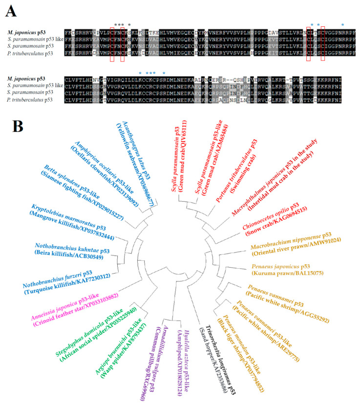
The partial sequence data of the Mjp53 gene were obtained from the GS-FLX transcriptome database of M. japonicus. Mjp53 was 477 bp long, including an open reading frame of 159 amino acids (Figure 1A). The alignment of the Mjp53 amino acid sequence with those of other crabs revealed that residues at functional sites, including DNA-binding sites, zinc-binding sites, and dimerization sites, are conserved (Figure 1A). This implies that the partial sequence from M. japonicus is a predicted p53. The deduced amino acid sequence of Mjp53 was 70% and 67% homologous to that of Scylla paramamosain (QDO16138) and Portunus trituberculatus (AZM65484), respectively. At the nucleotide level, there was no similarity with p53 or p53-like genes of other crabs. Phylogenetic analysis placed the Mjp53 sequence in the same clade with p53 homologs of other crabs (Figure 1B). In crustacean species, the Mjp53 gene formed a cluster with p53-homologous genes from S. paramamosain, P. trituberculatus, and Chionoecetes opilio. Another clade was composed of p53 homologs involving shrimps and prawns. Fish species formed one large clade with p53 and p53-like genes of various fishes.
Figure 1.
Characterization of Macrophthalmus japonicus p53 gene. (A) ClustalW multiple-sequence alignment of the deduced Mjp53 gene sequence with homologous p53 genes of various crabs. Shaded marks in black indicated completely conserved residues in all species. Dimerization site (polypeptide-binding site) and zinc-binding site (ion-binding site) are indicated by black asterisk mark and blue asterisk mark, respectively. DNA-binding site (nucleic-acid-binding site) is presented as a red rectangular box. (B) Phylogenetic circle tree of Mjp53 gene with other p53s. Neighbor-joining analysis showed a circle tree for phylogenetic relationships in p53 amino acid sequences using the MEGA v.4.0 software. Bootstrap values represent 1000 replicates.
3.2. Tissue Distribution of Mjp53 Expression
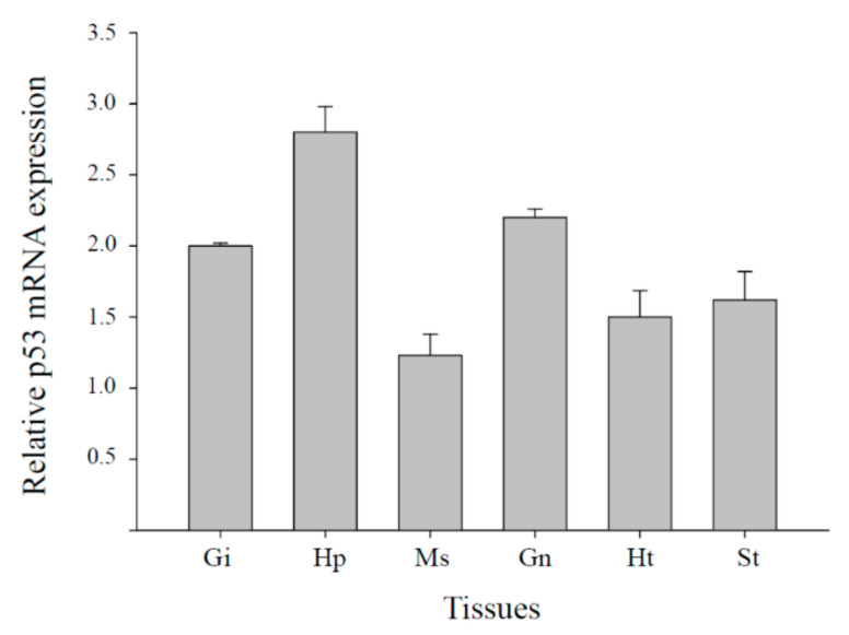
To investigate tissue-specific expression patterns, we measured Mjp53 mRNA expression in six tissues (gills, hepatopancreas, muscle, gonad, heart, and stomach) using real-time RT-qPCR. As shown in Figure 2, high levels of Mjp53 gene expression were observed in hepatopancreas tissues, whereas relatively low levels of Mjp53 mRNA expression were observed in muscle. Mjp53 gene expression was detected in all tested tissues.
Figure 2.
Basal transcriptional levels of M. japonicus p53 genes in various tissues (Gi, gills; Hp, hepatopancreas; Ht, heart; Gn, gonad; Ms, muscle; and St, stomach). All data are indicated as means ± standard deviation. Expression level of GAPDH transcripts was used for normalization of the relative transcriptional levels in each tissue from 10 crabs. The experiment was repeated three times.
3.3. Catalase Gene Expression in Oxidative Stress Responses to Exposure of HBCD or BDE-47
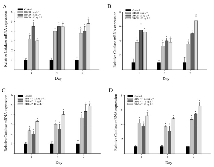
M. japonicus catalase expression was significantly induced in the gill and hepatopancreas in response to all concentrations of HBCD or BDE-47 tested (Figure 3). After HBCD exposure, significant expression of the catalase gene (p < 0.05) was continuously observed from day 1 to day 7 (Figure 3A). In the hepatopancreas, on day 1, catalase mRNA expression was significantly upregulated in M. japonicus at all concentrations of HBCD (p < 0.05). The increase in catalase gene expression was decreased by day 4 compared to the level on day 1, although catalase was more upregulated in HBCD-exposed groups than in the non-exposed control group (Figure 3B). Furthermore, HBCD exposure triggered a significant induction of catalase gene expression on day 7 (p < 0.05) in a dose-dependent manner. Upon HBCD exposure, the highest expression of catalase was observed at a relatively high concentration of 100 µg L−1 HBCD (elevated 4.8-fold in gills and 6.8-fold in hepatopancreas) on day 7.
Figure 3.
Relative transcriptional levels of catalase gene in M. japonicus gills (A,C) and hepatopancreas (B,D) after exposures to 1, 10, and 100 μg L−1 HBCD (A,B) and 0.1, 1, and 10 μg L−1 BDE-47 (C,D). Exposure periods were days 1, 4, and 7. GAPDH levels were used for normalization of the values. All values are indicated as mean ± SD. Statistically significant differences are presented by an asterisk mark (* p < 0.05 and ** p < 0.01) compared with the relative control value (catalase = 1).
After BDE-47 exposure, the significant upregulation of catalase gene expression was observed in M. japonicus gill tissue (Figure 3C). Catalase gene expression patterns in response to the range of HBCD concentrations were similar on day 1 and day 4. A significant increase in catalase mRNA was identified in gill tissue for all concentrations of BDE-47 and exposure times (p < 0.05) (Figure 3C) in an exposure time-dependent manner. In the hepatopancreas, catalase gene expression was also significantly increased on day 1 after BDE-47 exposure (p < 0.05) (Figure 3D). The level of catalase gene expression decreased slightly on day 4. The expression of catalase was significantly induced on day 7 in a dose-dependent manner. The U-shaped pattern of catalase gene expression was only observed in the hepatopancreas of M. japonicus exposed to both HBCD and BDE-47 POPs. The highest expression of catalase was observed at the relatively high concentration of 10 µg L−1 BDE-47 on day 7 in gills (4.9-fold) and the hepatopancreas (6.5-fold) (p < 0.05).
3.4. Mjp53 Gene Expression Responses to HBCD or BDE-47 Exposure
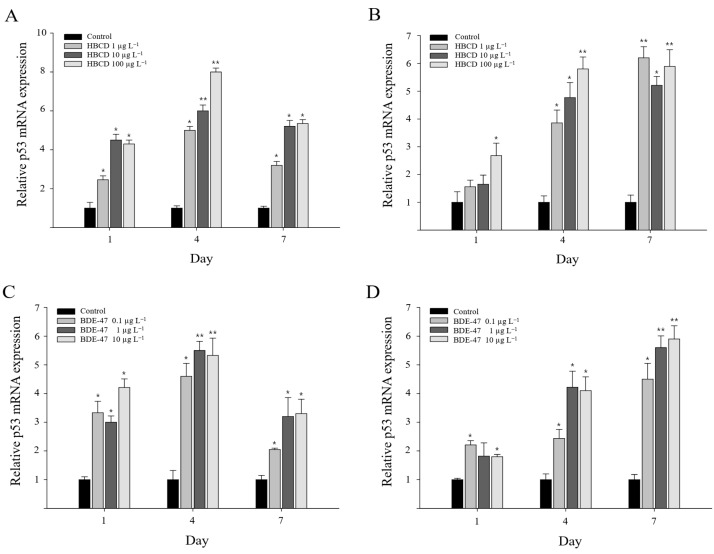
Mjp53 expression in response to POP (HBCD and BDE-47) exposure was also studied in gills and the hepatopancreas. On day 1, Mjp53 expression was significantly increased in gills after HBCD exposure (p < 0.05) (Figure 4A). A significant increase in Mjp53 gene expression was induced in gill tissue by exposure to all concentrations of HBCD on day 4 (p < 0.01). Its expression pattern was dose-dependent. Mjp53 expression continuously increased in the gills of M. japonicus crabs exposed to HBCD, although expressional levels were lower on day 7 than on day 4. In the hepatopancreas, the expression of Mjp53 mRNA on day 4 was significantly upregulated at all concentrations of HBCD in a dose-dependent manner, whereas Mjp53 gene expression was only increased at the high concentration of 100 µg L−1 HBCD on day 1 (Figure 4B). By day 7, the upregulation of Mjp53 was observed to be higher in the hepatopancreas than in gills. On HBCD exposure, the highest expression of Mjp53 was found after exposure to 100 µg L−1 HBCD (8.1-fold) on day 4 in gills and 1 µg L−1 HBCD (6.2-fold) on day 7 in the hepatopancreas (p < 0.01).
Figure 4.
Relative transcriptional levels of p53 gene in M. japonicus gills (A,C) and hepatopancreas (B,D) after exposures to 1, 10, and 100 μg L−1 HBCD (A,B) and 0.1, 1, and 10 μg L−1 BDE-47 (C,D). Exposure periods were days 1, 4, and 7. GAPDH levels were used for normalization of the values. All values are indicated as mean ± SD. Statistically significant differences are presented by an asterisk mark (* p < 0.05 and ** p < 0.01) compared with the relative control value (p53 = 1).
BDE-47 exposure also induced the upregulation of Mjp53 transcripts in gills and hepatopancreas of M. japonicus crabs (Figure 4C,D). On day 1, Mjp53 gene expression was significantly elevated in gills after exposure to all concentrations of BDE-47. A statistically significant increase in Mjp53 mRNA abundance was identified in gill tissue at all concentrations of BDE-47 and for all exposure times (p < 0.05) (Figure 4C). The highest expression of Mjp53 mRNA was observed at the relatively high concentrations of 1 and 10 µg L−1 BDE-47 on day 4 in gills. In the hepatopancreas, a significant induction of Mjp53 gene expression was observed with 0.1 and 10 µg L−1 BDE-47 on day 1. Mjp53 gene expression continuously increased through days 4 and 7 after BDE-47 exposure. The increase in Mjp53 transcript levels on day 7 was dose-dependent. The highest Mjp53 gene expression was found at the relatively high concentration of 10 µg L−1 BDE-47 (5.9-fold) on day 7 in the hepatopancreas (p < 0.01). The POP-exposed groups exhibited significant increases in their Mjp53 expression, indicating that the Mjp53 gene may be involved in POP-induced apoptotic responses.
4. Discussion
Due to their high flame retardant efficiency and good thermal stability, HBCD and BDE-47 are widely used in polystyrene foam and textiles [1,2,23]. POPs such as HBCD and BDE-47 continue to threaten aquatic environments and cause concern as serious global pollutants [2]. However, risk assessments are restricted by poor knowledge of the distribution and quantity of these substances in aquatic environments [24]. BDE-47 toxicity induces major perturbances in terms of the reproductive, immune, and neuronal systems of aquatic organisms [25,26]. The presence of HBCD also causes oxidative damage to the thyroid system, impacts neurodevelopment, and disrupts the endocrine system [1,20,27]. However, there are insufficient studies surrounding the potential effects of POPs such as HBCD and BDE-47 in p53-related apoptotic responses of marine invertebrates, including M. japonicus mud crabs.
In the present study, we provided supporting evidence for functional defense by apoptotic responses to HBCD or BDE-47 toxicity through the upregulation of Mjp53 transcription. M. japonicus, an intertidal mud crab, is an indicator species which is able to reflect sediment toxicity conditions through POP accumulation in marine environments. Exposure to POPs such as HBCD and BDE-47 significantly increased transcription levels of p53 in gills and hepatopancreas over all exposure periods. These results represent the first report of the induction of apoptosis through activated p53 transcript signaling in response to HBCD and BDE-47 toxicity in crabs, although there are some limited data regarding apoptotic responses induced by increasing p53 levels in crabs after exposure to ultraviolet radiation, Vibrio infection, and nitrites [28,29,30,31]. Our results are consistent with those concerning M. japonicus crabs in a study that indicated HBCD exposure causes oxidative DNA damage, thereby triggering the activation of p53 transcription in the marine copepod T. japonicus and medaka fish (O. melastigma) [15,20]. In addition, BDE-47 induces oxidative-stress-mediated DNA damage with the transcriptional regulation of the apoptosis-related p53 gene, resulting in developmental retardation in T. japonicus [3]. The tumor suppressor protein p53, as a “Guardian of the Genome”, plays an important role in cell cycle arrest, DNA repair, apoptosis, and genetic stability [12,31]. Concerning p53 gene functions, elevated p53 transcription might be related to the regulation of multiple cellular processes, including metabolism, antioxidant responses, and DNA repair, finally resulting in the modulation of development or decreased survival in M. japonicus crabs.
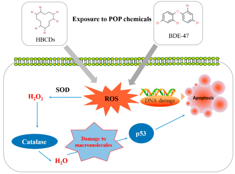
The hepatopancreas is the largest organ of the digestive tract and represents the main metabolic organ in crustaceans [32]. It serves for the absorption and metabolization of nutrients, storage of energy and minerals, detoxification, digestive functions, oxygen transport, and in immune defense [32,33]. In our study, the basal levels of p53 transcription were highest in M. japonicus hepatopancreas. p53 gene expression is also high in hepatopancreas tissue of the Chinese mitten crab Eriocheir sinensis [12]. Our results indicated different response patterns involving Mjp53 gene expression to HBCD and BDE-47 toxicity in gills and hepatopancreas tissue. After HBCD and BDE-47 exposure, significant p53 gene expression changes were observed with similar patterns on day 4 in both tested tissues. However, p53 transcriptional levels were significantly more upregulated in the hepatopancreas compared to gills exposed to HBCD or BDE-47 during long-term exposure on day 7. Environmental pollutants induce detoxification mechanisms or engage long-lasting functions involving cellular protection against antioxidants and for detoxification to diminish oxidative stress induced by exposure to POPs in the mud crab M. japonicus (Figure 5).
Figure 5.
Schematic summary of the suggested molecular process involving antioxidation and p53-mediated apoptosis in M. japonicus animals exposed to POPs (HBCD and BDE-47). SOD: Superoxide dismutase, ROS: Reactive oxygen species.
5. Conclusions
In this study, we provided transcriptional responses of p53 and catalase genes in M. japonicus crabs exposed to POPs such as HBCD or BDE-47. We characterized a partial sequence of the Mjp53 involving protein dimerization and DNA- and zinc-binding sites. The basal expression level of the p53 gene was high in the hepatopancreas among six tissues. HBCD or BDE-47 exposures induced an increase in antioxidant catalase gene expressions in the gills and hepatopancreas of M. japonicus. The highest expressions of p53 and catalase genes were generally observed at the relatively high concentrations of 100 µg L−1 HBCD and 10 µg L−1 BDE-47 on day 7. The BDE-47 exposure in gills was correlated with a linear increase in the catalase gene expression. In addition, the BDE-47 exposure in the hepatopancreas was correlated with a linear increase in the p53 gene expression in an exposure time-dependent manner. The result indicated the induction of oxidative stress via ROS production to POP exposure. The POPs-induced oxidative stress finally boosted apoptosis responses. The transcriptional level of the apoptotic p53 gene was significantly increased in the hepatopancreas and gills of M. japonicus after exposures to HBCD or BDE-47 for all exposure periods. These results suggest that apoptosis and DNA damage via long-lasting oxidative stress are found in the gills and hepatopancreas of M. japonicus after exposures to POPs such as HBCD or BDE-47.
Author Contributions
Conceptualization, K.P. and I.-S.K.; methodology, K.P. and I.-S.K.; software, K.P.; validation, K.P. and I.-S.K.; formal analysis, K.P. and I.-S.K.; investigation, K.P. and I.-S.K.; resources, K.P. and I.-S.K.; data curation, K.P. and I.-S.K.; writing—original draft preparation, K.P.; writing—review and editing, K.P. and I.-S.K.; visualization, K.P. and I.-S.K.; supervision, I.-S.K.; project administration, I.-S.K.; funding acquisition, I.-S.K. All authors have read and agreed to the published version of the manuscript.
Funding
This study was supported by the National Research Foundation of Korea, South Korea, which is funded by the Korean Government [NRF-2018-R1A6A1A-03024314], [NRF-2020-R1A2C-1013936].
Institutional Review Board Statement
The study was conducted according to the guidelines of the Chonnam National University Institutional Animal Care and Use Committee. The date of approval for the animal experiment was 20 October 2019 (ethical code: CNUIACUC-YS-2019-7C).
Informed Consent Statement
Not applicable.
Data Availability Statement
The data presented in this study are available on request from the corresponding author. The data are not publicly available due to reasons of privacy.
Conflicts of Interest
The authors declare no conflict of interest.
Footnotes
Publisher’s Note: MDPI stays neutral with regard to jurisdictional claims in published maps and institutional affiliations.
References
- 1.Wang X., Sun R., Chen Y., Zhang X., Cui Z. Temporal-spatial distribution and diastereoisomer pattern of hexabromocyclododecane in the vicinity of a chemical plant. J. Environ. Sci. 2019;82:203–212. doi: 10.1016/j.jes.2019.03.010. [DOI] [PubMed] [Google Scholar]
- 2.Liu Y., Feng Y., Li J., Zhou D., Guo R., Ji R., Chen J. The bioaccumulation, elimination, and trophic transfer of BDE-47 in the aquatic food chain of Chlorella pyrenoidosa-Daphnia magna. Environ. Pollut. 2020;258:113720. doi: 10.1016/j.envpol.2019.113720. [DOI] [PubMed] [Google Scholar]
- 3.Han J., Won E.J., Lee M.C., Seo J.S., Lee S.J., Lee J.S. Developmental retardation, reduced fecundity, and modulated expression of the defensome in the intertidal copepod Tigriopus japonicus exposed to BDE-47 and PFOS. Aquat. Toxicol. 2015;165:136–143. doi: 10.1016/j.aquatox.2015.05.022. [DOI] [PubMed] [Google Scholar]
- 4.Saquib Q., Siddiqui M.A., Ahmed J., Al-Salim A., Ansari S.M., Faisal M., Al-Khedhairy A.A., Musarrat J., AlWathnani H.A., Alatar A.A., et al. Hazards of low dose flame-retardants (BDE-47 and BDE-32): Influence on transcriptome regulation and cell death in human liver cells. J. Hazard. Mater. 2016;308:37–49. doi: 10.1016/j.jhazmat.2016.01.025. [DOI] [PubMed] [Google Scholar]
- 5.Gandhi N., Gewurtz S.B., Drouillard K.G., Kolic T., Macpherson K., Reiner E.J., Bhavsar S.P. Polybrominated diphenyl ethers (PBDEs) in Great Lakes fish: Levels, patterns, trends and implications for human exposure. Sci. Total Environ. 2017;576:907–916. doi: 10.1016/j.scitotenv.2016.10.043. [DOI] [PubMed] [Google Scholar]
- 6.Zhuang J., Pan Z.J., Li M., Hong F.S., Zhu C.K., Wu N., Chang G., Wang H., Zhao X.X. BDE-47 induced apoptosis in zebrafish embryos through mitochondrial ROS-mediated JNK signaling. Chemosphere. 2020;258:127385. doi: 10.1016/j.chemosphere.2020.127385. [DOI] [PubMed] [Google Scholar]
- 7.Chang C.J., Terrell M.L., Marcus M., Marder M.E., Panuwet P., Ryan P.B., Pearson M., Barton H., Barr D.B. Serum concentrations of polybrominated biphenyls (PBBs), polychlorinated biphenyls (PCBs) and polybrominated diphenyl ethers (PBDEs) in the Michigan PBB Registry 40 years after the PBB contamination incident. Environ. Int. 2020;137:105526. doi: 10.1016/j.envint.2020.105526. [DOI] [PMC free article] [PubMed] [Google Scholar]
- 8.Barghi M., Shin E.S., Son M.H., Choi S.D., Pyo H., Chang Y.S. Hexabromocyclododecane (HBCD) in the Korean food basket and estimation of dietary exposure. Environ. Pollut. 2016;213:268–277. doi: 10.1016/j.envpol.2016.02.026. [DOI] [PubMed] [Google Scholar]
- 9.Dong H., Lu G., Yan Z., Liu J., Yang H., Nkoom M. Bioconcentration and effects of hexabromocyclododecane exposure in crucian carp (Carassius auratus) Ecotoxicology. 2018;27:313–324. doi: 10.1007/s10646-018-1896-6. [DOI] [PubMed] [Google Scholar]
- 10.Zhang M., Zhao F., Zhang J., Shi J., Tao H., Ge H., Guo W., Liu D., Cai Z. Toxicity and accumulation of 6-OH-BDE-47 and newly synthesized 6,6′-diOH-BDE-47 in early life-stages of Zebrafish (Danio rerio) Sci. Total Environ. 2021;763:143036. doi: 10.1016/j.scitotenv.2020.143036. [DOI] [PubMed] [Google Scholar]
- 11.Zhang S., Kuang G., Zhao G., Wu X., Zhang C., Lei R., Xia T., Chen J., Wang Z., Ma R., et al. Involvement of the mitochondrial p53 pathway in PBDE-47-induced SH-SY5Y cells apoptosis and its underlying activation mechanism. Food Chem. Toxicol. 2013;62:699–706. doi: 10.1016/j.fct.2013.10.008. [DOI] [PubMed] [Google Scholar]
- 12.Hou C.C., Yang W.X. Characterization and expression pattern of p53 during spermatogenesis in the Chinese mitten crab Eriocheir sinensis. Mol. Biol. Rep. 2013;40:1043–1051. doi: 10.1007/s11033-012-2145-3. [DOI] [PubMed] [Google Scholar]
- 13.Qiu N., Wang R., Sun Y., Wang X., Jiang D., Meng Y., Zhou F. Toxic effects and mechanism of 2,2′,4,4′-tetrabromodiphenyl ether (BDE-47) on Lemna minor. Chemosphere. 2018;193:711–719. doi: 10.1016/j.chemosphere.2017.10.148. [DOI] [PubMed] [Google Scholar]
- 14.Deng J., Yu L., Liu C., Yu K., Shi X., Yeung L.W.Y., Lam P.K.S., Wu R.S.S., Zhou B. Hexabromocyclododecane-induced developmental toxicity and apoptosis in zebrafish embryos. Aquat. Toxicol. 2009;93:29–36. doi: 10.1016/j.aquatox.2009.03.001. [DOI] [PubMed] [Google Scholar]
- 15.Hong H., Li D., Shen R., Wang X., Shi D. Mechanisms of hexabromocyclododecanes induced developmental toxicity in marine medaka (Oryzias melastigma) embryos. Aquat. Toxicol. 2014;152:173–185. doi: 10.1016/j.aquatox.2014.04.010. [DOI] [PubMed] [Google Scholar]
- 16.Chon H.S., Ohandja D.G., Voulvoulis N. The role of sediments as a source of metals in river catchments. Chemosphere. 2012;88:1250–1256. doi: 10.1016/j.chemosphere.2012.03.104. [DOI] [PubMed] [Google Scholar]
- 17.Minor M.L., Mullarney J.C., Pilditch C.A., Huhn K. Crab burrow aspect ratio influences particle capture rates on intertidal sandflats. Geo-Maine Lett. 2020;40:197–216. doi: 10.1007/s00367-019-00630-x. [DOI] [Google Scholar]
- 18.Park K., Kim W.S., Choi B., Kwak I.S. Expression levels of the immune-related p38 mitogen-activated protein kinase transcript in response to environmental pollutants on Macrophthalmus japonicus crab. Genes. 2020;11:958. doi: 10.3390/genes11090958. [DOI] [PMC free article] [PubMed] [Google Scholar]
- 19.Hwang D.S., Lee J.S., Rhee J.S., Han J., Lee Y.M., Kim I.C., Park G.S., Lee J., Lee J.S. Modulation of p53 gene expression in the intertidal copepod Tigriopus japonicus exposed to alkylphenols. Mar. Environ. Res. 2010;69:S77–S80. doi: 10.1016/j.marenvres.2009.12.002. [DOI] [PubMed] [Google Scholar]
- 20.Shi D., Lv D., Liu W., Shen R., Li D., Hong H. Accumulation and developmental toxicity of hexabromocyclododecanes (HBCDs) on the marine copepod Tigriopus japonicus. Chemosphere. 2017;167:155–162. doi: 10.1016/j.chemosphere.2016.09.160. [DOI] [PubMed] [Google Scholar]
- 21.Park K., Kim W.S., Kwak I.S. Endocrine-disrupting chemicals impair the innate immune prophenoloxidase system in the intertidal mud crab, Macrophthalmus japonicus. Fish Shellfish Immunol. 2019;87:322–332. doi: 10.1016/j.fsi.2019.01.025. [DOI] [PubMed] [Google Scholar]
- 22.Park K., Nikapitiya C., Kwak I.S. Identification and expression of proteolysis response genes for Macrophthalmus japonicus exposure to irgarol toxicity. Ann. Limnol. Int. J. Limnol. 2016;52:65–74. doi: 10.1051/limn/2015023. [DOI] [Google Scholar]
- 23.Chai M., Li R., Cong S. Contamination of polybrominated diphenyl ethers (PBDEs) in urban mangroves of Southern China. Sci. Total Environ. 2019;646:390–399. doi: 10.1016/j.scitotenv.2018.07.278. [DOI] [PubMed] [Google Scholar]
- 24.Windsor F.M., Pereira M.G., Tyler C.R., Ormerod S.J. River organisms as indicators of the distribution and sources of persistent organic pollutants in contrasting catchments. Environ. Pollut. 2019;255:113144. doi: 10.1016/j.envpol.2019.113144. [DOI] [PubMed] [Google Scholar]
- 25.Liu Y., Guo R., Tang S., Zhu F., Zhang S., Yan Z., Chen J. Single and mixture toxicities of BDE-47, 6-OH-BDE-47 and 6-MeO-BDE-47 on the feeding activity of Daphnia magna: From behavior assessment to neurotoxicity. Chemosphere. 2018;195:542. doi: 10.1016/j.chemosphere.2017.12.045. [DOI] [PubMed] [Google Scholar]
- 26.Tanaka Y., Fujiwara M., Shindo A., Yin G., Teraoka H. Aroclor 1254 and BDE-47 inhibit dopaminergic function manifesting as changes in locomotion behaviors in zebrafish embryos. Chemosphere. 2018;193:1207–1215. doi: 10.1016/j.chemosphere.2017.11.138. [DOI] [PubMed] [Google Scholar]
- 27.Darnerud P.O. Toxic effects of brominated flame retardants in man and in wildlife. Environ. Int. 2003;29:841–853. doi: 10.1016/S0160-4120(03)00107-7. [DOI] [PubMed] [Google Scholar]
- 28.Miguel N.C.O., Wajsenzon I.J.R., Takiya C.M., Andrade L.R., Tortelote G.G., Einicker-Lamas M., Allodi S. Catalase, Bax and p53 expression in the visual system of the crab Ucides cordatus following exposure to ultraviolet radiation. Cell Tissue Res. 2007;329:159–168. doi: 10.1007/s00441-007-0410-x. [DOI] [PubMed] [Google Scholar]
- 29.Hollmann G., Linden R., Giangrande A., Allodi S. Increased p53 and decreased p21 accompany apoptosis induced by ultraviolet radiation in the nervous system of a crustacean. Aquat. Toxicol. 2016;173:1–8. doi: 10.1016/j.aquatox.2015.12.025. [DOI] [PubMed] [Google Scholar]
- 30.Cheng C.H., Su Y.L., Ma H.L., Deng Y.Q., Feng J., Chen X.L., Jie Y.K., Guo Z.X. Effect of nitrite exposure on oxidative stress, DNA damage and apoptosis in mud crab (Scylla paramamosain) Chemosphere. 2020;239:124668. doi: 10.1016/j.chemosphere.2019.124668. [DOI] [PubMed] [Google Scholar]
- 31.Cheng C.H., Ma H.L., Deng Y.Q., Feng J., Jie Y.K., Guo Z.X. Oxidative stress, cell cycle arrest, DNA damage and apoptosis in the mud crab (Scylla paramamosain) induced by cadmium exposure. Chemosphere. 2021;263:128277. doi: 10.1016/j.chemosphere.2020.128277. [DOI] [PubMed] [Google Scholar]
- 32.Vogt G. Functional cytology of the hepatopancreas of decapod crustaceans. J. Morphol. 2019;280:1405–1444. doi: 10.1002/jmor.21040. [DOI] [PubMed] [Google Scholar]
- 33.Yang J., Sun H., Qian Y., Yang J. Impairments of cadmium on vitellogenin accumulation in the hepatopancreas of freshwater crab Sinopotamon henanense. Environ. Sci. Pollut. Res. Int. 2017;24:18160–18167. doi: 10.1007/s11356-017-9491-6. [DOI] [PubMed] [Google Scholar]
Associated Data
This section collects any data citations, data availability statements, or supplementary materials included in this article.
Data Availability Statement
The data presented in this study are available on request from the corresponding author. The data are not publicly available due to reasons of privacy.


