Figure 6.
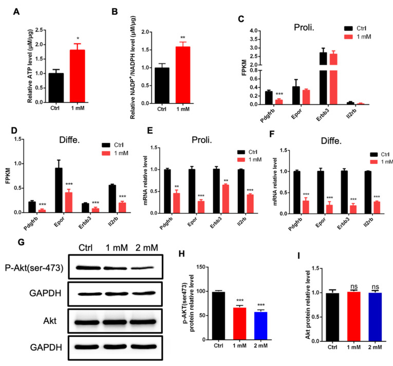
NAD+ exposure alters the PI3k-Akt signaling pathway. (A) ELISA results showing the increased level of ATP induced by NAD+ exposure compared to Ctrl group. Proliferating aNSPCs were cultured with 1 mM NAD+ supplemented for 48 h. Data are presented as the mean ± SEM. n = 3 independent experiments, unpaired t-test. (B) NAD+ exposure increased the levels of NADP+ and NADPH. Proliferating aNSPCs were cultured with 1 mM NAD+ supplemented for 48 h. Data are presented as the mean ± SEM. n = 3 independent experiments, unpaired t-test. (C) FPKM values of multiple genes relating to PI3k-Akt signaling pathway (proliferating condition). (D) FPKM values of multiple genes relating to PI3k-Akt signaling pathway (differentiation condition). (E) The relative mRNA levels of Pdgfrb, Epor, Erbb3 and Il2rb in aNSPCs treated with NAD+ (proliferating condition). Data are presented as the mean ± SEM. n = 3 independent experiments, unpaired t-test. (F) The relative mRNA levels of Pdgfrb, Epor, Erbb3, Il2rb in aNSPCs treated with NAD+ (differentiation condition). Data are presented as the mean ± SEM. n = 3 independent experiments, unpaired t-test. (G–I) Western blot assay and the quantification results show that NAD+ exposure significantly decreased the level of p-Akt (ser473) while the level of total Akt was not affected. GAPDH was used as an internal control. Data are presented as the mean ± SEM. n = 3 independent experiments, unpaired t-test; *, p < 0.05; **, p < 0.01; ***, p < 0.001.

