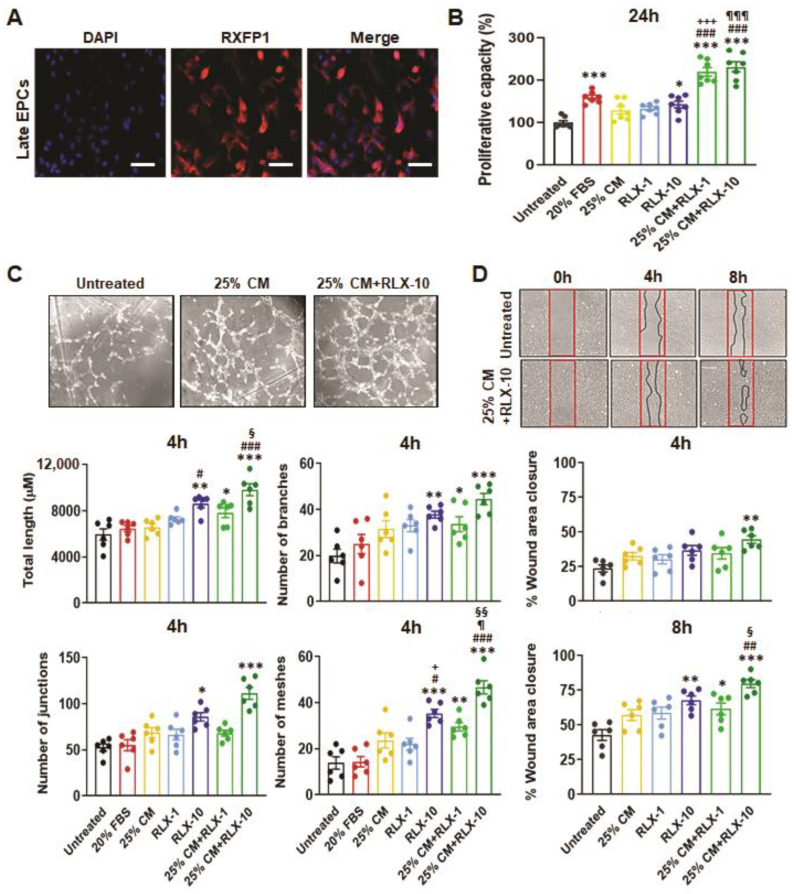
Figure 1.
The effects of BM-MSC-derived CM and/or RLX on healthy patient-derived EPC viability/proliferation, angiogenesis and wound healing. CD34+ late EPCs from the blood of healthy male patients were found to express the cognate RLX receptor, RXFP1; scale bar = 50 µm (A). EPC proliferation after 24 hours (24 h) from n = 7 patient-derived EPCs per group (B); EPC-mediated tube formation (determined from total tube length, number of branches, number of junctions and number of meshes) after 4 h from n = 6 patient-derived EPCs per group (C) and EPC-mediated wound healing after 4 h and 8 h from n = 6 patient-derived EPCs per group (D), was determined in response to 20% fetal bovine serum (FBS; positive control), 25% BM-MSC-CM (25% CM), RLX [1 ng/mL (RLX-1) or 10 ng/mL (RLX-10)] or the combined effects of 25% CM and RLX-1 or RLX-10. Complete wound closure was achieved by untreated EPCs by 24 h. Representative images of capillary tube formation from untreated EPCs and EPCs treated with 25% CM alone or in combination with RLX-10 (C) and of wound closure from untreated EPCs and those treated with 25% CM+RLX-10 (D) are shown. The coloured circles in each of the columns of each bar graph represent individual data points from each group. Data were analysed using a one-way ANOVA followed by a Tukey’s post-hoc test to allow for multiple comparisons between groups. * p < 0.05, ** p < 0.01, *** p < 0.001 vs. untreated cells; # p < 0.05, ## p < 0.01, ### p < 0.001 vs. 25% CM-treated cells, + p < 0.05, +++ p < 0.001 vs. RLX-1-treated cells; ¶ p < 0.05, ¶¶¶ p < 0.001 vs. RLX-10-treated cells; § p < 0.05, §§ p < 0.01 vs. 25% CM+RLX-1-treated cells.

