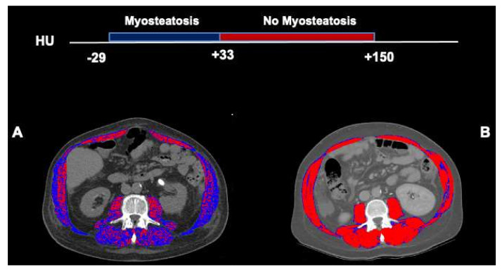
Figure 1.
Abdominal computed tomography images taken at the 3rd. lumbar vertebra to quantify muscle radiodensity in patients with cirrhosis. Comparison of two male patients with cirrhosis and similar skeletal muscle index (58 cm2/m2). Skeletal muscle areas with high radiodensity (33 to 150) are shown in red, and low radiodensity muscle (−29 to 32) is shown in dark blue. In a patient with low mean muscle radiodensity (24 HU) or myosteatosis (A), the majority of the muscle areas are composed of the low attenuation muscle whereas, in a patient with normal muscle radiodensity (51 HU, no-myosteatosis), areas with the normal attenuation range are predominant (B).

