Abstract
(1) Background: In recent years, several studies have described various and heterogenous methods to sensitize nanoparticles (NPs) to pH changes; therefore, in this current scoping review, we aimed to map current protocols for pH functionalization of NPs and analyze the outcomes of drug-loaded pH-functionalized NPs (pH-NPs) when delivered in vivo in tumoral tissue. (2) Methods: A systematic search of the PubMed database was performed for all published studies relating to in vivo models of anti-tumor drug delivery via pH-responsive NPs. Data on the type of NPs, the pH sensitization method, the in vivo model, the tumor cell line, the type and name of drug for targeted therapy, the type of in vivo imaging, and the method of delivery and outcomes were extracted in a separate database. (3) Results: One hundred and twenty eligible manuscripts were included. Interestingly, 45.8% of studies (n = 55) used polymers to construct nanoparticles, while others used other types, i.e., mesoporous silica (n = 15), metal (n = 8), lipids (n = 12), etc. The mean acidic pH value used in the current literature is 5.7. When exposed to in vitro acidic environment, without exception, pH-NPs released drugs inversely proportional to the pH value. pH-NPs showed an increase in tumor regression compared to controls, suggesting better targeted drug release. (4) Conclusions: pH-NPs were shown to improve drug delivery and enhance antitumoral effects in various experimental malignant cell lines.
Keywords: pH-responsive nanoparticles, drug delivery, cancer therapy, nanocarriers
1. Introduction
The advancements made in nanotechnology in recent years has led to an unprecedented interest in developing targeted therapies for cancer based on nanoparticles (NPs). NPs are defined as nano-sized particles with diameters ranging from 1 to 100 nm [1,2,3]. Although small, NPs have a large surface area and can be used as carriers for a wide range of peptides [4], antibodies [5], drugs [6], or contrast agents [7]. NPs are widely used as a platform for delivering drugs due to their stable high carrier capacity and their ability to accumulate in tumors through the enhanced permeation and retention effect (EPR) [8,9]. Because of the accelerated angiogenesis, tumors are supplied by immature blood vessels with a defective architecture with wide endothelial gaps through which molecules smaller than 700 nm can penetrate [10,11,12]. This characteristic represents the core which led to NPs becoming an important platform for research into cancer theranostics. Inversely, many tumors are heterogenous and possess a dense extracellular matrix which increases interstitial pressure by blocking the passive transport of NPs from the peritumoral vessels [9], which explains why NPs mostly accumulate in the peritumoral region but fail to penetrate the deep tumoral tissue in experimental applications.
Studies have described techniques to improve the penetration of NPs by using the tumor microenvironment as a targeting site for NPs. One of the constant distinct features of the tumoral microenvironment is the acidic pH, between 0.3 to 0.7 units lower than the pH of normal tissue [13]. Based on this trait, several studies have designed functionalized NPs, making them responsive to pH changes. Once the pH-functionalized NPs (pH-NPs) penetrate through the endothelium via the EPR effect, they respond to the acidic pH and may either disintegrate and release drugs or change their size and shape, thus enhancing their capacity to diffuse towards the tumors’ core. In recent years, several studies have described various and heterogenous methods to sensitize NPs to pH changes; thus, in this current scoping review, we aimed to map current protocols for pH functionalization and analyze the antitumoral outcomes of drug-loaded pH-NPs.
2. Materials and Methods
2.1. Literature Search and Study Selection
As previously described [14,15,16], a systematic search of the PubMed database was performed for all published studies relating to in vivo models of anti-tumor drug delivery via pH-responsive NPs using the following search algorithm: pH AND nanoparticles AND cancer AND delivery AND in vivo. The systematic search was carried out by adhering to the Preferred Reporting Items for Systematic Reviews and Meta-Analyses (PRISMA) guidelines which were adapted to experimental studies [17]. The PRISMA checklist was followed to conduct the methodology. Inclusion criteria were used according to the Problem/Population, Intervention, Comparison, and Outcome (PICO) formula (Table 1). All studies published in English from the 1st of January 2017 to the 31th of December 2021 describing drug-loaded pH-responsive NPs for targeted delivery in tumors were selected for full-text review. The experimental lot (population) consisted of pH-functionalized nanoparticles tested in vitro to assess pH responsiveness and in vivo to assess the antitumoral effects of pH-NPs loaded with chemotherapeutics. Embryos, cell cultures, tumor spheroids, and human studies were excluded. Nanogels or nano-emulsions were excluded. The intervention was defined as administration of pH-responsive conjugated NPs in tumor-bearing animals. Comparison criteria were further selected from subgroups of the included studies. Primary outcomes were tumor uptake of pH-NPs and tumor regression rate.
Table 1.
Overview of inclusion and exclusion criteria.
| Inclusion Criteria | Exclusion Criteria |
|---|---|
| Experimental studies | Clinical studies |
| Full text available in English | Full text not available/other language used |
| Testing of pH-NPs in vitro and in vivo (animal model) | In vitro/in vivo only |
| Descriptive data on type and synthesis of NPs | Type of NPs not named/method of synthesis not described |
| Descriptive data on pH functionalization method | No detailed data on how the NPs were functionalized |
| Data on animal model and malignant cell line used | No data on animal model/malignant cell line |
| pH-NPs used to deliver chemotherapeutics | Other use of pH-NPs (e.g., tumor imaging, hyperthermia) |
| Analysis of tumor uptake of pH-NPs and tumor regression | No data on tumoral response to pH-NPs |
| Detailed description of methodology (is the method reproducible?) | Methods not reproducible based on given data (requiring supplemental data from authors) |
| ARRIVE score ≥ 15 | ARRIVE score < 15 |
2.2. Data Analysis
The following data information regarding each included study was extracted: the author name, the year of publication, the type of NPs, the pH sensitization method, the in vivo model, the tumor cell line, the type and name of drug used for targeted therapy, the type of in vivo imaging, method of delivery, and the outcomes regarding the cellular uptake of NPs.
2.3. Quality Assessment
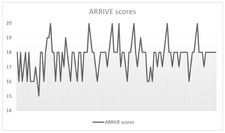
Two authors (SM and BCM) independently examined the title and abstract of citations, and the full texts of potentially eligible studies were obtained; disagreements were resolved by discussion. The Essential 10 ARRIVE guidelines were used to quantify the quality of included studies [18]. Each study was marked for each ARRIVE item with 0 if the data were lacking, 1 if the data were incomplete, and 2 if the data were complete; thus, the final score of each article could range from zero to a maximum of twenty. Only studies with a minimum ARRIVE score of 14 were included (Figure 1). The reference lists of retrieved papers were further screened for additional eligible publications.
Figure 1.
ARRIVE scores breakdown of included studies.
3. Results
3.1. Overview of Included Studies
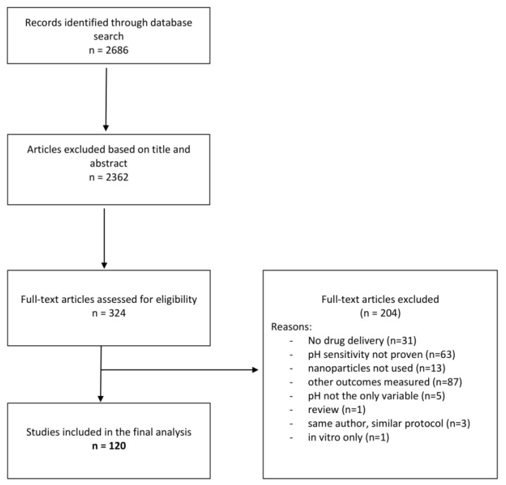
An initial search of PubMed database found 2686 articles. After triage of title and abstract, 324 full texts were assessed for inclusion. Records based on the title and abstract were excluded if they did not answer our research question: “Can pH functionalized NPs be used as drug carriers for targeted, in vivo, cancer therapy?”. Further, records were excluded if any of the exclusion criteria were obvious within the title or abstract. Eligible full texts were triaged according to the same principles (Table 1). The PRISMA flowchart shows a breakdown of excluded full texts (Figure 2). One hundred and twenty fully eligible manuscripts were included for in-depth analysis [19,20,21,22,23,24,25,26,27,28,29,30,31,32,33,34,35,36,37,38,39,40,41,42,43,44,45,46,47,48,49,50,51,52,53,54,55,56,57,58,59,60,61,62,63,64,65,66,67,68,69,70,71,72,73,74,75,76,77,78,79,80,81,82,83,84,85,86,87,88,89,90,91,92,93,94,95,96,97,98,99,100,101,102,103,104,105,106,107,108,109,110,111,112,113,114,115,116,117,118,119,120,121,122,123,124,125,126,127,128,129,130,131,132,133,134,135,136,137,138,139] (Table S1). Interestingly, 45.8% of studies (n = 55) used polymers to construct nanoparticles—either natural polymers (such as chitosan) or synthetic ones (Table 2 and Table 3). The most common pH sensitization method used acid-labile bounds (e.g., hydrazone, ester, imide) (Table 2, Table 3, Table 4, Table 5 and Table 6). BALB/c mice were part of the chosen experimental model in 98.3% (n = 118) of studies. pH-NPs were used in a wide array of malignancies, including breast carcinoma (40%, n = 48), hepatocarcinoma (14.1%, n = 17), lung cancer (11.6%, n = 14), colon carcinoma (6.6%, n = 8), cervical cancer (6.6%, n = 8), and melanoma cell lines (1.6%, n = 2) (Table 2, Table 3, Table 4, Table 5 and Table 6). Fluorescent imaging (70.8%, n = 85) and transmission electron microscopy (24.1%, n = 29) were used to quantify in vivo biodistribution of pH-NPs. Most studies (80.8%, n = 97) used control NPs which were not pH-sensitized to compare biodistribution and tumor penetration. Furthermore, almost all researchers (n = 119) compared cargo release from NPs in both physiological and acidic pH. Four studies proved that NPs increase in size when exposed to low pH, due to associated swelling and widening of membrane gaps, before drug release. The mean acidic pH value used in the current literature is 5.7 [5–6.8], which is significantly lower than that measured in tumor microenvironments, which can vary between 6.7 and 7.1, as previously reported.
Figure 2.
PRISMA flowchart.
Table 2.
Summary of methods used in studies.
|
Summary of Studies Overview of Common Methods |
||
|---|---|---|
| Type/Method | No. of Studies | |
| Type of NPs | Polymeric | 55 |
| Lipid | 12 | |
| MSN | 13 | |
| Metallic | 11 | |
| Other | 29 | |
| pH Sensitization Method | pH-labile linkers | 70 |
| pH-triggered structural changes | 35 | |
| pH-triggered hydrophobic to hydrophilic transition | 8 | |
| Other methods | 7 | |
| Cancer Model | Breast malignant cell lines (4T1, MCF-7, MDA-MB-231) |
48 |
| Cervical malignant cell lines (HeLA) |
8 | |
| Lung malignant cell lines (A549) |
14 | |
| Colorectal malignant cell lines (CT-26, HCT116, SW480) |
8 | |
| Liver malignant cell lines (H22, HepG2, SMMC 7721) |
17 | |
| Other | 25 | |
| Types of Chemotherapeutics | Doxorubicin | 69 |
| Paclitaxel | 9 | |
| Other | 42 | |
Table 3.
Overview of polymeric NPs: structure, pH sensitization method, tumor type, and delivered drug.
| First Author | Publication Year | Structure of NPs | pH Sensitization Method | Tumor Type | Drug |
|---|---|---|---|---|---|
| Adeyemi [19] | 2019 | FA-chitosan-PEG-polyethylenimine | pH-triggered structural changes | KYSE 30 scuamos cell carcinoma | Endostatin |
| Cao [21] | 2019 | TAT peptide-polyphosphoester | pH sensitive transactivator of transcription (TAT) | MDA-MB-231 breast carcinoma cell line | Doxorubicin |
| Chen [23] | 2018 | lactobionic acid-chitosan-lipoic acid | pH-labile amide linkers | HepG2 liver cancer | Doxorubicin |
| Chen [25] | 2020 | TPGS-HA polymer-PEG | hydrophobic to hydrophilic transition | PC3 prostate cancer | Docetaxel |
| Cheng [30] | 2019 | Poly(ortho ester urethanes) copolymers | pH-labile borate ester linkers | MCF-7 breast carcinoma cell line | Doxorubicin |
| Cheng [28] | 2018 | carboxymethyl chitosan | pH-labile hydrazone linkers | MCF-7 breast carcinoma cell line | Doxorubicin |
| Cui [31] | 2017 | transferrin-PEG | pH-labile hydrazone linkers | MCF-7 breast carcinoma cell line | Doxorubicin |
| Debele [32] | 2017 | PEG-methacrylamide-tocopheryl succinate-histidine | pH-labile imidazole linkers | HCT116 colon carcinoma | Doxorubicin |
| Deng [33] | 2019 | PEG-methylpropenoic acid-glycerol-cinnamaldehyde | pH-labile cinnamylaldehyde linkers | 4T1 breast carcinoma cell line | Doxorubicin |
| Du [36] | 2017 | PEG-PTTMA | PTTMA disassembly in acidic pH | HeLa cervival cancer | siRNA |
| Fan [39] | 2017 | polyethylenimine-PEG | pH-labile borate ester linkers | 4T1 breast carcinoma cell line | siRNA |
| Fang [40] | 2020 | chitosan-polysaccharide | pH-labile borate ester linkers | PANC-1 pancreatic cancer | Curcumin |
| Feng [41] | 2020 | PEG-PAH-DMA | pH-triggered structural changes | A549 NSLC cell line | Paclitaxel |
| Gao [44] | 2017 | poly (L-γ-glutamylcarbocistein-RBC membrane | pH-triggered structural changes | NCI-H460 cell line | Paclitaxel |
| Gibbens-Bandala [45] | 2019 | PLGA-polyvinyl alcohol | hydrophobic to hydrophilic transition | MDA-MB-231 breast carcinoma cell line | Paclitaxel |
| Gong [47] | 2018 | PEG-PPMT | hydrophobic to hydrophilic transition | CT-26 colon carcinoma | Docetaxel |
| Guo [49] | 2018 | PBLG-Sericin | pH-labile carboxyl linkers | A549 NSLC cell line | Methotrexate |
| Guo [51] | 2020 | DMA-PEG | pH-triggered structural changes | MCF-7 breast carcinoma cell line | Doxorubicin |
| Hong [53] | 2019 | U11 peptide-PLGA | pH-triggered structural changes | A549 NSLC cell line | Doxorubicin and Curcumin |
| Jin [57] | 2018 | PEI-PLA | pH-triggered structural changes | A549 NSLC cell line | Paclitaxel |
| Jung [58] | 2020 | PBA | pH-labile borate ester linkers | MG glioblastoma | Doxorubicin |
| Khan [61] | 2020 | PLGA | pH-triggered structural changes | MCF-7 breast carcinoma cell line | Doxorubicin |
| Kou [64] | 2017 | lactose myristoyl carboxymethyl chitosan | pH-triggered structural changes | Huh-7 hepatocellular carcinoma | Adriamycin |
| Lee [66] | 2018 | chitosan-PEG-acetyl histidine | pH-triggered structural changes | CT-26 Pulmonary Metastasis Model | Piperlongumine |
| Li [70] | 2018 | DGL-PEG-Tat-KK-DMA | pH-labile amide linkers | HepG2 liver cancer | Doxorubicin |
| Li [73] | 2020 | RGD-PEG-Arginine-SA | pH-labile hydrazone linkers | HN6 squamos cell carcinoma | GNA002 |
| Li [75] | 2021 | PDA-HA | pH-labile PDA coating | 4T1 breast carcinoma cell line | Cisplatin |
| Liu [79] | 2018 | polycarbonate-PEG | pH-labile acetal linkers | BT 474 breast carcinoma | Bortezomib |
| Luo [87] | 2021 | PEG-TAT-HA | pH-triggered structural changes | H22 hepatocellular carcinoma | Disulfiram |
| Mhatre [89] | 2021 | polydopamine | pH-triggered structural changes | MDA-MB-231 breast carcinoma cell line | Niclosamide |
| Palanikumar [96] | 2020 | ATRAM-BSA-PLGA | pH-labile ester bonds | MCF-7 breast carcinoma cell line | Doxorubicin |
| Qu [100] | 2018 | carboxymethyl chitosan | pH-labile phenylboronic acid pinacol ester | HepG2 liver cancer | Doxorubicin |
| Quadir [101] | 2017 | PEG-PPLG | pH-labile amine linkers | MCF-7 breast carcinoma cell line | Doxorubicin |
| Ray [102] | 2020 | PEG | pH-labile amine linkers | PANC-1 pancreatic cancer | Gemcitabine |
| Saravankumar [103] | 2019 | APT-PLGA-PVP-AS1411 aptamet | pH-triggered structural changes | A549 NSLC cell line | Doxorubicin |
| Shi [105] | 2018 | PEG-PLH | pH-labile PSD linker | A549 NSLC cell line | siRNA |
| Shi [106] | 2021 | PEG-PLL-DMA | pH-labile amide linkers | A549 NSLC cell line | siRNA |
| Soe [107] | 2019 | poloxamer-Tf-EDC-NHS | NR | MDA-MB-231 breast carcinoma cell line | Doxorubicin |
| Su [108] | 2020 | PEG-PMT | pH-labile tioether linkers | Colon26 cell line | Docetaxel |
| Wang [113] | 2017 | RGD-PLGA-PEG | pH-labile amine linkers | MCF-7 breast carcinoma cell line | Doxorubicin |
| Wang [115] | 2018 | chitosan-graphene oxide | pH-triggered structural changes (less electrostatic interaction | HepG2 liver cancer | Doxorubicin |
| Wei [118] | 2020 | PEG | pH-labile amine linkers (schiff base) | B16F10 melanoma | Doxorubicin |
| Xiong [122] | 2019 | TPGS-PEG | pH-labile hydrazone linkers | MCF-7 breast carcinoma cell line | Doxorubicin |
| Xu [123] | 2018 | DTPA-PEG-DMA | pH labine amine linkers | PC3 prostate cancer | Doxorubicin |
| Xu [124] | 2021 | chitosan | pH-labile ester linkers | HepG2 liver cancer | Doxorubicin |
| Yadav [125] | 2020 | RGD-chitosan-Cy5.5 | pH-labile amine linkers | MDA-MB-231 breast carcinoma cell line | Raloxifene |
| Yan [126] | 2017 | POEAd-galactose-LA | pH-labile ester linkers | HepG2 liver cancer | Doxorubicin |
| Yang [127] | 2018 | glycol Chitosan-PDPA | hydrophobic to hydrophilic transition (PDPA) | MCF-7 breast carcinoma cell line | Paclitaxel |
| Yu [128] | 2019 | PLGA-CPT-DMMA-PEI | pH-triggered structural changes | MCF-7 breast carcinoma cell line | Doxorubicin |
| Zhang [129] | 2017 | TPGS-MSN | pH-labile ester linkers | SMMC 7721 hepatocellular carcinoma | Doxorubicin |
| Zhang [131] | 2018 | DMA-Cystamine-PEG | pH-labile ester linkers | A549 NSLC cell line | Paclitaxel |
| Zhou [138] | 2020 | polyphosphazene | pH-labile hydrazone linkers | HeLa cervival cancer | Doxorubicin |
Legend: FA, folic acid; TPGS, tocopheryl polyethylene glycol 1000 succinate; HA, hyaluronic acid; PEG, polyethylene glycol; PTTMA, poly(2,4,6-trimethoxybenzylidene-1,1,1-tris(hydroxymethyl)ethane methacrylate; DMA, dimethylmaleic acid; PAH, polyallylamine; RBC, red blood cell; PLGA, poly(lactic-co-glycolic acid); PPMT, poly(o-pentadecalactone-co-N-methyldiethyleneamineco-3,30-thiodipropionate; PBLG, poly(c-benzyl-L-glutamate); U11 peptide, urokinase plasminogen activator receptor (uPAR) targeting peptide; PEI, polyethyleneimine; PLA, polylactic acid; PBA, phenylboronic acid; DGL, dendrigraft poly-L-lysine; TAT, tumor-associated antigens; RGD, arginine–glycine–aspartic peptide; DTPA, 3,3′-dithiodipropionic acid; Cy5.5, cyanine; SA, stearic acid; PDA, hydrochloride dopamine; ATRAM, acidity-triggered rational membrane peptide; BSA, bovine serum albumin; PPLG, poly (γ-propargyl L-glutamate); APT, aptamer; PVP, poly(N-vinylpyrrolidone); PLH, poly(L-histidine); PLL, poly-L-lysine; EDC, 1-ethyl-3-(3-dimethylaminopropyl)-carbodiimide hydrochloride; NHS, N-hydroxysuccinimide; PMT, poly(ω-pentadecalactone-co-N-methyldiethyleneaminesebacate-co-2,2’-thiodiethylene sebacate); DTPA, 3,3′-dithiodipropionic acid; POEAd, poly(ortho ester diamide); LA, lactobionic acid; PDPA, poly(2-(diisopropylamino)ethyl methacrylate); CPT, C18-PEG2000-TPP.
Table 4.
Overview of mesoporous silica NPs: structure, pH sensitization method, tumor type, and delivered drug.
| First Author | Publication Year | Structure of NPs | pH Sensitization Method | Tumor Type | Drug |
|---|---|---|---|---|---|
| Chen [24] | 2020 | MSN-citraconic-poly-L-lisine | acid-labile disulfide linkers | 4T1 breast carcinoma cell line | Doxorubicin |
| Cheng [27] | 2017 | Polydopamine-FA-PEG-MSN | pH-labile polydopamine coating | HeLa cervival cancer | Doxorubicin |
| Ding [34] | 2020 | MSN-carboxymethyl chitin-GRP78 peptide | pH-labile thioketal linkers | H22 hepatocellular carcinoma | Doxorubicin |
| Ding [35] | 2020 | MSN-lipidbilayer-TLS11a aptamer | pH-labile TAT peptide | 4T1 breast carcinoma cell line | Doxorubicin |
| Kundu [65] | 2020 | MSN-FA | pH-labile PAA linker | MCF-7 breast carcinoma cell line | Umbelliferone |
| Li [73] | 2020 | Gal-P123-MSN | pH-triggered structural changes (DC lipid) | Huh-7 hepatocellular carcinoma | Irinotecan |
| Li [68] | 2017 | DM1-MSN-PDA | pH-labile PDA coating | SW480 colorectal cancer cell line | EpCAM |
| Liao [76] | 2021 | Chitosan-MSN | pH-labile imidazole linkers | 4T1 breast carcinoma cell line | Doxorubicin |
| Liu [80] | 2019 | MSN | pH-labile calcium carbonate | LNCaP-AI prostate carcinoma | Doxorubicin |
| Mu [94] | 2017 | MSN-PLH-PEG | hydrophobic to hydrophilic transition | H22 hepatocellular carcinoma | Sorafenib |
| Saroj [104] | 2018 | MSN | pH-labile PAA linker | PC3 prostate cancer | Bicalutamide |
| Zhang [130] | 2017 | MSN-pH-responsive peptide | pH-responsive peptide | MCF-7 breast carcinoma cell line | Doxorubicin |
| Zhao [136] | 2018 | MSN-TPGS | pH-labile ester linkers | MCF-7 breast carcinoma cell line | Doxorubicin |
Legend: MSN, mesoporous silica nanoparticles; FA, folic acid; PEG, polyethylene glycol; GRP78P, glucose regulated protein 78 peptide; TAT, tumor-associated antigens; Gal, gala tosyl; DM1, maytansinoid conjugate; PDA, hydrochloride dopamine; PLH, D-alpha-tocopherol polyethylene glycol 1000-succinate; PAA, polyacrylic acid.
Table 5.
Overview of gold NPs: structure, pH sensitization method, tumor type, and delivered drug.
| First Author | Publication Year | Structure of NPs | pH Sensitization Method | Tumor Type | Drug |
|---|---|---|---|---|---|
| Aguilar [20] | 2021 | polycaffeic acid-FA-Au | pH-labile catechol-boronic acid linkers | SCC7 squamos cell carcinoma | Bortezomib |
| Essawy [38] | 2020 | Au-hydrazine | pH-labile hydrazone linkers | HBPC oral carcinoma | Doxorubicin |
| Guo [50] | 2018 | Au-Chitosan-AS1411 aptamer | pH-triggered structural changes | A549 lung cancer cell line | Methorexate |
| Kumar [63] | 2020 | Au | pH-labile peptide linker (Lys-Phe-Gly) | BT 474 breast carcinoma | Doxorubicin |
| Liu [81] | 2018 | Au-iron oxide-PEG | pH-labile oleic acid linkers | SGC-7901 gastric adenocarcinoma | Herceptin |
| Mahalunkar [91] | 2019 | Au-PVP-FA | pH-triggered structural changes | MCF-7 breast carcinoma cell line | Curcumin |
| Sun [110] | 2019 | Au-AS1411 aptamer | pH-triggered structural changes | HeLa cervival cancer | Doxorubicin |
Legend: FA, folic acid; Au, gold; PEG, polyethylene glycol; PVP, polyvinylpyrrolidone.
Table 6.
Overview of lipid-based NPs: structure, pH sensitization method, tumor type, and delivered drug.
| First Author | Publication Year | Structure of NPs | pH Sensitization Method | Tumor Type | Drug |
|---|---|---|---|---|---|
| Juang [59] | 2019 | lipid-PEG | pH-labile imide linkers | HCT116 colon carcinoma | Irinotecan and microRNA |
| Li [69] | 2017 | TF-PEG-GMS | pH-labile hydrazone linkers | A549/DTX lung cancer cell line | Docetaxel and Baicalin |
| Li [71] | 2019 | LDL-OA | pH-labile hydrazone linkers | 4T1 breast carcinoma cell line | Doxorubicin |
| Sun [111] | 2021 | DSPE-PEG | pH-triggered structural changes | LNCaP-AI prostate carcinoma | Doxorubicin |
| Tan [112] | 2017 | PAA-OA | pH-labile oleic acid linkers | A549 NSLC cell line | Erlotinib |
| Men [92] | 2020 | lipid-HA-PBAE | pH-triggered structural changes | A549 NSLC cell line | Doxorubicin |
| Cavalcante [22] | 2021 | DSPE-PEG-OA | pH-labile oleic acid linkers | 4T1 breast carcinoma cell line | Doxorubicin |
| Li [67] | 2017 | DSPE-PEG | pH-labile imine linkers | FTC-133 thyroid cancer | Doxorubicin |
| Lo [85] | 2020 | DSPE-PEG | pH-labile oleic acid linkers | SAS squamos carcinoma cell line | Daunorubicin and Irinotecan |
| Ma [90] | 2021 | DSPE-PEG | pH-triggered structural changes | HepG2 liver cancer | hydroxycamptothecin |
| Pang [98] | 2020 | lipid-polymeric NPs | pH-labile dihydrazide linkers | A549 NSLC cell line | Erlotinib |
| Xie [120] | 2018 | DSPE-PEG | pH-labile imine linkers | MCF-7 breast carcinoma cell line | Methotrexate |
Legend: PEG, polyethylene glycol; TF; transferrin; GMS, glyceryl monostearate; PAA, polyacrylic acid; HA, hyaluronic acid; PBAE, poly(b-amino ester; DSPE, 1,2-distearoyl-sn-glycero-3-phosphoethanolamine; OA, oleic acid.
3.2. Types of NPs Used
The sensitization of various NPs to acidic pH was measured. Those that were polymeric in nature were most common (Table 2 and Table 3); however, mesoporous silica nanoparticles (MSNPs) (Table 4), gold-based NPs (Table 5), or lipid-based NPs (Table 6) were other common options. Polymeric NPs were synthetized through emulsion–solvent evaporation methods or by nanoprecipitation. Polymers have the advantage of being biocompatible and biodegradable and can be designed to either incorporate drugs or simply attach drugs to their matrix via pH-labile linkers. Chitosan was commonly used to form nanocomposites because it is a positively charged biocompatible polymer with good stability in blood circulation which can form complexes with anionic peptides. Another way of using polymers in the design of pH-NPs is by coating the surface of other types of NPs to increase in vivo stability (e.g., PEGylated lipid NPs) (Table 6). Polyethylene glycol (PEG) is hydrophilic and biocompatible, thus coating the surface with PEG (e.g., PEGylation) ensured a longer and more stable intravascular circulation with low immunogenicity. MSN-NPs were another widely used platform for designing pH-responsive drug carriers (11.6%, n = 14) synthetized via the solution–gel method (Table 4). Their main advantage is their porous structure which allows inner encapsulation of drugs, but also the surface linkage of tumor-targeting peptides (e.g., folic acid, transferrin) and pH-responsive binders (e.g., imidazole, hydrazine) can prove useful too.
3.3. Outcomes of pH-NPs
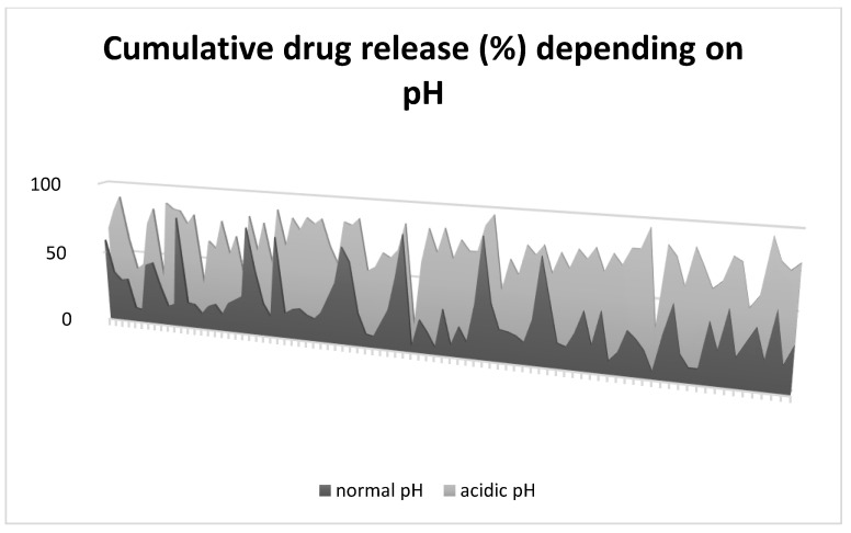
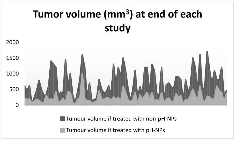
When exposed to in vitro acidic environment, without exception, pH-NPs released drugs inversely proportional to the pH value (Figure 3). In all scenarios, both control and pH-NPs showed similar biodistribution and good stability in vivo; however, pH-NPs showed an increase in tumor regression compared to controls, suggestive of better targeted drug release. As seen in Figure 4, the volume of tumors was lower in groups treated with pH-NPs compared to non-pH-NPs.
Figure 3.
Rate of cumulative drug release for each of the included studies. Dark gray area shows rate (%) of drug released at a physiological pH (7.4). Light gray shows rate (%) of drug released in acidic pH (lowest value used in each study).
Figure 4.
Volume of tumor (mm3) at the end of experiment for each of the included studies. Dark gray area shows the tumor volume for specimens treated with non-pH-NPs. Light gray area shows the tumor volume for specimens treated with pH-NPs.
4. Discussion
Our results show that NPs may be used as pH-responsive platforms with excellent results in tumor penetration and tumor regression rates. pH-NPs, regardless of being metallic or polymeric, were shown to have good tumor penetration in most experimental malignant cell lines in vivo.
Polymers were the most common nanomaterials used in the synthesis of pH-NPs. Besides being used for surface coating to increase the colloidal stability of NPs, polymers (e.g., PEG, PLGA, PHA) were used in the core structure of NPs, making polymeric NPs a widely used platform due to their key advantages: biocompatibility, high stability, non-toxicity, easy synthesis, and versatility. Chemotherapeutics can be linked onto or within the polymers via electrostatic interactions. Once assembled, polymeric NPs have high stability in blood circulation and can maintain the EPR effect, which allows them to escape in the tumoral microenvironment, where drugs are released in a controlled fashion [140]. Mesoporous silica nanoparticles (MSN NPs) were also commonly used to design pH-responsive nanocarriers. The main advantage of MSN NPs is their large surface area and large porous structure, in which a high volume of drugs can be encapsulated. Their surface can be also chemically modified to attach various linkers which react to pH changes [141]. Lipid NPs are usually spherical in shape and formed by a bilayer lipid membrane and an aqueous core. They are highly biocompatible and can transport hydrophilic, hydrophobic, and lipophilic drugs; however, lipid NPs can be cleared by the reticuloendothelial system. For this reason, their surface is usually coated with polymers (e.g., PEGylation) to increase their biostability [142]. Gold NPs can be pH-functionalized using surface pH-responsive linkers. Gold NPs have unique optical characteristics, making them suitable for cancer theranostics and photothermal therapy [143].
The tumor specificity of pH-NPs was further enhanced using tumor-targeting peptides linked to the surface of NPs which can target specific receptors commonly expressed by cancers. The folate receptor is known to be overexpressed in various tumors [144] and was used as a target for NPs coated with folic acid, which facilitates the receptor-mediated endocytosis of NPs, where drug cargo can be released in the acidic intracellular environment. Other studies used Fe ions attached to the surface of NPs, as many tumors use Fe for cellular proliferation [145]. Increased expression of transferrin on tumors promotes NPs attachment and internalization [146]. Xie et al. [120] used methotrexate as an antitumor agent and also as a tumor-targeting agent due to its structural similarity to folic acid and capacity to bind to folate expressed by tumors. Gong et al. [49] used arginine–glycine–aspartate triad (RGD peptide) which is a low-toxicity, highly stable peptide with increased affinity to integrins, which in turn are overexpressed by tumoral neo-vessels.
Doxorubicin is the most used chemotherapeutic in current experiments. Doxorubicin is an anthracycline with potent antimitotic and cytotoxic activity. Its mechanism of action involves intercalation between base pairs where it inhibits DNA synthesis and, in addition, inhibits topoisomerase II activity, thus reducing DNA replication [147,148]. Despite having excellent antitumor activity, its use is limited by important side effects, such as cardiotoxicity and myelosuppression [148]. In a conjugated form, incorporated in the hydrophobic core of nanocarriers, doxorubicin can be administered in higher doses, and can be released at the tumor site where nanoparticles accumulate through enhanced permeability release or by active tumor targeting through pH-dependent conversion, as demonstrated in the included studies.
Drugs are usually loaded into NPs either through core encapsulation or surface bounding. Core encapsulation refers to the organization of NPs around drugs, usually due to their amphipathic property, and the hydrophobic end safeguards the drugs in the center, while the hydrophilic end forms a protective shell, enabling a safe transport of cargo to the tumor. Another way is to attach drugs to the surface of NPs, especially when PEGylation is used to coat the surface. PEG is a stable carrier and binder, and various linkers can be used to attach drugs or tumor-targeting receptors to its surface.
Acid-labile Schiff base linkages were the core from which nanoparticles, regardless of type, were designed to respond to pH changes. Imine Schiff bases undergo hydrolyzation under acidic conditions and such are used as linkers when nanoparticles are assembled. Once the peritumoral acidic pH is sensed, the linkers break, causing disruption of the nanocarriers and release of drugs. In other scenarios, the nanocarriers were coated with tumor-targeting peptides (e.g., folic acid, AS1411 aptamer) which interacted with cancer cells and allowed for the nanocarriers to reach the intracellular environment, via endocytic pathways, where the drugs were released. Another pH sensitization method is the use of electrostatic interactions. pH-NPs were coated with a negative-charged surface which reverted to a positive charge in the acidic environment, leading to the release of positively charged peptides, which were linked to drugs [42].
Functionalized NPs may become a cornerstone in cancer treatment as they can overcome the barrier of systemic toxicity produced by non-targeted chemotherapeutics and can increase the amount of drug delivered to the tumor. Designing NPs responsive to acidic pH has proven to be a solid option. However, we must consider that, in most studies, the maximal effects of pH-NPs were at a pH lower than 6.5. To ensure similar outcomes in clinical studies, pH-NPs need to be ultra-sensitized to release similar amounts of drugs at pH values of 6.8–7.2, which is the usual pH value in the tumor microenvironment.
5. Conclusions
This scoping review mapped the current methods and outcomes of using pH-responsive nanoparticles to improve drug delivery and enhance antitumoral effects. Regardless of their type and structure, pH-responsive nanoparticles can increase tumor regression rates compared to the controls. Drug delivery, therefore, is dependent on the exposure of NPs to acidic pH.
Supplementary Materials
The following supporting information can be downloaded at: https://www.mdpi.com/article/10.3390/gels8040232/s1, Table S1: Detailed Overview of Included Studies.
Author Contributions
Conceptualization, S.M. and G.-M.D.; methodology S.M., B.C.M., R.G., A.C., C.T., R.I. and G.-M.D.; data extraction, S.M. and B.C.M.; data analysis, S.M., B.C.M. and G.-M.D.; draft preparation, S.M., B.C.M., R.G., A.C., C.T., R.I. and G.-M.D.; review and editing, S.M., B.C.M., R.G., A.C., C.T., R.I. and G.-M.D.; supervision, R.I. and G.-M.D. All authors have read and agreed to the published version of the manuscript.
Funding
This research was funded by the Iceland Liechtenstein Norway Grant EEA-RO-NO-2018-0246.
Institutional Review Board Statement
Not applicable.
Informed Consent Statement
Not applicable.
Data Availability Statement
Data can be made available at request.
Conflicts of Interest
We declare no conflicts of interest.
Footnotes
Publisher’s Note: MDPI stays neutral with regard to jurisdictional claims in published maps and institutional affiliations.
References
- 1.Najahi-Missaoui W., Arnold R.D., Cummings B.S. Safe nanoparticles: Are we there yet? Int. J. Mol. Sci. 2021;22:385. doi: 10.3390/ijms22010385. [DOI] [PMC free article] [PubMed] [Google Scholar]
- 2.Baetke S.C., Lammers T., Kiessling F. Applications of nanoparticles for diagnosis and therapy of cancer. Br. J. Radiol. 2015;88:20150207. doi: 10.1259/bjr.20150207. [DOI] [PMC free article] [PubMed] [Google Scholar]
- 3.Wolfram J., Zhu M., Yang Y., Shen J., Gentile E., Paolino D., Fresta M., Nie G., Chen C., Shen H., et al. Safety of nanoparticles in medicine. Curr. Drug Targets. 2015;16:1671–1681. doi: 10.2174/1389450115666140804124808. [DOI] [PMC free article] [PubMed] [Google Scholar]
- 4.Jeong W.J., Bu J., Kubiatowicz L.J., Chen S.S., Kim Y., Hong S. Peptide-nanoparticle conjugates: A next generation of diagnostic and therapeutic platforms? Nano Converg. 2018;5:38. doi: 10.1186/s40580-018-0170-1. [DOI] [PMC free article] [PubMed] [Google Scholar]
- 5.Johnston M.C., Scott C.J. Antibody conjugated nanoparticles as a novel form of antibody drug conjugate chemotherapy. Drug Discov. Today Technol. 2018;30:63–69. doi: 10.1016/j.ddtec.2018.10.003. [DOI] [PubMed] [Google Scholar]
- 6.Patra J.K., Das G., Fraceto L.F., Campos E.V.R., Rodriguez-Torres M.D.P., Acosta-Torres L.S., Diaz-Torres L.A., Grillo R., Swamy M.K., Sharma S., et al. Nano based drug delivery systems: Recent developments and future prospects. J. Nanobiotechnol. 2018;16:71. doi: 10.1186/s12951-018-0392-8. [DOI] [PMC free article] [PubMed] [Google Scholar]
- 7.Naseri N., Ajorlou E., Asghari F., Pilehvar-Soltanahmadi Y. An update on nanoparticle-based contrast agents in medical imaging. Artif. Cells Nanomed. Biotechnol. 2018;46:1111–1121. doi: 10.1080/21691401.2017.1379014. [DOI] [PubMed] [Google Scholar]
- 8.Fang J., Nakamura H., Maeda H. The EPR effect: Unique features of tumor blood vessels for drug delivery, factors involved, and limitations and augmentation of the effect. Adv. Drug Deliv. Rev. 2011;63:136–151. doi: 10.1016/j.addr.2010.04.009. [DOI] [PubMed] [Google Scholar]
- 9.Su Y.L., Hu S.H. Functional Nanoparticles for Tumor Penetration of Therapeutics. Pharmaceutics. 2018;10:193. doi: 10.3390/pharmaceutics10040193. [DOI] [PMC free article] [PubMed] [Google Scholar]
- 10.Shen Y., Tang H., Radosz M., Van Kirk E., Murdoch W.J. pH-responsive nanoparticles for cancer drug delivery. Methods Mol. Biol. 2008;437:183–216. doi: 10.1007/978-1-59745-210-6_10. [DOI] [PubMed] [Google Scholar]
- 11.Barua S., Mitragotri S. Challenges associated with Penetration of Nanoparticles across Cell and Tissue Barriers: A Review of Current Status and Future Prospects. Nano Today. 2014;9:223–243. doi: 10.1016/j.nantod.2014.04.008. [DOI] [PMC free article] [PubMed] [Google Scholar]
- 12.Zhang M., Gao S., Yang D., Fang Y., Lin X., Jin X., Liu Y., Liu X., Su K., Shi K. Influencing factors and strategies of enhancing nanoparticles into tumors in vivo. Acta Pharm. Sin. B. 2021;11:2265–2285. doi: 10.1016/j.apsb.2021.03.033. [DOI] [PMC free article] [PubMed] [Google Scholar]
- 13.Zhang X., Lin Y., Gillies R.J. Tumor pH and its measurement. J. Nucl. Med. 2010;51:1167–1170. doi: 10.2967/jnumed.109.068981. [DOI] [PMC free article] [PubMed] [Google Scholar]
- 14.Morărașu Ș., Iacob Ș., Tudorancea I., Luncă S., Dimofte M.-G. Targeted Chemotherapy Delivery via Gold Nanoparticles: A Scoping Review of In Vivo Studies. Crystals. 2021;11:1169. doi: 10.3390/cryst11101169. [DOI] [Google Scholar]
- 15.Roată C.-E., Iacob Ș., Morărașu Ș., Livadaru C., Tudorancea I., Luncă S., Dimofte M.-G. Collagen-Binding Nanoparticles: A Scoping Review of Methods and Outcomes. Crystals. 2021;11:1396. doi: 10.3390/cryst11111396. [DOI] [Google Scholar]
- 16.Morarasu S., Morarasu B.C., Ghețu N., Dimofte M.G., Iliescu R., Pieptu D. Experimental models for controlled burn injuries in rats: A systematic analysis of original methods and burn devices. J. Burn Care Res. 2021;9:irab234. doi: 10.1093/jbcr/irab234. [DOI] [PubMed] [Google Scholar]
- 17.Page M.J., McKenzie J.E., Bossuyt P.M., Boutron I., Hoffmann T.C., Mulrow C.D., Shamseer L., Tetzlaff J.M., Akl E.A., Brennan S.E., et al. The PRISMA 2020 statement: An updated guideline for reporting systematic reviews. BMJ. 2021;372:n71. doi: 10.1136/bmj.n71. [DOI] [PMC free article] [PubMed] [Google Scholar]
- 18.Percie du Sert N., Hurst V., Ahluwalia A., Alam S., Tvey M.T., Baker M., Browne W.J., Clark A., Cuthill I.C., Dirnagl U., et al. The ARRIVE guidelines 2.0: Updated guidelines for reporting animal research. PLoS Biol. 2020;18:e3000410. doi: 10.1371/journal.pbio.3000410. [DOI] [PMC free article] [PubMed] [Google Scholar]
- 19.Adeyemi S.A., Choonara Y.E., Kumar P., Du Toit L.C., Marimuthu T., Kondiah P.P.D., Pillay V. Folate-decorated, endostatin-loaded, nanoparticles for anti-proliferative chemotherapy in esophaegeal squamous cell carcinoma. Biomed. Pharmacother. 2019;119:109450. doi: 10.1016/j.biopha.2019.109450. [DOI] [PubMed] [Google Scholar]
- 20.Aguilar L.E., Chalony C., Kumar D., Park C.H., Kim C.S. Phenol-Boronic surface functionalization of gold nanoparticles; to induce ROS damage while inhibiting the survival mechanisms of cancer cells. Int. J. Pharm. 2021;596:120267. doi: 10.1016/j.ijpharm.2021.120267. [DOI] [PubMed] [Google Scholar]
- 21.Cao Z., Li D., Wang J., Xiong M., Yang X. Direct Nucleus-Targeted Drug Delivery Using Cascade pHe /Photo Dual-Sensitive Polymeric Nanocarrier for Cancer Therapy. Small. 2019;15:e1902022. doi: 10.1002/smll.201902022. [DOI] [PubMed] [Google Scholar]
- 22.Cavalcante C.H., Fernandes R.S., de Oliveira Silva J., Ramos Oda C.M., Leite E.A., Cassali D.G., Charlie-Silva I., Ventura Fernandes B.H., Miranda Ferreira L.A., De Barros A.L.B., et al. Doxorubicin-loaded pH-sensitive micelles: A promising alternative to enhance antitumor activity and reduce toxicity. Biomed. Pharmacother. 2021;134:111076. doi: 10.1016/j.biopha.2020.111076. [DOI] [PubMed] [Google Scholar]
- 23.Chen W.L., Yang S.D., Li F., Qu C., Liu Y., Wang D., Zhang X. Programmed pH/reduction-responsive nanoparticles for efficient delivery of antitumor agents in vivo. Acta Biomater. 2018;81:219–230. doi: 10.1016/j.actbio.2018.09.040. [DOI] [PubMed] [Google Scholar]
- 24.Chen Z., Wan L., Yuan Y., Kuang Y., Xu X., Liao T., Liu J., Xu Z.-Q., Jiang B., Li C. pH/GSH-Dual-Sensitive Hollow Mesoporous Silica Nanoparticle-Based Drug Delivery System for Targeted Cancer Therapy. ACS Biomater. Sci. Eng. 2020;6:3375–3387. doi: 10.1021/acsbiomaterials.0c00073. [DOI] [PubMed] [Google Scholar]
- 25.Chen M.L., Lai C.J., Lin Y.N., Huang C.M., Lin Y.H. Multifunctional nanoparticles for targeting the tumor microenvironment to improve synergistic drug combinations and cancer treatment effects. J. Mater. Chem. B. 2020;8:10416–10427. doi: 10.1039/D0TB01733G. [DOI] [PubMed] [Google Scholar]
- 26.Chen W., Li J., Xing Y., Wang X., Zhang H., Xia M., Wang D. Dual-pH Sensitive Charge-Reversal Drug Delivery System for Highly Precise and Penetrative Chemotherapy. Pharm. Res. 2020;37:134. doi: 10.1007/s11095-020-02852-6. [DOI] [PubMed] [Google Scholar]
- 27.Cheng W., Nie J., Xu L., Liang C., Peng Y., Liu G., Wang T., Mei L., Huang L., Zeng X. pH-Sensitive Delivery Vehicle Based on Folic Acid-Conjugated Polydopamine-Modified Mesoporous Silica Nanoparticles for Targeted Cancer Therapy. ACS Appl. Mater. Interfaces. 2017;9:18462–18473. doi: 10.1021/acsami.7b02457. [DOI] [PubMed] [Google Scholar]
- 28.Cheng C., Meng Y., Zhang Z., Li Y., Zhang Q. Tumoral Acidic pH-Responsive cis-Diaminodichloroplatinum-Incorporated Cy5.5-PEG- g-A-HA Nanoparticles for Targeting Delivery of CDDP against Cervical Cancer. ACS Appl. Mater. Interfaces. 2018;10:26882–26892. doi: 10.1021/acsami.8b07425. [DOI] [PubMed] [Google Scholar]
- 29.Cheng X., Zeng X., Zheng Y., Wang X., Tang R. Surface-fluorinated and pH-sensitive carboxymethyl chitosan nanoparticles to overcome biological barriers for improved drug delivery in vivo. Carbohydr. Polym. 2019;208:59–69. doi: 10.1016/j.carbpol.2018.12.063. [DOI] [PubMed] [Google Scholar]
- 30.Cheng X., Li D., Sun M., He L., Zheng Y., Wang X., Tang R. Co-delivery of DOX and PDTC by pH-sensitive nanoparticles to overcome multidrug resistance in breast cancer. Colloids Surf. B Biointerfaces. 2019;181:185–197. doi: 10.1016/j.colsurfb.2019.05.042. [DOI] [PubMed] [Google Scholar]
- 31.Cui T., Zhang S., Sun H. Co-delivery of doxorubicin and pH-sensitive curcumin prodrug by transferrin-targeted nanoparticles for breast cancer treatment. Oncol. Rep. 2017;37:1253–1260. doi: 10.3892/or.2017.5345. [DOI] [PubMed] [Google Scholar]
- 32.Debele T.A., Lee K.Y., Hsu N.Y., Chiang Y.T., Yu L.Y., Shen Y.A., Lo C.L. A pH sensitive polymeric micelle for co-delivery of doxorubicin and α-TOS for colon cancer therapy. J. Mater. Chem. B. 2017;5:5870–5880. doi: 10.1039/C7TB01031A. [DOI] [PubMed] [Google Scholar]
- 33.Deng L., Feng Z., Deng H., Jiang Y., Song K., Shi Y., Liu S., Zhang J., Bai S., Qin Z., et al. Rational Design of Nanoparticles to Overcome Poor Tumor Penetration and Hypoxia-Induced Chemotherapy Resistance: Combination of Optimizing Size and Self-Inducing High Level of Reactive Oxygen Species. ACS Appl. Mater. Interfaces. 2019;11:31743–31754. doi: 10.1021/acsami.9b12129. [DOI] [PubMed] [Google Scholar]
- 34.Ding X., Yu W., Wan Y., Yang M., Hua C., Peng N., Liu Y. A pH/ROS-responsive, tumor-targeted drug delivery system based on carboxymethyl chitin gated hollow mesoporous silica nanoparticles for anti-tumor chemotherapy. Carbohydr. Polym. 2020;245:116493. doi: 10.1016/j.carbpol.2020.116493. [DOI] [PubMed] [Google Scholar]
- 35.Ding Z., Wang D., Shi W., Yang X., Duan S., Mo F., Hou X., Liu A., Lu X. In vivo Targeting of Liver Cancer with Tissue- and Nuclei-Specific Mesoporous Silica Nanoparticle-Based Nanocarriers in mice. Int. J. Nanomed. 2020;15:8383–8400. doi: 10.2147/IJN.S272495. [DOI] [PMC free article] [PubMed] [Google Scholar]
- 36.Du L., Zhou J., Meng L., Wang X., Wang C., Huang Y., Zheng S. Deng, L.; Cao, H.; Liang, Z. et al. The pH-Triggered Triblock Nanocarrier Enabled Highly Efficient siRNA Delivery for Cancer Therapy. Theranostics. 2017;7:3432–3445. doi: 10.7150/thno.20297. [DOI] [PMC free article] [PubMed] [Google Scholar]
- 37.Duan F., Feng X., Yang X., Sun W., Jin Y., Liu H., Ge K., Li Z., Zhang J. A simple and powerful co-delivery system based on pH-responsive metal-organic frameworks for enhanced cancer immunotherapy. Biomaterials. 2017;122:23–33. doi: 10.1016/j.biomaterials.2017.01.017. [DOI] [PubMed] [Google Scholar]
- 38.Essawy M.M., El-Sheikh S.M., Raslan H.S., Ramadan H.S., Kang B., Talaat I.M., Afifi M.M. Function of gold nanoparticles in oral cancer beyond drug delivery: Implications in cell apoptosis. Oral Dis. 2021;27:251–265. doi: 10.1111/odi.13551. [DOI] [PubMed] [Google Scholar]
- 39.Fan B., Kang L., Chen L., Sun P., Jin M., Wang Q., Bae Y.H., Huang W., Gao Z. Systemic siRNA Delivery with a Dual pH-Responsive and Tumor-targeted Nanovector for Inhibiting Tumor Growth and Spontaneous Metastasis in Orthotopic Murine Model of Breast Carcinoma. Theranostics. 2017;7:357–376. doi: 10.7150/thno.16855. [DOI] [PMC free article] [PubMed] [Google Scholar]
- 40.Fang L., Lin H., Wu Z., Wang Z., Fan X., Cheng Z., Hou X., Chen D. In vitro/vivo evaluation of novel mitochondrial targeting charge-reversal polysaccharide-based antitumor nanoparticle. Carbohydr. Polym. 2020;234:115930. doi: 10.1016/j.carbpol.2020.115930. [DOI] [PubMed] [Google Scholar]
- 41.Feng W., Zong M., Wan L., Yu X., Yu W. pH/redox sequentially responsive nanoparticles with size shrinkage properties achieve deep tumor penetration and reversal of multidrug resistance. Biomater. Sci. 2020;8:4767–4778. doi: 10.1039/D0BM00695E. [DOI] [PubMed] [Google Scholar]
- 42.Mozar F.S., Chowdhury E.H. Surface-Modification of Carbonate Apatite Nanoparticles Enhances Delivery and Cytotoxicity of Gemcitabine and Anastrozole in Breast Cancer Cells. Pharmaceutics. 2017;9:21. doi: 10.3390/pharmaceutics9020021. [DOI] [PMC free article] [PubMed] [Google Scholar]
- 43.Gao C., Tang F., Gong G., Zhang J., Hoi M.P.M., Lee S.M.Y., Wang R. pH-Responsive prodrug nanoparticles based on a sodium alginate derivative for selective co-release of doxorubicin and curcumin into tumor cells. Nanoscale. 2017;9:12533–12542. doi: 10.1039/C7NR03611F. [DOI] [PubMed] [Google Scholar]
- 44.Gao L., Wang H., Nan L., Peng T., Sun L., Zhou J., Xiao Y., Wang J., Sun J., Liu W., et al. Erythrocyte Membrane-Wrapped pH Sensitive Polymeric Nanoparticles for Non-Small Cell Lung Cancer Therapy. Bioconjug. Chem. 2017;28:2591–2598. doi: 10.1021/acs.bioconjchem.7b00428. [DOI] [PubMed] [Google Scholar]
- 45.Gibbens-Bandala B., Morales-Avila E., Ferro-Flores G., Santos-Cuevas C., Melendez-Alafort L., Trujillo-Nolasco M., Ocampo-Garcia B. 177Lu-Bombesin-PLGA (paclitaxel): A targeted controlled-release nanomedicine for bimodal therapy of breast cancer. Mater. Sci. Eng. C Mater. Biol. Appl. 2019;105:110043. doi: 10.1016/j.msec.2019.110043. [DOI] [PubMed] [Google Scholar]
- 46.Gong T., Dong Z., Fu Y., Gong T., Deng L., Zhang Z. Hyaluronic acid modified doxorubicin loaded Fe3O4 nanoparticles effectively inhibit breast cancer metastasis. J. Mater. Chem. B. 2019;7:5861–5872. doi: 10.1039/C9TB01250H. [DOI] [PubMed] [Google Scholar]
- 47.Gong Y.H., Shu M., Xie J.H., Zhang C., Cao Z., Jiang Z.Z., Liu J. Enzymatic synthesis of PEG-poly(amine-co-thioether esters) as highly efficient pH and ROS dual-responsive nanocarriers for anticancer drug delivery. J. Mater. Chem. B. 2019;7:651–664. doi: 10.1039/C8TB02882F. [DOI] [PubMed] [Google Scholar]
- 48.Gong Z., Liu X., Zhou B., Wang G., Guan X., Xu Y., Zhang J., Hong Z., Cao J., Sun X., et al. Tumor acidic microenvironment-induced drug release of RGD peptide nanoparticles for cellular uptake and cancer therapy. Colloids Surf. B Biointerfaces. 2021;202:111673. doi: 10.1016/j.colsurfb.2021.111673. [DOI] [PubMed] [Google Scholar]
- 49.Guo W., Deng L., Yu J., Chen Z., Woo Y., Liu H., Li T., Lin T., Chen H., Zhao M., et al. Sericin nanomicelles with enhanced cellular uptake and pH-triggered release of doxorubicin reverse cancer drug resistance. Drug Deliv. 2018;25:1103–1116. doi: 10.1080/10717544.2018.1469686. [DOI] [PMC free article] [PubMed] [Google Scholar]
- 50.Guo X., Zhuang Q., Ji T., Yinlong Z., Changjian L., Wang Y., Li H., Jia H., Liu Y., Du L. Multi-functionalized chitosan nanoparticles for enhanced chemotherapy in lung cancer. Carbohydr. Polym. 2018;195:311–320. doi: 10.1016/j.carbpol.2018.04.087. [DOI] [PubMed] [Google Scholar]
- 51.Guo Z., Sui J., Ma M., Hu J., Sun Y., Yang L., Fan Y., Zhang X. pH-Responsive charge switchable PEGylated ε-poly-l-lysine polymeric nanoparticles-assisted combination therapy for improving breast cancer treatment. J. Control. Release. 2020;326:350–364. doi: 10.1016/j.jconrel.2020.07.030. [DOI] [PubMed] [Google Scholar]
- 52.Hao Y., Zheng C., Wang L., Hu Y., Guo H., Song Q., Zhang H., Zhang Z., Zhang Y. Covalent self-assembled nanoparticles with pH-dependent enhanced tumor retention and drug release for improving tumor therapeutic efficiency. J. Mater. Chem. B. 2017;5:2133–2144. doi: 10.1039/C6TB02833K. [DOI] [PubMed] [Google Scholar]
- 53.Hong Y., Che S., Hui B., Yang Y., Wang X., Zhang X., Qiang Y., Ma H. Lung cancer therapy using doxorubicin and curcumin combination: Targeted prodrug based, pH sensitive nanomedicine. Biomed. Pharmacother. 2019;112:108614. doi: 10.1016/j.biopha.2019.108614. [DOI] [PubMed] [Google Scholar]
- 54.Hu H., Steinmetz N.F. Cisplatin Prodrug-Loaded Nanoparticles Based on Physalis Mottle Virus for Cancer Therapy. Mol. Pharm. 2020;17:4629–4636. doi: 10.1021/acs.molpharmaceut.0c00834. [DOI] [PMC free article] [PubMed] [Google Scholar]
- 55.Hu H., Steinmetz N.F. Doxorubicin-Loaded Physalis Mottle Virus Particles Function as a pH-Responsive Prodrug Enabling Cancer Therapy. Biotechnol. J. 2020;15:e2000077. doi: 10.1002/biot.202000077. [DOI] [PMC free article] [PubMed] [Google Scholar]
- 56.Huang H., Yang D.P., Liu M., Wang X., Zhang Z., Zhou G., Liu W., Cao Y., Zhang W.J., Wang X. pH-sensitive Au-BSA-DOX-FA nanocomposites for combined CT imaging and targeted drug delivery. Int. J. Nanomed. 2017;12:2829–2843. doi: 10.2147/IJN.S128270. [DOI] [PMC free article] [PubMed] [Google Scholar]
- 57.Jin M., Jin G., Kang L., Chen L., Gao Z., Huang W. Smart polymeric nanoparticles with pH-responsive and PEG-detachable properties for co-delivering paclitaxel and survivin siRNA to enhance antitumor outcomes. Int. J. Nanomed. 2018;13:2405–2426. doi: 10.2147/IJN.S161426. [DOI] [PMC free article] [PubMed] [Google Scholar]
- 58.Jung S., Lee J., Lim J., Suh J., Kim T., Ahn J., Kim W.J., Kim Y.T. Polymeric Nanoparticles Controlled by On-Chip Self-Assembly Enhance Cancer Treatment Effectiveness. Adv. Healthc. Mater. 2020;9:e2001633. doi: 10.1002/adhm.202001633. [DOI] [PMC free article] [PubMed] [Google Scholar]
- 59.Juang V., Chang C.H., Wang C.S., Wang H.E., Lo Y.L. pH-Responsive PEG-Shedding and Targeting Peptide-Modified Nanoparticles for Dual-Delivery of Irinotecan and microRNA to Enhance Tumor-Specific Therapy. Small. 2019;15:e1903296. doi: 10.1002/smll.201903296. [DOI] [PubMed] [Google Scholar]
- 60.Kang M.S., Singh R.K., Kim T.H., Kim J.H., Patel K.D., Kim H.W. Optical imaging and anticancer chemotherapy through carbon dot created hollow mesoporous silica nanoparticles. Acta Biomater. 2017;55:466–480. doi: 10.1016/j.actbio.2017.03.054. [DOI] [PubMed] [Google Scholar]
- 61.Khan I., Joshi G., Sarkar B., Nakhate K.T., Ajazuddin, Mantha A.K., Kumar R., Kaul A., Chaturvedi S., Mishra A.K., et al. Doxorubicin and Crocin Co-delivery by Polymeric Nanoparticles for Enhanced Anticancer Potential In Vitro and In Vivo. ACS Appl. Bio. Mater. 2020;3:7789–7799. doi: 10.1021/acsabm.0c00974. [DOI] [PubMed] [Google Scholar]
- 62.Kou C.H., Han J., Han X.L., Zhuang H.J., Zhao Z.M. Preparation and characterization of the Adriamycin-loaded amphiphilic chitosan nanoparticles and their application in the treatment of liver cancer. Oncol. Lett. 2017;14:7833–7841. doi: 10.3892/ol.2017.7210. [DOI] [PMC free article] [PubMed] [Google Scholar]
- 63.Kumar K., Moitra P., Bashir M., Kondaiah P., Bhattacharya S. Natural tripeptide capped pH-sensitive gold nanoparticles for efficacious doxorubicin delivery both in vitro and in vivo. Nanoscale. 2020;12:1067–1074. doi: 10.1039/C9NR08475D. [DOI] [PubMed] [Google Scholar]
- 64.Kundu M., Sadhukhan P., Ghosh N., Chatterjee S., Manna P., Das J., Sil P.C. pH-responsive and targeted delivery of curcumin via phenylboronic acid-functionalized ZnO nanoparticles for breast cancer therapy. J. Adv. Res. 2019;18:161–172. doi: 10.1016/j.jare.2019.02.036. [DOI] [PMC free article] [PubMed] [Google Scholar]
- 65.Kundu M., Chatterjee S., Ghosh N., Manna P., Das J., Sil P.C. Tumor targeted delivery of umbelliferone via a smart mesoporous silica nanoparticles controlled-release drug delivery system for increased anticancer efficiency. Mater. Sci. Eng. C Mater. Biol. Appl. 2020;116:111239. doi: 10.1016/j.msec.2020.111239. [DOI] [PubMed] [Google Scholar]
- 66.Lee H.L., Hwang S.C., Nah J.W., Kim J., Cha B., Kang D.H., Jeong Y.I. Redox- and pH-Responsive Nanoparticles Release Piperlongumine in a Stimuli-Sensitive Manner to Inhibit Pulmonary Metastasis of Colorectal Carcinoma Cells. J. Pharm. Sci. 2018;107:2702–2712. doi: 10.1016/j.xphs.2018.06.011. [DOI] [PubMed] [Google Scholar]
- 67.Li Y., Song L., Lin J., Ma J., Pan Z., Zhang Y., Su G., Ye S., Luo F., Zhu X., et al. Programmed Nanococktail Based on pH-Responsive Function Switch for Self-Synergistic Tumor-Targeting Therapy. ACS Appl. Mater. Interfaces. 2017;9:39127–39142. doi: 10.1021/acsami.7b08218. [DOI] [PubMed] [Google Scholar]
- 68.Li Y., Duo Y., Bao S., He L., Kai L., Luo J., Zhang Y., Huang H., Zhang H., Yu X. EpCAM aptamer-functionalized polydopamine-coated mesoporous silica nanoparticles loaded with DM1 for targeted therapy in colorectal cancer. Int. J. Nanomed. 2017;12:6239–6257. doi: 10.2147/IJN.S143293. [DOI] [PMC free article] [PubMed] [Google Scholar]
- 69.Li S., Wang L., Li N., Liu Y., Su H. Combination lung cancer chemotherapy: Design of a pH-sensitive transferrin-PEG-Hz-lipid conjugate for the co-delivery of docetaxel and baicalin. Biomed. Pharmacother. 2017;95:548–555. doi: 10.1016/j.biopha.2017.08.090. [DOI] [PubMed] [Google Scholar]
- 70.Li X.X., Chen J., Shen J.M., Zhuang R., Zhang S.Q., Zhu Z.Y., Ma J.B. pH-Sensitive nanoparticles as smart carriers for selective intracellular drug delivery to tumor. Int. J. Pharm. 2018;545:274–285. doi: 10.1016/j.ijpharm.2018.05.012. [DOI] [PubMed] [Google Scholar]
- 71.Li W., Fu J., Ding Y., Liu D., Jia N., Chen D., Hu H. Low density lipoprotein-inspired nanostructured lipid nanoparticles containing pro-doxorubicin to enhance tumor-targeted therapeutic efficiency. Acta Biomater. 2019;96:456–467. doi: 10.1016/j.actbio.2019.06.051. [DOI] [PubMed] [Google Scholar]
- 72.Li F., Xu X., Liang Y., Li Y., Wang M., Zhao F., Wang X., Sun Y., Chen W. Nuclear-targeted nanocarriers based on pH-sensitive amphiphiles for enhanced GNA002 delivery and chemotherapy. Nanoscale. 2021;13:4774–4784. doi: 10.1039/D0NR07239G. [DOI] [PubMed] [Google Scholar]
- 73.Li Y., Miao Y., Chen M., Chen X., Li F., Zhang X., Gan Y. Stepwise targeting and responsive lipid-coated nanoparticles for enhanced tumor cell sensitivity and hepatocellular carcinoma therapy. Theranostics. 2020;10:3722–3736. doi: 10.7150/thno.42008. [DOI] [PMC free article] [PubMed] [Google Scholar]
- 74.Li Y., Niu Y., Zhu J., Gao C., Xu Q., He Z., Chen D., Xu M., Liu Y. Tailor-made legumain/pH dual-responsive doxorubicin prodrug-embedded nanoparticles for efficient anticancer drug delivery and in situ monitoring of drug release. Nanoscale. 2020;12:2673–2685. doi: 10.1039/C9NR08558K. [DOI] [PubMed] [Google Scholar]
- 75.Li Y., Xiong J., Guo W., Jin Y., Miao W., Wang C., Zhang H., Hu Y., Huang H. Decomposable black phosphorus nano-assembly for controlled delivery of cisplatin and inhibition of breast cancer metastasis. J. Control. Release. 2021;335:59–74. doi: 10.1016/j.jconrel.2021.05.013. [DOI] [PubMed] [Google Scholar]
- 76.Liao T., Liu C., Ren J., Chen H., Kuang Y., Jiang B., Chen J., Sun Z., Li C. A chitosan/mesoporous silica nanoparticle-based anticancer drug delivery system with a “tumor-triggered targeting” property. Int. J. Biol. Macromol. 2021;183:2017–2029. doi: 10.1016/j.ijbiomac.2021.06.004. [DOI] [PubMed] [Google Scholar]
- 77.Lin Y., Zhong Y., Chen Y., Li L., Chen G., Zhang J., Li P., Zhou C., Sun Y., Ma Y., et al. Ligand-Modified Erythrocyte Membrane-Cloaked Metal-Organic Framework Nanoparticles for Targeted Antitumor Therapy. Mol. Pharm. 2020;17:3328–3341. doi: 10.1021/acs.molpharmaceut.0c00421. [DOI] [PubMed] [Google Scholar]
- 78.Liu L., Yi H., He H., Pan H., Cai L., Ma Y. Tumor associated macrophage-targeted microRNA delivery with dual-responsive polypeptide nanovectors for anti-cancer therapy. Biomaterials. 2017;134:166–179. doi: 10.1016/j.biomaterials.2017.04.043. [DOI] [PubMed] [Google Scholar]
- 79.Liu Y., Li L., Li L., Zhou Z., Wang F., Xiong X., Zhou R., Huang Y. Programmed drug delivery system based on optimized “size decrease and hydrophilicity/hydrophobicity transformation” for enhanced hepatocellular carcinoma therapy of doxorubicin. Nanomedicine. 2018;14:1111–1122. doi: 10.1016/j.nano.2018.02.006. [DOI] [PubMed] [Google Scholar]
- 80.Liu S., Luo X., Liu S., Xu P., Wang J., Hu Y. Acetazolamide-Loaded pH-Responsive Nanoparticles Alleviating Tumor Acidosis to Enhance Chemotherapy Effects. Macromol. Biosci. 2019;19:e1800366. doi: 10.1002/mabi.201800366. [DOI] [PubMed] [Google Scholar]
- 81.Liu D., Li X., Chen C., Li C., Zhou C., Zhang W., Zhao J., Fan J., Cheng K., Chen L. Target-specific delivery of oxaliplatin to HER2-positive gastric cancer cells in vivo using oxaliplatin-au-fe3o4-herceptin nanoparticles. Oncol. Lett. 2018;15:8079–8087. doi: 10.3892/ol.2018.8323. [DOI] [PMC free article] [PubMed] [Google Scholar]
- 82.Liu S., Ono R.J., Yang C., Gao S., Ming Tan J.Y., Hedrick J.L., Yang Y.Y. Dual pH-Responsive Shell-Cleavable Polycarbonate Micellar Nanoparticles for in Vivo Anticancer Drug Delivery. ACS Appl. Mater. Interfaces. 2018;10:19355–19364. doi: 10.1021/acsami.8b01954. [DOI] [PubMed] [Google Scholar]
- 83.Liu P., Wu Q., Li Y., Li P., Yuan J., Meng X., Xiao Y. DOX-Conjugated keratin nanoparticles for pH-Sensitive drug delivery. Colloids Surf. B Biointerfaces. 2019;181:1012–1018. doi: 10.1016/j.colsurfb.2019.06.057. [DOI] [PubMed] [Google Scholar]
- 84.Liu C.M., Chen G.B., Chen H.H., Zhang J.B., Li H.Z., Sheng M.X., Weng W.B., Guo S.M. Cancer cell membrane-cloaked mesoporous silica nanoparticles with a pH-sensitive gatekeeper for cancer treatment. Colloids Surf. B Biointerfaces. 2019;175:477–486. doi: 10.1016/j.colsurfb.2018.12.038. [DOI] [PubMed] [Google Scholar]
- 85.Lo Y.L., Chang C.H., Wang C.S., Yang M.H., Lin A.M.Y., Hong C.J., Tseng W.H. PEG-coated nanoparticles detachable in acidic microenvironments for the tumor-directed delivery of chemo- and gene therapies for head and neck cancer. Theranostics. 2020;10:6695–6714. doi: 10.7150/thno.45164. [DOI] [PMC free article] [PubMed] [Google Scholar]
- 86.Lu Z., Douek A.M., Rozario A.M., Tabor R.F., Kaslin J., Follink B., Teo B.M. Bioinspired polynorepinephrine nanoparticles as an efficient vehicle for enhanced drug delivery. J. Mater. Chem. B. 2020;8:961–968. doi: 10.1039/C9TB02375E. [DOI] [PubMed] [Google Scholar]
- 87.Luo Q., Shi W., Wang P., Zhang Y., Meng J., Zhang L. Tumor Microenvironment-Responsive Shell/Core Composite Nanoparticles for Enhanced Stability and Antitumor Efficiency Based on a pH-Triggered Charge-Reversal Mechanism. Pharmaceutics. 2021;13:895. doi: 10.3390/pharmaceutics13060895. [DOI] [PMC free article] [PubMed] [Google Scholar]
- 88.Ma J., Kang K., Zhang Y., Yi Q., Gu Z. Detachable Polyzwitterion-Coated Ternary Nanoparticles Based on Peptide Dendritic Carbon Dots for Efficient Drug Delivery in Cancer Therapy. ACS Appl. Mater. Interfaces. 2018;10:43923–43935. doi: 10.1021/acsami.8b17041. [DOI] [PubMed] [Google Scholar]
- 89.Xiaoyu M., Xiuling D., Chunyu Z., Yi S., Jiangchao Q., Yuan Y., Changsheng L. Polyglutamic acid-coordinated assembly of hydroxyapatite nanoparticles for synergistic tumor-specific therapy. Nanoscale. 2019;11:15312–15325. doi: 10.1039/C9NR03176F. [DOI] [PubMed] [Google Scholar]
- 90.Ma Z., Liu J., Li X., Xu Y., Liu D., He H., Wang Y., Tang X. Hydroxycamptothecin (HCPT)-loaded PEGlated lipid-polymer hybrid nanoparticles for effective delivery of HCPT: QbD-based development and evaluation. Drug Deliv. Transl. Res. 2022;12:306–324. doi: 10.1007/s13346-021-00939-0. [DOI] [PubMed] [Google Scholar]
- 91.Mahalunkar S., Yadav A.S., Gorain M., Pawar V., Braathen R., Weiss S., Bogen B., Gosavi S.W., Kundu G.C. Functional design of pH-responsive folate-targeted polymer-coated gold nanoparticles for drug delivery and in vivo therapy in breast cancer. Int. J. Nanomed. 2019;14:8285–8302. doi: 10.2147/IJN.S215142. [DOI] [PMC free article] [PubMed] [Google Scholar]
- 92.Men W., Zhu P., Dong S., Liu W., Zhou K., Bai Y., Liu X., Gong S., Zhang S. Layer-by-layer pH-sensitive nanoparticles for drug delivery and controlled release with improved therapeutic efficacy in vivo. Drug Deliv. 2020;27:180–190. doi: 10.1080/10717544.2019.1709922. [DOI] [PMC free article] [PubMed] [Google Scholar]
- 93.Mhatre O., Reddy B.P.K., Patnaik C., Chakrabarty S., Ingle A., De A., Srivastava R. pH-responsive delivery of anti-metastatic niclosamide using mussel inspired polydopamine nanoparticles. Int. J. Pharm. 2021;597:120278. doi: 10.1016/j.ijpharm.2021.120278. [DOI] [PubMed] [Google Scholar]
- 94.Mu S., Liu Y., Wang T., Zhang J., Jiang D., Yu X., Zhang N. Unsaturated nitrogen-rich polymer poly(l-histidine) gated reversibly switchable mesoporous silica nanoparticles using “graft to” strategy for drug controlled release. Acta Biomater. 2017;63:150–162. doi: 10.1016/j.actbio.2017.08.050. [DOI] [PubMed] [Google Scholar]
- 95.Nguyen H.T., Soe Z.C., Yang K.Y., Phung C.D., Nguyen L.T.-T., Jeong J.-H., Jin S.G., Choi H.-G., Ku S.K., Yong C.S., et al. Transferrin-conjugated pH-sensitive platform for effective delivery of porous palladium nanoparticles and paclitaxel in cancer treatment. Colloids Surf. B Biointerfaces. 2019;176:265–275. doi: 10.1016/j.colsurfb.2019.01.010. [DOI] [PubMed] [Google Scholar]
- 96.Palanikumar L., Al-Hosani S., Kalmouni M., Nguyen V.P., Ali L., Pasricha R., Barrera F.N., Magzoub M. pH-responsive high stability polymeric nanoparticles for targeted delivery of anticancer therapeutics. Commun. Biol. 2020;3:95. doi: 10.1038/s42003-020-0817-4. [DOI] [PMC free article] [PubMed] [Google Scholar]
- 97.Pan C., Liu Y., Zhou M., Wang W., Shi M., Xing M., Liao W. Theranostic pH-sensitive nanoparticles for highly efficient targeted delivery of doxorubicin for breast tumor treatment. Int. J. Nanomed. 2018;13:1119–1137. doi: 10.2147/IJN.S147464. [DOI] [PMC free article] [PubMed] [Google Scholar]
- 98.Pang J., Xing H., Sun Y., Feng S., Wang S. Non-small cell lung cancer combination therapy: Hyaluronic acid modified, epidermal growth factor receptor targeted, pH sensitive lipid-polymer hybrid nanoparticles for the delivery of erlotinib plus bevacizumab. Biomed. Pharmacother. 2020;125:109861. doi: 10.1016/j.biopha.2020.109861. [DOI] [PubMed] [Google Scholar]
- 99.Peng J.Q., Fumoto S., Suga T., Miyamoto H., Kuroda N., Kawakami S., Nishida K. Targeted co-delivery of protein and drug to a tumor in vivo by sophisticated RGD-modified lipid-calcium carbonate nanoparticles. J. Control. Release. 2019;302:42–53. doi: 10.1016/j.jconrel.2019.03.021. [DOI] [PubMed] [Google Scholar]
- 100.Qu C., Li J., Zhou Y., Yang S., Chen W., Li F., You B., Liu Y., Zhang X. Targeted Delivery of Doxorubicin via CD147-Mediated ROS/pH Dual-Sensitive Nanomicelles for the Efficient Therapy of Hepatocellular Carcinoma. AAPS J. 2018;20:34. doi: 10.1208/s12248-018-0195-8. [DOI] [PubMed] [Google Scholar]
- 101.Quadir M.A., Morton S.W., Mensah L.B., Shopsowitz K., Dobbelaar J., Effenberger N., Hammond P.T. Ligand-decorated click polypeptide derived nanoparticles for targeted drug delivery applications. Nanomedicine. 2017;13:1797–1808. doi: 10.1016/j.nano.2017.02.010. [DOI] [PMC free article] [PubMed] [Google Scholar]
- 102.Ray P., Dutta D., Haque I., Nair G., Mohammed J., Parmer M., Kale N., Orr M., Jain P., Banerjee S., et al. pH-Sensitive Nanodrug Carriers for Codelivery of ERK Inhibitor and Gemcitabine Enhance the Inhibition of Tumor Growth in Pancreatic Cancer. Mol. Pharm. 2021;18:87–100. doi: 10.1021/acs.molpharmaceut.0c00499. [DOI] [PubMed] [Google Scholar]
- 103.Saravanakumar K., Hu X., Shanmugam S., Chelliah R., Sekar P., Oh D.H., Vijayakumar S., Kathiresan K., Wang M.H. Enhanced cancer therapy with pH-dependent and aptamer functionalized doxorubicin loaded polymeric (poly, D.; L-lactic-co-glycolic acid) nanoparticles. Arch. Biochem. Biophys. 2019;671:143–151. doi: 10.1016/j.abb.2019.07.004. [DOI] [PubMed] [Google Scholar]
- 104.Saroj S., Rajput S.J. Facile development, characterization, and evaluation of novel bicalutamide loaded pH-sensitive mesoporous silica nanoparticles for enhanced prostate cancer therapy. Drug Dev. Ind. Pharm. 2019;45:532–547. doi: 10.1080/03639045.2018.1562463. [DOI] [PubMed] [Google Scholar]
- 105.Shi M., Zhao X., Zhang J., Pan S., Yang C., Wei Y., Hu H., Qiao M., Chen D., Zhao X. pH-responsive hybrid nanoparticle with enhanced dissociation characteristic for siRNA delivery. Int. J. Nanomed. 2018;13:6885–6902. doi: 10.2147/IJN.S180119. [DOI] [PMC free article] [PubMed] [Google Scholar]
- 106.Shi M., Wang Y., Zhao X., Zhang J., Hu H., Qiao M., Zhao X., Chen D. Stimuli-Responsive and Highly Penetrable Nanoparticles as a Multifunctional Nanoplatform for Boosting Nonsmall Cell Lung Cancer siRNA Therapy. ACS Biomater. Sci. Eng. 2021;7:3141–3155. doi: 10.1021/acsbiomaterials.1c00582. [DOI] [PubMed] [Google Scholar]
- 107.Soe Z.C., Kwon J.B., Thapa R.K., Ou W., Nguyen H.T., Gautam M., Oh T.K., Choi H.-G., Ku S.K., Yong C.S., et al. Transferrin-Conjugated Polymeric Nanoparticle for Receptor-Mediated Delivery of Doxorubicin in Doxorubicin-Resistant Breast Cancer Cells. Pharmaceutics. 2019;11:63. doi: 10.3390/pharmaceutics11020063. [DOI] [PMC free article] [PubMed] [Google Scholar]
- 108.Su M., Xiao S., Shu M., Lu Y., Zeng Q., Xie J., Jiang Z., Liu J. Enzymatic multifunctional biodegradable polymers for pH- and ROS-responsive anticancer drug delivery. Colloids Surf. B Biointerfaces. 2020;193:111067. doi: 10.1016/j.colsurfb.2020.111067. [DOI] [PubMed] [Google Scholar]
- 109.Sui J., Cui Y., Cai H., Bian S., Xu Z., Zhou L., Sun Y., Liang J., Fan Y., Zhang X. Synergistic chemotherapeutic effect of sorafenib-loaded pullulan-Dox conjugate nanoparticles against murine breast carcinoma. Nanoscale. 2017;9:2755–2767. doi: 10.1039/C6NR09639E. [DOI] [PubMed] [Google Scholar]
- 110.Sun G.Y., Du Y.C., Cui Y.X., Wang J., Li X.Y., Tang A.N., Kong D.M. Terminal Deoxynucleotidyl Transferase-Catalyzed Preparation of pH-Responsive DNA Nanocarriers for Tumor-Targeted Drug Delivery and Therapy. ACS Appl. Mater. Interfaces. 2019;11:14684–14692. doi: 10.1021/acsami.9b05358. [DOI] [PubMed] [Google Scholar]
- 111.Sun G., Sun K., Sun J. Combination prostate cancer therapy: Prostate-specific membranes antigen targeted, pH-sensitive nanoparticles loaded with doxorubicin and tanshinone. Drug Deliv. 2021;28:1132–1140. doi: 10.1080/10717544.2021.1931559. [DOI] [PMC free article] [PubMed] [Google Scholar]
- 112.Tan S., Wang G. Redox-responsive and pH-sensitive nanoparticles enhanced stability and anticancer ability of erlotinib to treat lung cancer in vivo. Drug Des. Dev. Ther. 2017;11:3519–3529. doi: 10.2147/DDDT.S151422. [DOI] [PMC free article] [PubMed] [Google Scholar]
- 113.Wang H., Zhu W., Huang Y., Li Z., Jiang Y., Xie Q. Facile encapsulation of hydroxycamptothecin nanocrystals into zein-based nanocomplexes for active targeting in drug delivery and cell imaging. Acta Biomater. 2017;61:88–100. doi: 10.1016/j.actbio.2017.04.017. [DOI] [PubMed] [Google Scholar]
- 114.Wang T., Wang D., Liu J., Feng B., Zhou F., Zhao H., Zhou L., Yin Q., Zhang Z., Cao Z., et al. Acidity-Triggered Ligand-Presenting Nanoparticles to Overcome Sequential Drug Delivery Barriers to Tumors. Nano Lett. 2017;17:5429–5436. doi: 10.1021/acs.nanolett.7b02031. [DOI] [PubMed] [Google Scholar]
- 115.Wang C., Zhang Z., Chen B., Gu L., Li Y., Yu S. Design and evaluation of galactosylated chitosan/graphene oxide nanoparticles as a drug delivery system. J. Colloid Interface Sci. 2018;516:332–341. doi: 10.1016/j.jcis.2018.01.073. [DOI] [PubMed] [Google Scholar]
- 116.Wang X., Cheng X., He L., Zeng X., Zheng Y., Tang R. Self-Assembled Indomethacin Dimer Nanoparticles Loaded with Doxorubicin for Combination Therapy in Resistant Breast Cancer. ACS Appl. Mater. Interfaces. 2019;11:28597–28609. doi: 10.1021/acsami.9b05855. [DOI] [PubMed] [Google Scholar]
- 117.Wang X., Xu J., Xu X., Fang Q., Tang R. pH-sensitive bromelain nanoparticles by ortho ester crosslinkage for enhanced doxorubicin penetration in solid tumor. Mater. Sci. Eng. C Mater. Biol. Appl. 2020;113:111004. doi: 10.1016/j.msec.2020.111004. [DOI] [PubMed] [Google Scholar]
- 118.Wei Z., Wang H., Xin G., Zeng Z., Li S., Ming Y., Zhang X., Xing Z., Li L., Li Y., et al. A pH-Sensitive Prodrug Nanocarrier Based on Diosgenin for Doxorubicin Delivery to Efficiently Inhibit Tumor Metastasis. Int. J. Nanomed. 2020;15:6545–6560. doi: 10.2147/IJN.S250549. [DOI] [PMC free article] [PubMed] [Google Scholar]
- 119.Xia Y., Xiao M., Zhao M., Xu T., Guo M., Wang C., Li Y., Zhu B., Liu H. Doxorubicin-loaded functionalized selenium nanoparticles for enhanced antitumor efficacy in cervical carcinoma therapy. Mater. Sci. Eng. C Mater. Biol. Appl. 2020;106:110100. doi: 10.1016/j.msec.2019.110100. [DOI] [PubMed] [Google Scholar]
- 120.Xie J., Fan Z., Li Y., Zhang Y., Yu F., Su G., Xie L., Hou Z. Design of pH-sensitive methotrexate prodrug-targeted curcumin nanoparticles for efficient dual-drug delivery and combination cancer therapy. Int. J. Nanomed. 2018;13:1381–1398. doi: 10.2147/IJN.S152312. [DOI] [PMC free article] [PubMed] [Google Scholar]
- 121.Xiong H., Wu Y., Jiang Z., Zhou J., Yang M., Yao J. pH-activatable polymeric nanodrugs enhanced tumor chemo/antiangiogenic combination therapy through improving targeting drug release. J. Colloid Interface Sci. 2019;536:135–148. doi: 10.1016/j.jcis.2018.10.039. [DOI] [PubMed] [Google Scholar]
- 122.Xiong S., Wang Z., Liu J., Deng X., Xiong R., Cao X., Xie Z., Lei X., Chen Y., Tang G. A pH-sensitive prodrug strategy to co-deliver DOX and TOS in TPGS nanomicelles for tumor therapy. Colloids Surf. B Biointerfaces. 2019;173:346–355. doi: 10.1016/j.colsurfb.2018.10.012. [DOI] [PubMed] [Google Scholar]
- 123.Xu C., Song R.J., Lu P., Chen J.C., Zhou Y.Q., Shen G., Jiang M.J., Zhang W. pH-triggered charge-reversal and redox-sensitive drug-release polymer micelles codeliver doxorubicin and triptolide for prostate tumor therapy. Int. J. Nanomed. 2018;13:7229–7249. doi: 10.2147/IJN.S182197. [DOI] [PMC free article] [PubMed] [Google Scholar]
- 124.Xu X., Xue Y., Fang Q., Qiao Z., Liu S., Wang X., Tang R. Hybrid nanoparticles based on ortho ester-modified pluronic L61 and chitosan for efficient doxorubicin delivery. Int. J. Biol. Macromol. 2021;183:1596–1606. doi: 10.1016/j.ijbiomac.2021.05.096. [DOI] [PubMed] [Google Scholar]
- 125.Yadav A.S., Radharani N.N.V., Gorain M., Bulbule A., Shetti D., Roy G., Baby T., Kundu G.C. RGD functionalized chitosan nanoparticle mediated targeted delivery of raloxifene selectively suppresses angiogenesis and tumor growth in breast cancer. Nanoscale. 2020;12:10664–10684. doi: 10.1039/C9NR10673A. [DOI] [PubMed] [Google Scholar]
- 126.Yan G., Wang J., Hu L., Wang X., Yang G., Fu S., Cheng X., Zhang P., Tang R. Stepwise targeted drug delivery to liver cancer cells for enhanced therapeutic efficacy by galactose-grafted, ultra-pH-sensitive micelles. Acta Biomater. 2017;51:363–373. doi: 10.1016/j.actbio.2017.01.031. [DOI] [PubMed] [Google Scholar]
- 127.Yang H., Tang C., Yin C. Estrone-modified pH-sensitive glycol chitosan nanoparticles for drug delivery in breast cancer. Acta Biomater. 2018;73:400–411. doi: 10.1016/j.actbio.2018.04.020. [DOI] [PubMed] [Google Scholar]
- 128.Yu H., Li J.M., Deng K., Zhou W., Wang C.X., Wang Q., Li K.H., Zhao H.Y., Huang S.W. Tumor acidity activated triphenylphosphonium-based mitochondrial targeting nanocarriers for overcoming drug resistance of cancer therapy. Theranostics. 2019;9:7033–7050. doi: 10.7150/thno.35748. [DOI] [PMC free article] [PubMed] [Google Scholar]
- 129.Zhang J., Li J., Shi Z., Yang Y., Xie X., Lee S.M., Wang Y., Leong K.W., Chen M. pH-sensitive polymeric nanoparticles for co-delivery of doxorubicin and curcumin to treat cancer via enhanced pro-apoptotic and anti-angiogenic activities. Acta Biomater. 2017;58:349–364. doi: 10.1016/j.actbio.2017.04.029. [DOI] [PubMed] [Google Scholar]
- 130.Zhang Y., Dang M., Tian Y., Zhu Y., Liu W., Tian W., Su Y., Ni Q., Xu C., Lu N., et al. Tumor Acidic Microenvironment Targeted Drug Delivery Based on pHLIP-Modified Mesoporous Organosilica Nanoparticles. ACS Appl. Mater. Interfaces. 2017;9:30543–30552. doi: 10.1021/acsami.7b10840. [DOI] [PubMed] [Google Scholar]
- 131.Zhang B., Wu Q. pH and redox dual-responsive copolymer micelles with surface charge reversal for co-delivery of all-trans-retinoic acid and paclitaxel for cancer combination chemotherapy. Int. J. Nanomed. 2018;13:6499–6515. doi: 10.2147/IJN.S179046. [DOI] [PMC free article] [PubMed] [Google Scholar]
- 132.Zhang X., Zhao M., Cao N., Qin W., Zhao M., Wu J., Lin D. Construction of a tumor microenvironment pH-responsive cleavable PEGylated hyaluronic acid nano-drug delivery system for colorectal cancer treatment. Biomater. Sci. 2020;8:1885–1896. doi: 10.1039/C9BM01927H. [DOI] [PubMed] [Google Scholar]
- 133.Zhang Y., Zeng X., Wang H., Fan R., Hu Y., Hu X., Li J. Dasatinib self-assembled nanoparticles decorated with hyaluronic acid for targeted treatment of tumors to overcome multidrug resistance. Drug Deliv. 2021;28:670–679. doi: 10.1080/10717544.2021.1905751. [DOI] [PMC free article] [PubMed] [Google Scholar]
- 134.Zhao Y.P., Ye W.L., Liu D.Z., Cui H., Cheng Y., Liu M., Zhang B., Mei Q., Zhou S. Redox and pH dual sensitive bone targeting nanoparticles to treat breast cancer bone metastases and inhibit bone resorption. Nanoscale. 2017;9:6264–6277. doi: 10.1039/C7NR00962C. [DOI] [PubMed] [Google Scholar]
- 135.Zhao H., Yuan X., Yu J., Huang Y., Shao C., Xiao F., Lin L., Li Y., Tian L. Magnesium-Stabilized Multifunctional DNA Nanoparticles for Tumor-Targeted and pH-Responsive Drug Delivery. ACS Appl. Mater. Interfaces. 2018;10:15418–15427. doi: 10.1021/acsami.8b01932. [DOI] [PubMed] [Google Scholar]
- 136.Zhao P., Li L., Zhou S., Qiu L., Qian Z., Liu X., Cao X., Zhang H. TPGS functionalized mesoporous silica nanoparticles for anticancer drug delivery to overcome multidrug resistance. Mater. Sci. Eng. C Mater. Biol. Appl. 2018;84:108–117. doi: 10.1016/j.msec.2017.11.040. [DOI] [PubMed] [Google Scholar]
- 137.Zhou S., Wu D., Yin X., Jin X., Zhang X., Zheng S., Wang C., Liu Y. Intracellular pH-responsive and rituximab-conjugated mesoporous silica nanoparticles for targeted drug delivery to lymphoma B cells. J. Exp. Clin. Cancer Res. 2017;36:24. doi: 10.1186/s13046-017-0492-6. [DOI] [PMC free article] [PubMed] [Google Scholar]
- 138.Zhou N., Zhi Z., Liu D., Wang D., Shao Y., Yan K., Meng L., Yu D. Acid-Responsive and Biologically Degradable Polyphosphazene Nanodrugs for Efficient Drug Delivery. ACS Biomater. Sci. Eng. 2020;6:4285–4293. doi: 10.1021/acsbiomaterials.0c00378. [DOI] [PubMed] [Google Scholar]
- 139.Zou X., Jiang Z., Li L., Huang Z. Selenium nanoparticles coated with pH responsive silk fibroin complex for fingolimod release and enhanced targeting in thyroid cancer. Artif. Cells Nanomed. Biotechnol. 2021;49:83–95. doi: 10.1080/21691401.2021.1871620. [DOI] [PubMed] [Google Scholar]
- 140.Masood F. Polymeric nanoparticles for targeted drug delivery system for cancer therapy. Mater. Sci. Eng. C Mater. Biol. Appl. 2016;60:569–578. doi: 10.1016/j.msec.2015.11.067. [DOI] [PubMed] [Google Scholar]
- 141.Gao Y., Gao D., Shen J., Wang Q. A Review of Mesoporous Silica Nanoparticle Delivery Systems in Chemo-Based Combination Cancer Therapies. Front. Chem. 2020;8:598722. doi: 10.3389/fchem.2020.598722. [DOI] [PMC free article] [PubMed] [Google Scholar]
- 142.Mitchell M.J., Billingsley M.M., Haley R.M., Wechsler M.E., Peppas N.A., Langer R. Engineering precision nanoparticles for drug delivery. Nat. Rev. Drug Discov. 2021;20:101–124. doi: 10.1038/s41573-020-0090-8. [DOI] [PMC free article] [PubMed] [Google Scholar]
- 143.Arvizo R., Bhattacharya R., Mukherjee P. Gold nanoparticles: Opportunities and challenges in nanomedicine. Expert Opin. Drug Deliv. 2010;7:753–763. doi: 10.1517/17425241003777010. [DOI] [PMC free article] [PubMed] [Google Scholar]
- 144.Fernández M., Javaid F., Chudasama V. Advances in targeting the folate receptor in the treatment/imaging of cancers. Chem. Sci. 2017;9:790–810. doi: 10.1039/C7SC04004K. [DOI] [PMC free article] [PubMed] [Google Scholar]
- 145.Morales M., Xue X. Targeting iron metabolism in cancer therapy. Theranostics. 2021;11:8412–8429. doi: 10.7150/thno.59092. [DOI] [PMC free article] [PubMed] [Google Scholar]
- 146.Daniels T.R., Bernabeu E., Rodríguez J.A., Patel S., Kozman M., Chiappetta D.A., Holler E., Ljubimova J.Y., Helguera G., Penichet M.L. The transferrin receptor and the targeted delivery of therapeutic agents against cancer. Biochim. Biophys. Acta. 2012;1820:291–317. doi: 10.1016/j.bbagen.2011.07.016. [DOI] [PMC free article] [PubMed] [Google Scholar]
- 147.Zhao N., Woodle M.C., Mixson A.J. Advances in delivery systems for doxorubicin. J. Nanomed. Nanotechnol. 2018;9:519. doi: 10.4172/2157-7439.1000519. [DOI] [PMC free article] [PubMed] [Google Scholar]
- 148.Thorn C.F., Oshiro C., Marsh S., Hernandez-Boussard T., McLeod H., Klein T.E., Altman R.B. Doxorubicin pathways: Pharmacodynamics and adverse effects. Pharm. Genom. 2011;21:440–446. doi: 10.1097/FPC.0b013e32833ffb56. [DOI] [PMC free article] [PubMed] [Google Scholar]
Associated Data
This section collects any data citations, data availability statements, or supplementary materials included in this article.
Supplementary Materials
Data Availability Statement
Data can be made available at request.


