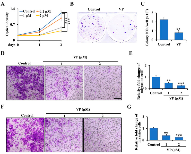
Figure 3.
VP inhibits proliferation, migration, and invasion in Trp53/Rb1-mutant Ctsk-expressing osteosarcoma cells. (A) The primary osteosarcoma cells from Ctsk-Cre;Trp53f/f/Rb1f/f mice were collected and seeded in a 96-well plate. After being cultured for the indicated time, the cell proliferation was determined by WST-1 Cell Proliferation Assay Kit. (B,C) Soft agar assay (B). The primary osteosarcoma cells from Ctsk-Cre;Trp53f/f/Rb1f/f mice were incubated with 0 or 2 μM VP for 3 weeks, and then the colony numbers were counted (C). (D,E) The primary osteosarcoma cells were isolated from Ctsk-Cre;Trp53f/f/Rb1f/f mice, and treated with indicated doses of VP in transwell plates. After incubation for 48 h, the migrated cells were stained by crystal violet and viewed under microscope (D). Scale bars, 100 μm. The corresponding quantification was identified (E). (F,G) Cell invasion and quantification as indicated. Scale bars, 100 μm. Error bars were the means ± SEM from three independent experiments. ** p < 0.01, *** p < 0.001.

