Abstract
Ion channels and transporters typically consist of biomolecules that play key roles in a large variety of physiological and pathological processes. Traditional therapies include many ion-channel blockers, and some activators, although the exact biochemical pathways and mechanisms that regulate ion homeostasis are yet to be fully elucidated. An emerging area of research with great innovative potential in biomedicine pertains the design and development of synthetic ion channels and transporters, which may provide unexplored therapeutic opportunities. However, most studies in this challenging and multidisciplinary area are still at a fundamental level. In this review, we discuss the progress that has been made over the last five years on ion channels and transporters, touching upon biomolecules and synthetic supramolecules that are relevant to biological use. We conclude with the identification of therapeutic opportunities for future exploration.
Keywords: ion channels, ion transporters, ionophores, ion carriers, channelopathies, cystic fibrosis, AMPs, peptides, supramolecular medicinal chemistry, nanotubes
1. Introduction
1.1. Ion Channels and Ion Transporters as Therapeutic Targets
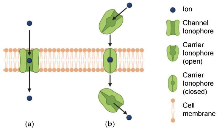
Natural ion channels are composed of large proteins that form pores spanning through lipid membranes (Figure 1a), and they can be grouped based on ion selectivity (e.g., Na+, Cl−) [1], and type of gating mechanism (e.g., activation mediated by voltage [2], ligand [3], mechanical [4,5], or light stimuli [6]). They are involved in a plethora of physiological and pathological processes, for which they have been attracting increasing interest for therapeutic intervention [7]. Remarkably, 19% of drugs that have been approved by the U.S. Food and Drug Administration (FDA) target ion-channel proteins, both gate-activated and ligand-activated [8]. Research in ion-channel modulation has become a hot topic, and encompasses both synthetic molecules [9] and biomolecules, such as lipids [10,11,12], toxins [13], antibodies and nanobodies [14], and venom-derived peptides [15,16]. Dysfunction of ion channels is linked to several pathologies that are generally termed channelopathies, which comprise a plethora of diverse diseases, of the central nervous system, the renal system, and cardiac tissue, as recently reviewed [17], and many others, affecting for instance the skeletal muscle [18], the immune response [19], and glucose levels [20]. The reasons for channel dysfunction are diverse, and include mutations [21,22,23] and post-translational defects [24,25].
Figure 1.
(a) Ion channels and (b) ion transporters. Reproduced with permission from [26]. Copyright Elsevier 2022.
Ion transporters are considered a separate class that plays a role in the regulation of ion homeostasis. They are mobile and they can travel through cell membranes. They transiently bind ions to enable their crossing through the lipid barrier (Figure 1b). They have been proposed as anticancer agents and sensitizers, and they include both natural biomolecules (e.g., peptides and antibiotics) and synthetic molecules (e.g., polyethers, crown ethers) [27,28]. They have been proposed as antimicrobial (AM) agents [26,29,30,31], with the added benefit of immunomodulation activity [26]. However, their successful use in therapy suffers from unsolved challenges in formulation, and in targeting, thus resulting in sub-optimal performance and side-effects. A potential solution could be their selective activation at the pathological site [28].
In therapy, channel blockers are used to treat numerous diseases. As a well-known example, selective Ca2+-channel blockers are anti-hypertensive drugs [32]. Several types of ion transporters are being considered as therapeutic targets for ischemic stroke [33]. They include transient receptor potential (TRP) channels, which are an inhibition target to treat not only stroke, but also depression, epilepsy, and neurodegenerative forms [34]. In fact, Ca2+-channel blockers used to treat hypertension were found to exert a neuroprotective effect against Alzheimer’s disease and depression [35]. Dysregulation of Ca2+ homeostasis was found to occur ubiquitously in Alzheimer’s disease, and thus it has been proposed as a therapeutic target for neurodegeneration [36,37].
Na+ and Ca2+ channel blockers find use in the treatment of bipolar disorders too, and another ion-related therapy that is effective for some patients includes administration of lithium salts [38]. Ca2+ channel blockers attenuate cellular hyperactivity, and for this reason they are considered in psychiatry to treat various disorders [39]. Na-K-Cl cotransporters are therapeutic targets for many diseases, including pain, epilepsy, brain edema, and hypertension [40]. Furthermore, Ca2+ and Na+ channel blockers are used to treat chronic and neurogenic pain [41]. Acid-sensing ion channels have been proposed as therapeutic target for migraine [42].
Recently, however, disruption of ion channel function was found to play a role in the etiology of type-2 diabetes [43]. Several types of ion channels are dysregulated in diabetes and could serve as therapeutic targets, although a key limitation is the risk of side effects arising from sub-optimal selectivity of channel blockers [44]. Furthermore, insulin resistance and altered Ca2+ homeostasis are associated with non-alcoholic liver steatosis, which is another important pathology that would benefit from Ca2+-channel targeting, as long as it is specific for pathologically relevant channel isoforms that are expressed in the liver [45].
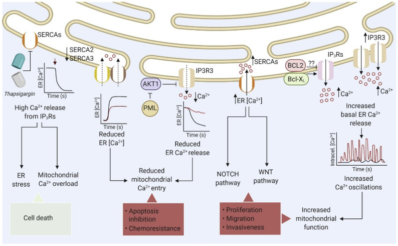
Overall, selectivity is indeed key, and there is a recent trend to employ biologics to achieve it, including engineered antibodies, nanobodies, and venom-derived peptides [46]. Besides organ-specificity (e.g., cardiac [47], cerebellar [48], and liver [45] tissues), organelle-specificity is also relevant, for instance to target ion channels of mitochondria [49] or lysosomes [50]. In particular, mitochondrial-associated endoplasmic reticulum membranes (MAMs) contain specific proteins and ion channels that control key cellular processes, such as redox homeostasis and Ca2+ signaling, and their alteration has been linked to several pathologies, including cancer (Figure 2) [51]. Likewise, lysosomes are underestimated regulators of Ca2+ levels that could be interesting targets for anticancer agents [51]. Several ion-channel blockers are being considered for the treatment of cancer metastases (Table 1), which are accompanied by ion alterations and are characterized by unique protein expression patterns that are different from the originating tumors and target host tissue [52].
Figure 2.
Cancer-associated defects of endoplasmic reticulum Ca2+ homeostasis. Reproduced with permission from ref. [51]. Copyright Elsevier 2020.
Table 1.
Ion-channel blockers and chelators that are drug candidates in clinical trials against cancer. The table includes drugs that are being considered or in clinical trials as anti-metastatic agents, which may counteract the ionic imbalance in disseminating or disseminated cancer cells. Adapted from [52] under a Creative Commons license.
| Ion | Function | Drug Candidate | Feature | Target Cancer | Phase |
|---|---|---|---|---|---|
| Ca2+ | Blocker | Amlodipine besylate | Selective for L-type Ca2+ channels, antihypertensive drug | Metastatic triple negative breast cancer |
1,2 |
| Blocker | Verapamil | Antihypertensive drug | Brain Cancer | 2 | |
| K+ | Blocker | Imipramine | Targets voltage-gated channels, drug against depression | HER2 Positive Breast Carcinoma |
0 |
| Cu2+ | Chelator | Trientine | Anti-angiogenesis, normally used to treat Wilson disease | Fallopian Tube Cancer Ovarian Neoplasms Malignant/primary peritoneal cancer |
1,2 |
| Salicylaldehyde pyrazole hydrazone | Anti-angiogenesis | - | - | ||
| Tetrathiomolybdate | Drug used against primary biliary cholangitis, Wilson Disease | Prostate cancer, carcinoma, colorectal cancer non-small cell lung cancer |
1,2 | ||
| Penicillamine | Drug against cystine renal calculi | Brain and CNS tumors | 2 | ||
| Disulfiram | Drug against alcohol dependency | Metastatic breast cancer Metastatic pancreatic cancer |
2 | ||
| Clioquinol | Drug against dermatitis and eczema | Acute lymphocytic leukemia Acute myeloid leukemia Chronic lymphocytic leukemia |
1 * | ||
| Fe2+/Fe3+ | Chelator | Ciclopirox olamine | Drug against onychomycosis, foot dermatoses | Hematologic malignancy, acute lymphocytic leukemia, advanced solid tumors |
1 |
| Thiosemicarbazones | Drug against renal failure, renal artery stenosis | Unspecified adult solid tumor, protocol specific, prostate cancer/metastatic well differentiated neuroendocrine neoplasm | 1 | ||
| Deferiprone | Drug against cardiomyopathy, iron overload, deteriorating renal function | Colon cancer, breast cancer, rectal cancer, urethral carcinoma | 2 | ||
| Deferasirox | It suppresses N-Cadherin; drug against acute undifferentiated leukemia/ iron overload |
Breast cancer, leukemia | 2 * | ||
| desferrioxamine | restores E-Cadherin localization. Drug against cardiomyopathy/iron overload |
Acute myeloid leukemia/acute lymphoblastic leukemia/ myelodysplastic syndrome |
* | ||
| Many | Blocker | Chlorotoxin | NKCC channel blocker | Breast cancer/non-small cell lung cancer/melanoma/ brain neoplasm | 1,2 |
| Na+ | Blocker | Propranolol | Targets VGSC 1, used for post-traumatic stress disorder, brain injuries | Invasive epithelial ovarian cancer, primary peritoneal carcinoma, fallopian tube cancer, cervical cancer, pediatric cancer/breast cancer | 1,2 |
| Ranolazine | Targets VGSC 1, used for pulmonary hypertension, angina | Adenocarcinoma of the prostate, bone metastases, soft tissue metastases | - | ||
| Phenytoin | Targets VGSC 1, used for acute kidney injury/impaired renal function/kidney failure | Pancreatic cancer, locally advanced breast cancer and large operable breast cancer/metastatic breast cancer, metastatic pancreatic cancer | 2,3 | ||
| Carbamazepine | Targets VGSC 1, used for bipolar disorder (bd), epilepsy, erythromelalgia | Brain and central nervous system tumors, glioblastoma | 1,2 | ||
| Valproate | Targets VGSC 1, used for acute kidney injury/impaired renal function/kidney failure | Advanced cancer/prostate cancer, breast cancer, pancreatic cancer | 1,2 | ||
| Lamotrigine | Targets VGSC 1, used for bipolar disorder | Brain and central nervous system tumors/malignant glioma | 2,4 | ||
| Ranolazine | Targets VGSC 1 | Adenocarcinoma of the prostate, bone metastases, soft tissue metastases | - | ||
| Ropivacaine | Targets VGSC 1, used for anesthesia, conduction/ arthroplasty, replacement/ postoperative pain |
Malignant neoplasm of breast | 3 | ||
| Lidocaine | Targets VGSC 1, used for anesthesia | Lung cancers, unspecified adult solid tumor, prostate cancer | 1,2 | ||
| Riluzole | Targts VGSC 1 | Breast cancer/metastatic cancer | 1 * |
* Denotes terminated clinical trials. 1 VGSC = voltage gated sodium channels.
Detailed knowledge of specific expression of the various types of ion channels is thus of paramount importance to develop effective therapy and reduce side-effects [53]. Alternatively, organism selectivity could be exploited to develop antiviral agents targeting ion channels (i.e., viroporins), which play key roles in the virus lifecycle and are immunomodulatory [54].
Besides cationophores, anionophores could also be interesting targets for therapeutic treatment. In particular, Cl− ion transporters are relatively underexplored to treat various diseases, such as constipation and secretory diarrheas, kidney stones, and polycystic kidney disease, but also dry eye disorders, hypertension, and even osteoporosis [55].
1.2. Therapeutic Activation of Ion Channels and Ion Transporters
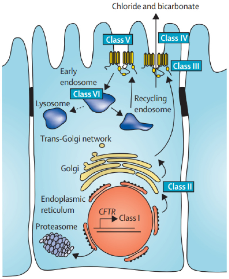
The pathology that is mostly known for the therapeutic effects of ion-channel activation is cystic fibrosis (CF). This disease has been ascribed to mutations of the CF transmembrane conductance regulator (CFTR) gene (Figure 3), which encodes for the epithelial anionic channel that transports Cl− and HCO3−, resulting in defective mucus hydration and clearance. The resulting clinical manifestations are diverse and multi-organ, affecting especially the lungs, and the gastrointestinal and the endocrine systems. Management strategies have been traditionally aimed at treating symptoms, while interventions at the root of the problem through modulation of ion-channel activity have just opened a whole new scenario for improved life-quality of affected patients. In particular, the identification of mutations in CFTR in CF patients has enabled the development of targeted therapies to restore the function of the ion channel [56]. Besides Cl− and HCO3− channel activators [57], several biologics are used to restore the channel function, i.e., gene therapy and editing, RNA therapy and micro RNAs [58]. Readers interested in further details pertaining this pathology and treatment strategies are referred to an excellent and comprehensive recent review [59].
Figure 3.
Cystic fibrosis (CF) arises from defective anion channels on epithelial cells, due to CFTR mutations that are grouped into several classes, depending on the cellular process that results impaired. Reproduced with permission from [59]. Copyright Elsevier 2021.
The therapeutic potential of ion channel activation goes well beyond CF and represents an unexplored avenue for new therapies. For instance, potentiation of specific sub-types of TRP channels has been proposed to prevent and treat obesity [60]. Importantly, dysregulation of Na+, K+, Ca2+, or Cl− intracellular levels can lead to programmed cell death, and for this reason several ion-channel modulators have been proposed against cancer [61]. It is worth noting that increasing cytosolic Ca2+ concentration can have opposite downstream effects in cancer cells, depending on the biochemical activation pathway [62]. Therefore, not only channel blockers, but also activators or artificial channels could pave the way to new therapeutic approaches, as long as cancer cell selectivity is achieved.
Restoration of cation transport is another underexplored avenue that can be of therapeutic relevance. For instance, impaired Na+ transport was recently identified as an upstream pathogenic factor in inflammatory bowel disease [63]. Numerous enteric pathogens target the expression and/or function of ion transporters, causing diarrhea [64]. Another type of cation transport that is neglected pertains Mg++ channels. Mg++ homeostasis is regulated by several ion channels that are either downregulated or upregulated in many digestive cancers, thus providing an opportunity for innovative treatments [65]. Zn++ transporters were also found to ameliorate oxidative stress and insulin resistance, and were thus identified as new therapeutic targets [66].
2. Biomolecular Ion Channels and Transporters as Therapeutic Agents
2.1. Ion-Channel Biomolecules as Therapeutic Agents
There are several ion-channel biomolecules that have attracted interest not only for their therapeutic potential, but also as models to design artificial channels. The majority consist of polypeptides or proteins. They have enabled the development of synthetic 3-in-1 transporters for cations, anions, and zwitterions [67]. Here we will provide a brief overview of the most studied channels.
Ion-transporting rhodopsins are fascinating light-activated proteins that were firstly identified in photo-responsive chemotactic bacteria. Nowadays, they are mainly used in optogenetics, and they have inspired the design of biomimetics. The two main groups are light-driven ion-pumps, and light-gated ion channels. The former use light energy to transport specific ions (H+, Na+, Cl−) against electrophysiological potential, while the latter are less specific and employ passive transport. Light-activation of rhodopsin pumps results in hyperpolarization of membrane potential that effectively inhibits neuron firing, thus exerting an inhibitory role [68]. Many variants have been described, and the detailed mechanisms of their activity were recently reviewed [69,70]. Their therapeutic application has been envisaged mainly in gene therapy to restore vision in patients with degenerative retinal diseases, which otherwise can progress to blindness [71].
Natural KcsA K+ channels have inspired the biomimetic design of highly selective K+ transporters that require their dehydration to enable transport [72]. A columnar wire that alternates K+ ions and water molecules allows to overcome the electrostatic destabilization through the channel, and it was reproduced in supramolecular biomimetics [73]. Interestingly, inclusion of light-driven rotary molecular motors boosted ion transport, thanks to the continuous rotation that is beneficial to the mass-transport, offering a mechanism of action that is different to the common conformational switches [74].
Another interesting class of ion-channel therapeutics falls within the category of antimicrobial peptides (AMPs). They typically exert their bioactivity through the formation of pores in bacterial membranes, and some AMPs actually form ion channels. Of these, the most studied is gramicidin. Gramicidin A presents features alternating L- and D-amino acids, which increase proteolytic resistance and they enable the formation of an amphipathic transmembrane ion channel through dimerization. Several analogues are being continuously developed to reduce its hemolytic side-effects that limit systemic use [75]. Others are being optimized to induce apoptosis in cancer cells [76]. Gramicidin A has inspired the design of cation transporters [77], and recent biomimetics composed of helical foldamers could match gramicidin’s speed of proton transport [78].
The alternating D- and L-amino acid design is also effective to attain cyclopeptides that stack into nanotubular ion channels, as recently reviewed [79,80]. Interestingly, both homochiral [81,82] and heterochiral linear peptides as short as two amino acids can form supramolecular nanotubes that gel [83,84]. These systems could be interesting to develop smart AMs, so that the bioactive supramolecular channels are formed when needed [85], and presence of D-amino acids could improve AMPs’ pharmacokinetics [86].
Amongst non-peptidic natural ion channels, amphotericin B is a well-known antifungal agent produced by bacteria that forms non-selective channels, which are permeable to both cations and anions. In cultured cells derived from CF patients, it restored HCO3− secretion, and, overall, host defenses. The process was independent from CFTR and it required functional interfacing with Na+, K+-ATPase [87]. This work offered an important proof of concept that biomolecule-based ion channels can compensate for deficient ion transport in human disease models, and it opened up new avenues for intervention [88].
2.2. Biomolecular Ion Transporters as Therapeutic Agents
There are several AMs that exert their activity through ion transport, as recently reviewed [26]. In many cases, the AM activity was well-known, and only later the ion transport was identified. Among peptides, daptomycin is one of the few AMPs that has been approved by the U.S. FDA for clinical use, with gramicidin, colistin, and vancomycin and its derivatives [89]. All of them derive from Gram-positive bacteria in the soil and disrupt lipid membrane organization. Daptomycin was recently found to bind Ca2+ and form transient ion carriers, leading to ion leakage [90]. Among the non-peptidic natural molecules, several polyethers act as ion transporters, such as salinomycin and monesin, which will not be discussed here since they have been recently reviewed in detail [30].
3. Artificial Ion Channels and Transporters as Therapeutic Agents
Self-assembly can be a powerful tool to use relatively simple (bio)molecules that organize into supramolecular ion channels for membrane insertion to mimic proteins [91]. There are diverse types of artificial ion channels based on supramolecular chemistry that can be grouped in cages and capsules, stacks of macrocycles, nanotubes, and helical structures [92,93]. The concept of self-assembly into chiral supramolecular structures, such as nanotubes, has attracted great attention for the inherent properties of molecular recognition [94], which are at the basis of medicinal chemistry to develop drugs.
The majority of artificial ionophores are designed for selective ion transport, and they can be divided in cationic and anionic transporters or channels [95]. Anion carriers are the most studied class, and readers interested in an overview of their biological use are referred to an excellent recent review [96]. Readers interested in the chemical details of non-covalent interactions pertaining the design of anion channels, especially for chloride anion transport to address CF, are referred to existing tutorial reviews covering aspects of supramolecular medicinal chemistry [97]. A common limitation regards the undesired concomitant proton transport, although recent research efforts successfully addressed this side effect [98]. The careful insertion of positive charges along the channel interior, for instance, ensures that only anions are transported [99].
While the majority of supramolecular anion transporters target chloride, bicarbonate is attracting interest for therapeutic applications too [100]. Iodide is also selectively transported [101]. Finally, macrocycles have been devised as ion-pair receptors for the concomitant transport of opposite-charged ions, but this topic will not be covered here since it has been comprehensively covered in detailed and excellent recent reviews [102,103].
Several biomolecule classes have inspired the design of artificial ion channels. For instance, cyclodextrins have attracted great interest for their biocompatibility [104]. Rhodopsin has inspired the design of supramolecular Cl− channels that can be regulated by visible light, offering means to potentially control the circadian clock [105]. The field of light-activated ion channels is making progress. As an example, Cl− transporters were recently designed to respond to red and blue light to control the transmembrane ion-transport rate [106]. More traditional systems rely on UV-light triggers to enable cis–trans conformational switches [107].
Light is not the only stimulus that has attracted interest to regulate ionophores’ activity. Metal–organic supramolecular Cl− channels were designed to be switched off upon ligand binding [108]. Voltage-dependent H+/Cl− symport, without uniport activity, was envisaged for new channel-design strategies [109]. Biomimetic approaches have recently produced dual-responsive ion channels that can be controlled by voltage and ligand binding [110], although the research in this area is for the majority still at a fundamental level. Below we discuss more in-detail recent examples of artificial channels and transporters designed for biological applications, mainly as anticancer or AM agents, and that were tested on cells or animal models.
3.1. Artificial Anion Channels as Anticancer Agents
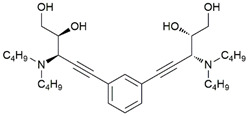
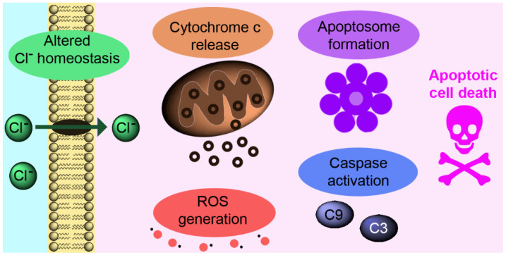
Talukdar’s research group has been very active on the development of artificial ion channels. Bis-diols were reported to self-assemble into amphipathic nanotubes via hydrogen bonds [111]. The diols were tethered at two alkyne ends of a central rigid 1,3-diethynylbenzene moiety and functionalized with dialkylamino groups with different lengths of the alkyl chain to modulate the lipophilicity of the systems. Among the members of the family, compound C1 (Table 2) had the optimal lipophilicity to allow its translocation across the phospholipid membrane of egg-yolk phosphatidylcholine large unilamellar vesicles. A detailed study allowed to define the selectivity towards Cl− over other anions, such as Br−, ClO4−, F−, NO3−, and I−, and the preferred transport mechanism as a Cl−/NO3− antiport. The Cl− transport ability across cell membranes was also successfully tested, by monitoring the dose-dependent fluorescent quenching of the cell-permeable Cl−-selective dye N-(ethoxycarbonylmethyl)-6-methoxyquinolinium bromide (MQAE) after incubation of HeLa cells with C1 for 24 h. The impact of Cl− transport mediated by C1 on cell viability was studied by MTT assay on different cell lines. The increased Cl− level inside the cells resulted in an increase in cell death. The disruption of Cl− homeostasis caused a change in the mitochondrial membrane potential that was accompanied by an increase in ROS production and release of cytochrome c. A detailed study of the cell death mechanism demonstrated that it was caspase-mediated (Figure 4).
Table 2.
Cl− selective artificial channels recently developed to induce apoptosis as anticancer agents.
Figure 4.
Schematic representation of caspase-mediated apoptosis.
The same group reported C2 (Table 2) forming channels for Cl− transport [113,114]. C2 could be obtained upon activation of a precursor, mediated either by an esterase or by glutathione (GSH). The latter is particularly appealing for two reasons. Firstly, GSH is an antioxidant that helps preventing oxidative stress linked to apoptosis in cancer cells [117]. Secondly, high levels of GSH in cancer cells can cause resistance to various chemotherapeutics [118]. The nanotube formed similarly to C1 through non-covalent interactions. The preferred transport mechanism was a M+/Cl− symport, and the channel formation was demonstrated by measuring the ionic conductance across planar lipid bilayer membrane [119] and molecular modelling. The study of cell viability demonstrated that the precursor was more toxic (IC50 = 0.5–1.0 μM) than C2 (IC50 = 50 μM). This difference was attributed to the presence of the sulfonyl group that increases cell membrane permeability. Fluorescence measurements demonstrated that cancer cells treated with the precursor had high levels of GSH that effectively produced C2. Cytotoxicity studies in the presence of Cl− demonstrated that cell death was connected to Cl− transport, with an effect also on the symport transport of Na+ and K+. Similarly to C1, also in this case the apoptotic pathway was caspase-mediated. Interestingly, the authors showed that 3D cultures of breast cancer cells treated with the C2 precursor displayed similar effects to those treated with doxorubicin, a potent inhibitor of their growth [120].
A similar compound (C3, Table 2) formed a channel capable of M+/Cl− symport across unilamellar vesicles and cancer-cell membranes, thus leading to apoptosis. The intrinsic fluorescence of C3 allowed to live-image cancer cells, to reveal the ability of the system to permeate the cytosol. Real-time analysis showed that the permeation occurred in a few seconds, and in a few minutes the cell morphology changed, and the cell volumes were decreased. Apoptosis was confirmed by mitochondrial membrane depolarization, reactive oxygen species generation with consequent cytochrome c release, activation of the caspase 9 pathway, poly (ADP-ribose) polymerase cleavage, and staining of nuclear contents with propidium iodide [115].
Another example of a Cl−-selective transmembrane channel was reported by Zeng and co-workers [116]. The authors described a family of receptors containing an amino acid-based scaffold functionalized with a Cl−-binding group. These receptors were able to self-assemble and stack by means of halogen and hydrogen bonding. In particular, the alanine-substituted C4 (Table 2) showed a good capability for OH−/Cl− antiport. C4 inhibited breast-cancer cell growth with an ic50 value of 20 μm, which was lower than that of the anticancer drug cisplatin (IC50 = 37 μm) [121].
3.2. Artificial Anion Channels as Antimicrobial (AM) Agents
Synthetic transmembrane channels have found applications also as AM agents, thanks to their ability to interact with the membrane of prokaryotic cells, similarly to certain antibiotics and AMPs. Hou and co-workers described the ability of tubular pillar [5]arene macrocycles (C5A–D, Scheme 1) functionalized with amino acidic sidechains to form channels and insert into the lipid bilayers of Gram-positive bacteria [122]. All the channels were selective for K+. C5B–D showed AM activity on Gram-positive S. aureus that was higher than that of gramicidin A, with C5B exhibiting the highest activity (IC50 = 0.3 μM). However, in mammalian-cell membrane models, C5A showed a weaker insertion ability than C5B–D, suggesting that the length of the sidechain played a role in the interaction with membrane lipids. In Gram-positive bacteria S. epidermidis, red-fluorescent derivatives could be imaged as rings in the bacterial wall. The authors hypothesized the formation of H-bonds and dipole interactions between the indole residues of the Trp and the membrane lipids, in contrast with the classic electrostatic interactions between positively charged AMPs and negatively charged membrane [123,124]. Conversely, when C5A–D were incubated with erythrocytes, the red fluorescence rings were not observed only for C5A, with very low hemolytic activity (HC50 > 100 μM).
Scheme 1.
Chemical structures of artificial K+ channels recently developed as AM agents [122,126].
An example of ionophores as AMs and as adjuvants for FDA-approved antibiotics has been reported by Gokel, Patel and co-workers [125]. In previous works, they had described the ability of the crown-ether amphiphile C6 (Scheme 1) to form ion-conducting pores in liposomal membranes and to improve the antibiotic activity of erythromycin, kanamycin, rifampicin, and tetracycline against Gram-negative bacteria (E. coli and P. aeruginosa), probably by enhancing the permeability of the membrane [126,127]. Recently, [125], they demonstrated that C6 can improve the AM activity of tetracycline with ciprofloxacin (i.e., norfloxacin), towards antibiotic-resistant K. pneumoniae. The effect was ascribed to a combination of C6 K+ channels abilities to inhibit bacterial growth and to enhance the bacterial membrane permeability that inhibited the efflux pump, a classic mechanism used by Gram-negative bacteria for pumping out AMs.
3.3. Artificial Anion Transporters as Anticancer Agents
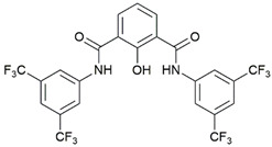
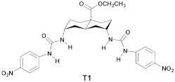
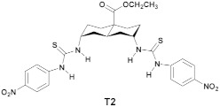
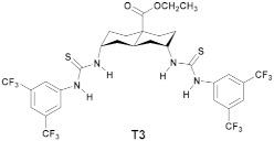
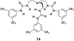
Gale, Davis, and Sheppard have recently showed that some anionophores can be active in cells [128]. They previously presented an assay employing the genetically-encoded halide-sensitive yellow fluorescent protein (YFP) with encouraging results for several anionophores. They reported an implementation of the YFP-based assay using a standard plate reader. The assay was applied to a set of 22 anionophores including several compound classes, such as trans-decalins, substituted cyclohexanes, and the more ideal candidates squaramides, anthracenes, ureas, and thioureas. The data indicated that compounds T2–T4 exhibited levels of activity comparable to the previously reported T1 [129]. Hence, the four compounds (Table 3) were selected for further studies.
Table 3.
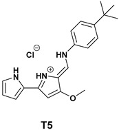
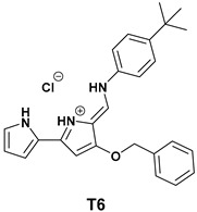
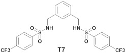
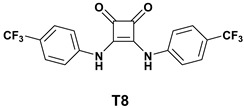
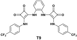
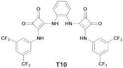
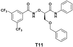
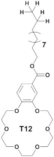
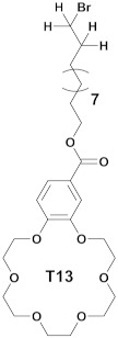
Artificial ion transporters that were recently developed as anticancer agents. T1–T10 are selective for Cl− transport, while T11 and T12 for K+.
| Compound | LogP | EC50 | n | Cell Line | Ref. |
|---|---|---|---|---|---|
|
4.5 | - | - | YFP-CSBE | [129] |
|
4.8 | - | - | YFP-CSBE | [128] |
|
9.3 | - | - | YFP-CSBE | [128] |
|
10.4 | - | - | YFP-CSBE | [128] |
|
4.13 | 50 ± 8 nM | 1.14 ± 0.3 | A549 | [130] |
|
5.58 | 60 ± 3 nM | 1.20 ± 0.09 | A549 | [130] |
|
4.84 | 5.89 ± 0.15 μM | - | MCF-7, U2OS, A549, NIH3T3 |
[131] |
|
3.87 | 1.5 nM | 1.2(0.09) | HeLa, A549 | [132] |
|
- | 2.12 nM | 1.22 (0.05) | HeLa, A549 | [132] |
|
- | 4.1 nM | 0.7 (0.2) | HeLa, A549 | [132] |
|
- | 4.04 mol % | 0.84 | HeLa, HEYA8, SKOV3, CSCs | [133] |
|
- | 0.22 ± 0.02 μM | - | PC3 | [134] |
|
- | 0.17 ± 0.01 μM | - | PC3 | [135] |

|
- | 0.15 ± 0.01 μM | - | PC3 | [135] |
T1–T4 mediated anion transport in CF cells (using CF bronchial epithelial cell line, YFP-CSBE), with T1 and T2 eliciting a similar response, and T4 being the least efficient. Cell treatment with purinergic G protein-coupled (p2y) receptor agonist UTP (uridine triphosphate) elevated the intracellular Ca2+ levels, thus activating Ca2+-dependent Cl− channels, which in turn did not affect the anion transport mediated by T1–T3. Conversely, it enhanced T4-mediated ion transport. Finally, XTT assays in YFP-CSBE cells revealed the cytotoxicity of T2, T3, and especially T4.
Soto-Cerrato, Pérez Tomàs et al. analyzed in detail the cellular and molecular mechanisms of action of two marine alkaloids (i.e., tambjamines), bearing aromatic enamine moieties T5 and T6 (Table 3) [130]. Both promoted transmembrane Cl− and HCO3− transport in liposomes. MTT assays on lung cancer epithelial cells A549 revealed a 50% decrease in cell viability within 24 h. It was demonstrated that the IC50 for these two compounds was much lower than that observed for the anticancer drug cisplatin (CDDP), i.e., 3.38 ± 0.98 and 1.67 ± 0.29 μM, respectively, vs. 200 μM for CDDP. Furthermore, lysosomal pH modifications in the same cell lines were evaluated using acridine orange, a pH-dependent dye. By treating cancer cells with T5 and T6 for 1 h at IC50 values, the orange fluorescence in vesicle compartments disappeared, indicating an increase in pH of these organelles and lysosomal alkalization. This may be ascribed to a lysosomal membrane permeabilization induced by T5 and T6 that could inactivate its hydrolytic enzymes, thus blocking autophagy. Overall, cell death was caused by a combination of caspase-mediated apoptosis, mitochondrial dysfunctions, and lysosome deacidification promoted by a disruption of the cell homeostasis, all triggered by T5 and T6.
Talukdar et al. conducted transmembrane ion transport studies over a set of bis(sulfonamides) derivatives [131]. The anion binding studies and model, along with the anion transport activity (Cl−/NO3− antiport mechanism) studies, suggested that T7 (Table 3) was the most efficient transporter thanks to appropriate lipophilicity and strong anion-binding ability. Cancer-cell and normal-cell viabilities were tested to evaluate how the influx of Cl− into the cell can induce apoptosis. T7 led to higher cancer cell death relative to the untreated controls, whereas no cytotoxicity was observed in non-cancerous cell lines. Moreover, the correlation between the increased extracellular Cl− levels and caspase-mediated cell death was demonstrated. This pattern is well known to trigger apoptosis [136,137,138].
Shin, Gale, Sessler et al. reported a family of squaramides able to cause a malfunctioning of the ion homeostasis and then induce cell death [139]. Compounds T8–T10 (Table 3) were the most active Cl− transporters (T8 > T9 > T10) in liposomes. The experimental evidence highlighted that Cl− was transported via antiport mechanism (Cl−/NO3−, Cl−/HCO3−, Cl−/SO42−, or Cl−/OH−), along with a symport mechanism Cl−/H+. Fischer rat thyroid epithelial cells (FRT) were incubated with each compound and the Cl− transport activity was found to be similar to that observed in liposomes. MTT assays on HeLa and A549 cancer cell lines revealed an IC50 in the range 2–6 μM for T8–T10. The apoptotic effect of T8–T10 was compared against carbonyl cyanide-4-(trifluoromethoxy)phenylhydrazone (FCCP), which is an apoptosis inducer that depolarizes mitochondrial membranes. Cell death was found to be caspase-mediated, rather than necrosis-promoted. The cell death mechanism was further elucidated in another contribution from the same authors [132]. T8 and T9 were studied by Kroemer, Zamzami et al. too, on a CFBE cell line expressing the most frequent CFTR mutation [140]. Cell treatment for 24 h with T8 or T9 revealed they inhibited the autophagic flux, which would have a negative effect on the disease [141,142], thus rendering them more suitable as anticancer agents [142].
Another interesting strategy was presented by Zhang et al. who have successfully constructed an ATP-regulated ion transporter nanosystem for homeostatic perturbation therapy (HTP) and sensitized photodynamic therapy (PDT) [143]. The smart nanotransporter SQU@PCN (porphyrinic porous coordination network incorporated with squaramide T8), accumulated in tumor sites avoiding metabolic clearance and side effects. The affinity of phosphates towards metal ions is well known [144], thus the interaction between the nanosystem and ATP was studied. The decomposition of the nanosystem along with the release of T8 inside cells was observed. T8 triggered Cl− transport across the cell membrane, increasing the intracellular ion concentration, which disrupted ion homeostasis and further induced tumor cell apoptosis.
The viability of HeLa cells after SQU@PCN treatment was assessed via MTT assay. The high cytotoxicity (IC50 = 1.36 mg L−1) was attributed to the release of T8 inside cells, suggesting the selectivity of ATP-SQU@PCN interaction in HeLa cancer cells. Irradiation with a 660-nm laser further enhanced cytotoxicity and lowered the IC50, suggesting the synergic effect of HPT and PDT in killing tumor cells. The excellent cancer-cell toxicity in vitro was confirmed in vivo. In particular, after 12 h of SQU@PCN intravenous injection, fluorescence intensity reached the maximum, suggesting the accumulation of SQU@PCN, especially in the tumor region (Figure 5). Hence, when mice were subjected to light irradiation at 660 nm for 8 min after 12 h of the SQU@PCN injection, the tumor growth was suppressed, confirming what already observed in vitro [143].
Figure 5.
Fluorescence imaging in vivo after intravenous injection with SQU@PCN. Reproduced from ref. [143]. Copyright © 2019 American Chemical Society.
3.4. Artificial Cation Transporters as Anticancer Agents
Yang et al. reported a family of synthetic K+ transporters [135]. They found that T11 (Table 3), an α-aminoxy acid derivative, exhibited the greatest K+ transport ability at the concentration of 10 μM, through a 1:1 carrier mechanism. Anionic T11 was able to bind K+ through electrostatic interaction and ion coordination by the aminoxy oxygen atom and the two carbonyl groups. When T11 entered liposomes with pH~6.8, it could be protonated to release K+ and freely diffuse through the membrane to complete the carrier cycle. T11 was found to be extremely selective towards K+ over other alkaline metal ions with no Cl− transport across membranes. However, by conducting the HPTS assay in the presence of valinomycin and FCCP, the electrogenic transport mediated by T11 displayed H+ > K+, suggesting how T11 could promote the transport of H+ and K+ independently.
T11 was also tested on human ovarian cancer HEYA8 cells, and no ion transport across the plasma membrane was detected. The authors hypothesized that the pH gradient in the intermembrane space (IMS) could be a driving force for the transport of K+ and H+. T11 could freely move through the IMS, in which T11 is present in its anionic form and entraps K+. The exchange K+/H+ finally concluded the cycle. This hypothesis was then confirmed in HeLa cells and in human ovarian SKOV3 cells. The authors further evaluated the mitochondrial ROS production, respiration, and mitochondrial morphology in HEYA8 cells. It was demonstrated that the transport mediated by T11 caused the damage of the mitochondrial functions, due to ion homeostasis dysfunction. Moreover, T11 mediated K+ transport also in ovarian cancer stem cells (CSCs) and it inhibited their growth at 5 μM. This behavior was not observed in the other cancerous and not-cancerous cell lines tested. The high selectivity towards CSCs prompted the authors to evaluate the effect of T11 on tumor growth in nude mice. CSCs were thus incubated ex vivo with T11 or paclitaxel (PTX) and then re-injected into nude mice after 10 days, to reveal a significantly decreased ability to form tumors, relative to controls [135].
Zeng et al. reported the novel class of cation transporters T12–T14 (Table 2), which were characterized by three modular components (i.e., a headgroup, a flexible alkyl-chain derived body, and a crown-ether derived foot for anion binding) [133]. The selective and efficient transport of K+ ions across large unilamellar vesicles promoted by T12–T14 was demonstrated, as suggested by the EC50 = 0.18–0.41 mol % relative to the lipid. Moreover, the most active transporter displayed a potent anticancer activity with low values of IC50 towards HeLa and prostate cancer cells PC3.
3.5. Artificial Ion Transporters as Antimicrobial (AM) Agents
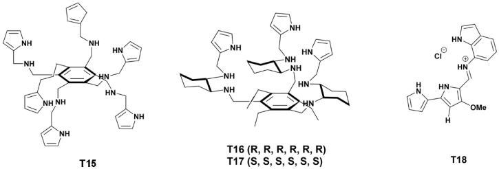
Quesada et al. used 6-indol-7-yl-decorated tambjamine-like compounds T15–T18 (Scheme 2) against Gram-positive and Gram-negative bacterial strains, as well as clinical isolates [145]. In particular, only T18 inhibited the growth of Gram-negatives, and it was also the most potent anionophore of the series, with good hemocompatibility.
Scheme 2.
Chemical structures of T15–T18 [145].
4. Conclusions and Future Perspectives
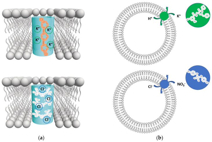
In this Review, we focused on the latest developments in the design of ion channels and transporters for therapeutic use. Firstly, we mentioned a few well-known natural examples based on biomolecules that have inspired biomimetic design, and then we have analyzed more in detail the case of synthetic ionophores (Figure 6) that were tested on biological models, including cells and animals.
Figure 6.
Examples of artificial ion (a) channels and (b) transporters for membrane insertion that derive from supramolecular-chemistry design.
There are many challenges to overcome, to bring artificial ionophores to the clinic. Besides biocompatibility and selectivity for the target cells, ion selectivity is key. This is not always straightforward, for instance for cation transport. However, good K+ selectivity over Na+ can be attained [146,147,148]. Interestingly, simple structural changes can reverse the selectivity and favor Na+ transport [149].
Sometimes the ionophore performance can be improved through the combination of different therapeutic agents or functional molecular components. For instance, addition of biomolecules, such as the K+ transporter valinomycin, to an anion transporter proved to be an effective strategy [150]. Combination with responsive polymers is another approach that is being explored to gain control over transport [151]. Moreover, external stimuli are very attractive to modulate the gating mechanisms, and amongst them, light is the new frontier for therapeutic approaches, especially to control neuronal ion channels [6].
Besides ion channels, also many other types of transmembrane channels offer unexplored therapeutic opportunities. Aquaporins selectively transport water and are impaired in several water balance disorders, such as nephrogenic diabetes insipidus [152]. Biomimetic approaches to develop artificial water channels capable of excluding ions and protons is indeed another area of great innovation potential [153,154]. New characterization methods to assess the transport across membranes are continuously being developed and are envisaged to assist research towards new therapies [155]. Furthermore, they could serve also for diagnosis and consequent development of personalized therapies, as demonstrated for the intestinal current measurements in the case of CF [156]. Overall, as we advance our understanding of artificial ion channels’ structure-activity relationship, and of the biochemical pathways involving ion transport in physiological and pathological contexts, we will witness a bright future at the interface between supramolecular chemistry and biomimetic design to innovate therapy.
Author Contributions
Writing—original draft preparation, G.P.; writing—review and editing, S.M. and C.C.; visualization, G.P., S.M. and C.C. All authors have read and agreed to the published version of the manuscript.
Funding
Financial support from MIUR (PRIN 2017 project 2017EKCS35), Fondazione di Sardegna (FdS Progetti Biennali di Ateneo, annualità 2020) is gratefully acknowledged.
Institutional Review Board Statement
Not applicable.
Informed Consent Statement
Not applicable.
Data Availability Statement
Not applicable.
Conflicts of Interest
The authors declare no conflict of interest.
Footnotes
Publisher’s Note: MDPI stays neutral with regard to jurisdictional claims in published maps and institutional affiliations.
References
- 1.Roux B. Ion channels and ion selectivity. Essays Biochem. 2017;61:201–209. doi: 10.1042/ebc20160074. [DOI] [PMC free article] [PubMed] [Google Scholar]
- 2.Zhang J., Chen X., Xue Y., Gamper N., Zhang X. Beyond voltage-gated ion channels: Voltage-operated membrane proteins and cellular processes. J. Cell. Physiol. 2018;233:6377–6385. doi: 10.1002/jcp.26555. [DOI] [PubMed] [Google Scholar]
- 3.Phillips M.B., Nigam A., Johnson J.W. Interplay between Gating and Block of Ligand-Gated Ion Channels. Brain Sci. 2020;10:928. doi: 10.3390/brainsci10120928. [DOI] [PMC free article] [PubMed] [Google Scholar]
- 4.Murthy S.E., Dubin A.E., Patapoutian A. Piezos thrive under pressure: Mechanically activated ion channels in health and disease. Nat. Rev. Mol. Cell Biol. 2017;18:771–783. doi: 10.1038/nrm.2017.92. [DOI] [PubMed] [Google Scholar]
- 5.Kefauver J.M., Ward A.B., Patapoutian A. Discoveries in structure and physiology of mechanically activated ion channels. Nature. 2020;587:567–576. doi: 10.1038/s41586-020-2933-1. [DOI] [PMC free article] [PubMed] [Google Scholar]
- 6.Paoletti P., Ellis-Davies G.C.R., Mourot A. Optical control of neuronal ion channels and receptors. Nat. Rev. Neurosci. 2019;20:514–532. doi: 10.1038/s41583-019-0197-2. [DOI] [PMC free article] [PubMed] [Google Scholar]
- 7.Liu Y., Wang K. Exploiting the Diversity of Ion Channels: Modulation of Ion Channels for Therapeutic Indications. Handb. Exp. Pharmacol. 2019;260:187–205. doi: 10.1007/164_2019_333. [DOI] [PubMed] [Google Scholar]
- 8.Santos R., Ursu O., Gaulton A., Bento A.P., Donadi R.S., Bologa C.G., Karlsson A., Al-Lazikani B., Hersey A., Oprea T.I., et al. A comprehensive map of molecular drug targets. Nat. Rev. Drug Discov. 2017;16:19–34. doi: 10.1038/nrd.2016.230. [DOI] [PMC free article] [PubMed] [Google Scholar]
- 9.Zhang Y., Wang K., Yu Z. Drug Development in Channelopathies: Allosteric Modulation of Ligand-Gated and Voltage-Gated Ion Channels. J. Med. Chem. 2020;63:15258–15278. doi: 10.1021/acs.jmedchem.0c01304. [DOI] [PubMed] [Google Scholar]
- 10.Poveda J.A., Marcela Giudici A., Lourdes Renart M., Morales A., Gonzalez-Ros J.M. Towards understanding the molecular basis of ion channel modulation by lipids: Mechanistic models and current paradigms. Biochim. Biophys. Acta Biomembr. 2017;1859:1507–1516. doi: 10.1016/j.bbamem.2017.04.003. [DOI] [PubMed] [Google Scholar]
- 11.Thompson M.J., Baenziger J.E. Ion channels as lipid sensors: From structures to mechanisms. Nat. Chem. Biol. 2020;16:1331–1342. doi: 10.1038/s41589-020-00693-3. [DOI] [PubMed] [Google Scholar]
- 12.Harraz O.F., Hill-Eubanks D., Nelson M.T. PIP2: A critical regulator of vascular ion channels hiding in plain sight. Proc. Natl. Acad. Sci. USA. 2020;117:20378–20389. doi: 10.1073/pnas.2006737117. [DOI] [PMC free article] [PubMed] [Google Scholar]
- 13.Kozlov S. Animal toxins for channelopathy treatment. Neuropharmacology. 2018;132:83–97. doi: 10.1016/j.neuropharm.2017.10.031. [DOI] [PubMed] [Google Scholar]
- 14.Stortelers C., Pinto-Espinoza C., Van Hoorick D., Koch-Nolte F. Modulating ion channel function with antibodies and nanobodies. Curr. Opin. Immunol. 2018;52:18–26. doi: 10.1016/j.coi.2018.02.003. [DOI] [PubMed] [Google Scholar]
- 15.Norton R.S., Chandy K.G. Venom-derived peptide inhibitors of voltage-gated potassium channels. Neuropharmacology. 2017;127:124–138. doi: 10.1016/j.neuropharm.2017.07.002. [DOI] [PubMed] [Google Scholar]
- 16.Chow C.Y., Absalom N., Biggs K., King G.F., Ma L. Venom-derived modulators of epilepsy-related ion channels. Biochem. Pharmacol. 2020;181:114043. doi: 10.1016/j.bcp.2020.114043. [DOI] [PubMed] [Google Scholar]
- 17.Sawarkar R., Bhandarkar S., Mendhi S., More S. Channelopathies an approach to elevate level of cure- a review. Int. J. Pharm. Sci. Rev. Res. 2021;70:65–74. doi: 10.47583/ijpsrr.2021.v70i02.010. [DOI] [Google Scholar]
- 18.Matthews E., Holmes S., Fialho D. Skeletal muscle channelopathies: A guide to diagnosis and management. Pract. Neurol. 2021;21:196–204. doi: 10.1136/practneurol-2020-002576. [DOI] [PubMed] [Google Scholar]
- 19.Vaeth M., Feske S. Ion channelopathies of the immune system. Curr. Opin. Immunol. 2018;52:39–50. doi: 10.1016/j.coi.2018.03.021. [DOI] [PMC free article] [PubMed] [Google Scholar]
- 20.Demirbilek H., Galcheva S., Vuralli D., Al-Khawaga S., Hussain K. Ion Transporters, Channelopathies, and Glucose Disorders. Int. J. Mol. Sci. 2019;20:2590. doi: 10.3390/ijms20102590. [DOI] [PMC free article] [PubMed] [Google Scholar]
- 21.Meisler M.H., Hill S.F., Yu W. Sodium channelopathies in neurodevelopmental disorders. Nat. Rev. Neurosci. 2021;22:152–166. doi: 10.1038/s41583-020-00418-4. [DOI] [PMC free article] [PubMed] [Google Scholar]
- 22.Fonseca D.J., Vaz da Silva M.J. Cardiac channelopathies: The role of sodium channel mutations. Rev. Port. Cardiol. 2018;37:179–199. doi: 10.1016/j.repc.2017.11.007. [DOI] [PubMed] [Google Scholar]
- 23.Albury C.L., Stuart S., Haupt L.M., Griffiths L.R. Ion channelopathies and migraine pathogenesis. Mol. Genet. Genom. 2017;292:729–739. doi: 10.1007/s00438-017-1317-1. [DOI] [PubMed] [Google Scholar]
- 24.Terragni B., Scalmani P., Franceschetti S., Cestele S., Mantegazza M. Post-translational dysfunctions in channelopathies of the nervous system. Neuropharmacology. 2018;132:31–42. doi: 10.1016/j.neuropharm.2017.05.028. [DOI] [PubMed] [Google Scholar]
- 25.Curran J., Mohlr P.J. Alternative paradigms for ion channelopathies: Disorders of ion channel membrane trafficking and posttranslational modification. Annu. Rev. Physiol. 2015;77:505–524. doi: 10.1146/annurev-physiol-021014-071838. [DOI] [PubMed] [Google Scholar]
- 26.Li G., De Oliveira D.M.P., Walker M.J. The antimicrobial and immunomodulatory effects of ionophores for the treatment of human infection. J. Inorg. Biochem. 2022;227:111661. doi: 10.1016/j.jinorgbio.2021.111661. [DOI] [PubMed] [Google Scholar]
- 27.Kaushik V., Yakisich J.S., Kumar A., Azad N., Iyer A.K.V. Ionophores: Potential Use as Anticancer Drugs and Chemosensitizers. Cancers. 2018;10:360. doi: 10.3390/cancers10100360. [DOI] [PMC free article] [PubMed] [Google Scholar]
- 28.Steinbrueck A., Sedgwick A.C., Brewster J.T., 2nd, Yan K.C., Shang Y., Knoll D.M., Vargas-Zúñiga G.I., He X.P., Tian H., Sessler J.L. Transition metal chelators, pro-chelators, and ionophores as small molecule cancer chemotherapeutic agents. Chem. Soc. Rev. 2020;49:3726–3747. doi: 10.1039/C9CS00373H. [DOI] [PubMed] [Google Scholar]
- 29.Bharti H., Singal A., Raza M., Ghosh P.C., Nag A. Ionophores as Potent Anti-malarials: A Miracle in the Making. Curr. Top. Med. Chem. 2019;18:2029–2041. doi: 10.2174/1568026619666181129125950. [DOI] [PubMed] [Google Scholar]
- 30.Antoszczak M., Steverding D., Huczyński A. Anti-parasitic activity of polyether ionophores. Eur. J. Med. Chem. 2019;166:32–47. doi: 10.1016/j.ejmech.2019.01.035. [DOI] [PubMed] [Google Scholar]
- 31.Prabhakar P.K. Bacterial Siderophores and Their Potential Applications: A Review. Curr. Mol. Pharmacl. 2020;13:295–305. doi: 10.2174/1874467213666200518094445. [DOI] [PubMed] [Google Scholar]
- 32.Bhullar S.K., Shah A.K., Dhalla N.S. Store-operated calcium channels: Potential target for the therapy of hypertension. Rev. Cardiovasc. Med. 2019;20:139–151. doi: 10.31083/j.rcm.2019.03.522. [DOI] [PubMed] [Google Scholar]
- 33.Hu H.J., Song M. Disrupted Ionic Homeostasis in Ischemic Stroke and New Therapeutic Targets. J. Stroke Cerebrovasc. Dis. 2017;26:2706–2719. doi: 10.1016/j.jstrokecerebrovasdis.2017.09.011. [DOI] [PubMed] [Google Scholar]
- 34.Thapak P., Vaidya B., Joshi H.C., Singh J.N., Sharma S.S. Therapeutic potential of pharmacological agents targeting TRP channels in CNS disorders. Pharmacol. Res. 2020;159:105026. doi: 10.1016/j.phrs.2020.105026. [DOI] [PubMed] [Google Scholar]
- 35.Bergantin L.B. The Interactions Between Alzheimer’s Disease and Major Depression: Role of Ca(2+) Channel Blockers and Ca(2+)/cAMP Signalling. Curr. Drug Res. Rev. 2020;12:97–102. doi: 10.2174/2589977512666200217093356. [DOI] [PubMed] [Google Scholar]
- 36.Tong B.C., Wu A.J., Li M., Cheung K.H. Calcium signaling in Alzheimer’s disease & therapies. Biochim. Biophys. Acta. Mol. Cell Res. 2018;1865:1745–1760. doi: 10.1016/j.bbamcr.2018.07.018. [DOI] [PubMed] [Google Scholar]
- 37.Popugaeva E., Pchitskaya E., Bezprozvanny I. Dysregulation of neuronal calcium homeostasis in Alzheimer’s disease—A therapeutic opportunity? Biochem. Biophys. Res. Commun. 2017;483:998–1004. doi: 10.1016/j.bbrc.2016.09.053. [DOI] [PMC free article] [PubMed] [Google Scholar]
- 38.Kato T. Current understanding of bipolar disorder: Toward integration of biological basis and treatment strategies. Psychiatry Clin. Neurosci. 2019;73:526–540. doi: 10.1111/pcn.12852. [DOI] [PubMed] [Google Scholar]
- 39.Dubovsky S.L. Applications of calcium channel blockers in psychiatry: Pharmacokinetic and pharmacodynamic aspects of treatment of bipolar disorder. Exp. Opin. Drug Metab. Toxicol. 2019;15:35–47. doi: 10.1080/17425255.2019.1558206. [DOI] [PubMed] [Google Scholar]
- 40.Garneau A.P., Slimani S., Fiola M.J., Tremblay L.E., Isenring P. Multiple Facets and Roles of Na(+)-K(+)-Cl(−) Cotransport: Mechanisms and Therapeutic Implications. Physiology. 2020;35:415–429. doi: 10.1152/physiol.00012.2020. [DOI] [PubMed] [Google Scholar]
- 41.Viswanath O., Urits I., Jones M.R., Peck J.M., Kochanski J., Hasegawa M., Anyama B., Kaye A.D. Membrane Stabilizer Medications in the Treatment of Chronic Neuropathic Pain: A Comprehensive Review. Curr. Pain Headache Rep. 2019;23:37. doi: 10.1007/s11916-019-0774-0. [DOI] [PubMed] [Google Scholar]
- 42.Karsan N., Gonzales E.B., Dussor G. Targeted Acid-Sensing Ion Channel Therapies for Migraine. Neurotherapeutics. 2018;15:402–414. doi: 10.1007/s13311-018-0619-2. [DOI] [PMC free article] [PubMed] [Google Scholar]
- 43.Jacobson D.A., Shyng S.L. Ion Channels of the Islets in Type 2 Diabetes. J. Mol. Biol. 2020;432:1326–1346. doi: 10.1016/j.jmb.2019.08.014. [DOI] [PMC free article] [PubMed] [Google Scholar]
- 44.Selvaraj C., Selvaraj G., Kaliamurthi S., Cho W.C., Wei D.Q., Singh S.K. Ion Channels as Therapeutic Targets for Type 1 Diabetes Mellitus. Curr. Drug Targets. 2020;21:132–147. doi: 10.2174/1389450119666190920152249. [DOI] [PubMed] [Google Scholar]
- 45.Ali E.S., Petrovsky N. Calcium Signaling As a Therapeutic Target for Liver Steatosis. Trends Endocrinol. Metab. 2019;30:270–281. doi: 10.1016/j.tem.2019.02.005. [DOI] [PubMed] [Google Scholar]
- 46.Wulff H., Christophersen P., Colussi P., Chandy K.G., Yarov-Yarovoy V. Antibodies and venom peptides: New modalities for ion channels. Nat. Rev. Drug Discov. 2019;18:339–357. doi: 10.1038/s41573-019-0013-8. [DOI] [PMC free article] [PubMed] [Google Scholar]
- 47.Jeevaratnam K., Chadda K.R., Huang C.L.H., Camm A.J. Cardiac Potassium Channels: Physiological Insights for Targeted Therapy. J. Cardiovasc. Pharmacol. Ther. 2018;23:119–129. doi: 10.1177/1074248417729880. [DOI] [PMC free article] [PubMed] [Google Scholar]
- 48.Bushart D.D., Shakkottai V.G. Ion channel dysfunction in cerebellar ataxia. Neurosci. Lett. 2019;688:41–48. doi: 10.1016/j.neulet.2018.02.005. [DOI] [PMC free article] [PubMed] [Google Scholar]
- 49.Szabo I., Zoratti M., Biasutto L. Targeting mitochondrial ion channels for cancer therapy. Redox Biol. 2021;42:101846. doi: 10.1016/j.redox.2020.101846. [DOI] [PMC free article] [PubMed] [Google Scholar]
- 50.Sterea A.M., Almasi S., El Hiani Y. The hidden potential of lysosomal ion channels: A new era of oncogenes. Cell Calcium. 2018;72:91–103. doi: 10.1016/j.ceca.2018.02.006. [DOI] [PubMed] [Google Scholar]
- 51.Marchi S., Giorgi C., Galluzzi L., Pinton P. Ca(2+) Fluxes and Cancer. Mol. Cell. 2020;78:1055–1069. doi: 10.1016/j.molcel.2020.04.017. [DOI] [PubMed] [Google Scholar]
- 52.Fnu G., Weber G.F. Alterations of Ion Homeostasis in Cancer Metastasis: Implications for Treatment. Front. Oncol. 2021;11:765329. doi: 10.3389/fonc.2021.765329. [DOI] [PMC free article] [PubMed] [Google Scholar]
- 53.Seitter H., Koschak A. Relevance of tissue specific subunit expression in channelopathies. Neuropharmacol. 2018;132:58–70. doi: 10.1016/j.neuropharm.2017.06.029. [DOI] [PMC free article] [PubMed] [Google Scholar]
- 54.Gargan S., Stevenson N.J. Unravelling the Immunomodulatory Effects of Viral Ion Channels, towards the Treatment of Disease. Viruses. 2021;13:2165. doi: 10.3390/v13112165. [DOI] [PMC free article] [PubMed] [Google Scholar]
- 55.Verkman A.S., Galietta L.J.V. Chloride transport modulators as drug candidates. Am. J. Physiol. Cell Physiol. 2021;321:C932–C946. doi: 10.1152/ajpcell.00334.2021. [DOI] [PMC free article] [PubMed] [Google Scholar]
- 56.Bergeron C., Cantin A.M. New Therapies to Correct the Cystic Fibrosis Basic Defect. Int. J. Mol. Sci. 2021;22:6193. doi: 10.3390/ijms22126193. [DOI] [PMC free article] [PubMed] [Google Scholar]
- 57.Laselva O., Guerra L., Castellani S., Favia M., Di Gioia S., Conese M. Small-molecule drugs for cystic fibrosis: Where are we now? Pulm. Pharmacol. Ther. 2021;72:102098. doi: 10.1016/j.pupt.2021.102098. [DOI] [PubMed] [Google Scholar]
- 58.Fonseca C., Bicker J., Alves G., Falcão A., Fortuna A. Cystic fibrosis: Physiopathology and the latest pharmacological treatments. Pharmacol. Res. 2020;162:105267. doi: 10.1016/j.phrs.2020.105267. [DOI] [PubMed] [Google Scholar]
- 59.Shteinberg M., Haq I.J., Polineni D., Davies J.C. Cystic fibrosis. Lancet. 2021;397:2195–2211. doi: 10.1016/S0140-6736(20)32542-3. [DOI] [PubMed] [Google Scholar]
- 60.Bishnoi M., Khare P., Brown L., Panchal S.K. Transient receptor potential (TRP) channels: A metabolic TR(i)P to obesity prevention and therapy. Obes. Rev. 2018;19:1269–1292. doi: 10.1111/obr.12703. [DOI] [PubMed] [Google Scholar]
- 61.Dueñas-Cuellar R.A., Santana C.J.C., Magalhães A.C.M., Pires O.R., Jr., Fontes W., Castro M.S. Scorpion Toxins and Ion Channels: Potential Applications in Cancer Therapy. Toxins. 2020;12:326. doi: 10.3390/toxins12050326. [DOI] [PMC free article] [PubMed] [Google Scholar]
- 62.Yang X., Lou J., Shan W., Hu Y., Du Q., Liao Q., Xie R., Xu J. Pathogenic roles of altered calcium channels and transporters in colon tumorogenesis. Life Sci. 2019;239:116909. doi: 10.1016/j.lfs.2019.116909. [DOI] [PubMed] [Google Scholar]
- 63.Prasad H., Visweswariah S.S. Impaired Intestinal Sodium Transport in Inflammatory Bowel Disease: From the Passenger to the Driver’s Seat. Cell. Mol. Gastroenterol. 2021;12:277–292. doi: 10.1016/j.jcmgh.2021.03.005. [DOI] [PMC free article] [PubMed] [Google Scholar]
- 64.Das S., Jayaratne R., Barrett K.E. The Role of Ion Transporters in the Pathophysiology of Infectious Diarrhea. Cell. Mol. Gastroenterol. 2018;6:33–45. doi: 10.1016/j.jcmgh.2018.02.009. [DOI] [PMC free article] [PubMed] [Google Scholar]
- 65.Auwercx J., Rybarczyk P., Kischel P., Dhennin-Duthille I., Chatelain D., Sevestre H., Van Seuningen I., Ouadid-Ahidouch H., Jonckheere N., Gautier M. Mg(2+) Transporters in Digestive Cancers. Nutrients. 2021;13:210. doi: 10.3390/nu13010210. [DOI] [PMC free article] [PubMed] [Google Scholar]
- 66.Adulcikas J., Sonda S., Norouzi S., Sohal S.S., Myers S. Targeting the Zinc Transporter ZIP7 in the Treatment of Insulin Resistance and Type 2 Diabetes. Nutrients. 2019;11:408. doi: 10.3390/nu11020408. [DOI] [PMC free article] [PubMed] [Google Scholar]
- 67.Shen J., Ye R., Zeng H. Crystal Packing-Guided Construction of Hetero-Oligomeric Peptidic Ensembles as Synthetic 3-in-1 Transporters. Angew. Chem. Int. Ed. 2021;60:12924–12930. doi: 10.1002/anie.202101489. [DOI] [PubMed] [Google Scholar]
- 68.Inoue K. Diversity, Mechanism, and Optogenetic Application of Light-Driven Ion Pump Rhodopsins. Adv. Exp. Med. Biol. 2021;1293:89–126. doi: 10.1007/978-981-15-8763-4_6. [DOI] [PubMed] [Google Scholar]
- 69.Kandori H. History and Perspectives of Ion-Transporting Rhodopsins. Adv. Exp. Med. Biol. 2021;1293:3–19. doi: 10.1007/978-981-15-8763-4_1. [DOI] [PubMed] [Google Scholar]
- 70.Engelhard C., Chizhov I., Siebert F., Engelhard M. Microbial Halorhodopsins: Light-Driven Chloride Pumps. Chem. Rev. 2018;118:10629–10645. doi: 10.1021/acs.chemrev.7b00715. [DOI] [PubMed] [Google Scholar]
- 71.Lu Q., Pan Z.H. Optogenetic Strategies for Vision Restoration. Adv. Exp. Med. Biol. 2021;1293:545–555. doi: 10.1007/978-981-15-8763-4_38. [DOI] [PubMed] [Google Scholar]
- 72.Barboiu M. Encapsulation versus Self-Aggregation toward Highly Selective Artificial K(+) Channels. Acc. Chem. Res. 2018;51:2711–2718. doi: 10.1021/acs.accounts.8b00311. [DOI] [PubMed] [Google Scholar]
- 73.Schneider S., Licsandru E.D., Kocsis I., Gilles A., Dumitru F., Moulin E., Tan J., Lehn J.M., Giuseppone N., Barboiu M. Columnar Self-Assemblies of Triarylamines as Scaffolds for Artificial Biomimetic Channels for Ion and for Water Transport. J. Am. Chem. Soc. 2017;139:3721–3727. doi: 10.1021/jacs.6b12094. [DOI] [PubMed] [Google Scholar]
- 74.Wang W.Z., Huang L.B., Zheng S.P., Moulin E., Gavat O., Barboiu M., Giuseppone N. Light-Driven Molecular Motors Boost the Selective Transport of Alkali Metal Ions through Phospholipid Bilayers. J. Am. Chem. Soc. 2021;143:15653–15660. doi: 10.1021/jacs.1c05750. [DOI] [PubMed] [Google Scholar]
- 75.Takada Y., Itoh H., Paudel A., Panthee S., Hamamoto H., Sekimizu K., Inoue M. Discovery of gramicidin A analogues with altered activities by multidimensional screening of a one-bead-one-compound library. Nat. Commun. 2020;11:4935. doi: 10.1038/s41467-020-18711-2. [DOI] [PMC free article] [PubMed] [Google Scholar]
- 76.Haoyang W.W., Xiao Q., Ye Z., Fu Y., Zhang D.W., Li J., Xiao L., Li Z.T., Hou J.L. Gramicidin A-based unimolecular channel: Cancer cell-targeting behavior and ion transport-induced apoptosis. Chem. Commun. 2021;57:1097–1100. doi: 10.1039/D0CC08073J. [DOI] [PubMed] [Google Scholar]
- 77.Ren C., Chen F., Ye R., Ong Y.S., Lu H., Lee S.S., Ying J.Y., Zeng H. Molecular Swings as Highly Active Ion Transporters. Angew. Chem. Int. Ed. 2019;58:8034–8038. doi: 10.1002/anie.201901833. [DOI] [PubMed] [Google Scholar]
- 78.Shen J., Fan J., Ye R., Li N., Mu Y., Zeng H. Polypyridine-Based Helical Amide Foldamer Channels: Rapid Transport of Water and Protons with High Ion Rejection. Angew. Chem. Int. Ed. 2020;59:13328–13334. doi: 10.1002/anie.202003512. [DOI] [PubMed] [Google Scholar]
- 79.Rodríguez-Vázquez N., Amorín M., Granja J.R. Recent advances in controlling the internal and external properties of self-assembling cyclic peptide nanotubes and dimers. Org. Biomol. Chem. 2017;15:4490–4505. doi: 10.1039/C7OB00351J. [DOI] [PubMed] [Google Scholar]
- 80.Fuertes A., Juanes M., Granja J.R., Montenegro J. Supramolecular functional assemblies: Dynamic membrane transporters and peptide nanotubular composites. Chem. Commun. 2017;53:7861–7871. doi: 10.1039/C7CC02997G. [DOI] [PubMed] [Google Scholar]
- 81.Görbitz C.H. Microporous organic materials from hydrophobic dipeptides. Chem. Eur. J. 2007;13:1022–1031. doi: 10.1002/chem.200601427. [DOI] [PubMed] [Google Scholar]
- 82.Bellotto O., Kralj S., De Zorzi R., Geremia S., Marchesan S. Supramolecular hydrogels from unprotected dipeptides: A comparative study on stereoisomers and structural isomers. Soft Matter. 2020;16:10151–10157. doi: 10.1039/D0SM01191F. [DOI] [PubMed] [Google Scholar]
- 83.Bellotto O., Kralj S., Melchionna M., Pengo P., Kisovec M., Podobnik M., De Zorzi R., Marchesan S. Self-Assembly of Unprotected Dipeptides into Hydrogels: Water-Channels Make the Difference. Chembiochem. 2022;23:e202100518. doi: 10.1002/cbic.202100518. [DOI] [PMC free article] [PubMed] [Google Scholar]
- 84.Kralj S., Bellotto O., Parisi E., Garcia A.M., Iglesias D., Semeraro S., Deganutti C., D’Andrea P., Vargiu A.V., Geremia S., et al. Heterochirality and Halogenation Control Phe-Phe Hierarchical Assembly. ACS Nano. 2020;14:16951–16961. doi: 10.1021/acsnano.0c06041. [DOI] [PMC free article] [PubMed] [Google Scholar]
- 85.Kurbasic M., Parisi E., Garcia A.M., Marchesan S. Self-Assembling, Ultrashort Peptide Gels as Antimicrobial Biomaterials. Curr. Top. Med. Chem. 2020;20:1300–1309. doi: 10.2174/1568026620666200316150221. [DOI] [PubMed] [Google Scholar]
- 86.Bellotto O., Semeraro S., Bandiera A., Tramer F., Pavan N., Marchesan S. Polymer Conjugates of Antimicrobial Peptides (AMPs) with d-Amino Acids (d-aa): State of the Art and Future Opportunities. Pharmaceutics. 2022;14:446. doi: 10.3390/pharmaceutics14020446. [DOI] [PMC free article] [PubMed] [Google Scholar]
- 87.Muraglia K.A., Chorghade R.S., Kim B.R., Tang X.X., Shah V.S., Grillo A.S., Daniels P.N., Cioffi A.G., Karp P.H., Zhu L., et al. Small-molecule ion channels increase host defences in cystic fibrosis airway epithelia. Nature. 2019;567:405–408. doi: 10.1038/s41586-019-1018-5. [DOI] [PMC free article] [PubMed] [Google Scholar]
- 88.Sheppard D.N., Davis A.P. Pore-forming small molecules offer a promising way to tackle cystic fibrosis. Nature. 2019;567:315–317. doi: 10.1038/d41586-019-00781-y. [DOI] [PubMed] [Google Scholar]
- 89.Chen C.H., Lu T.K. Development and Challenges of Antimicrobial Peptides for Therapeutic Applications. Antibiotics. 2020;9:24. doi: 10.3390/antibiotics9010024. [DOI] [PMC free article] [PubMed] [Google Scholar]
- 90.Huang H.W. DAPTOMYCIN, its membrane-active mechanism vs. that of other antimicrobial peptides. Biochim. Biophys. Acta Biomembr. 2020;1862:183395. doi: 10.1016/j.bbamem.2020.183395. [DOI] [PubMed] [Google Scholar]
- 91.Malla J.A., Ahmad M., Talukdar P. Molecular Self-Assembly as a Tool to Construct Transmembrane Supramolecular Ion Channels. Chem. Rec. 2021;22:e202100225. doi: 10.1002/tcr.202100225. [DOI] [PubMed] [Google Scholar]
- 92.Zheng S.P., Huang L.B., Sun Z., Barboiu M. Self-Assembled Artificial Ion-Channels toward Natural Selection of Functions. Angew. Chem. Int. Ed. 2021;60:566–597. doi: 10.1002/anie.201915287. [DOI] [PubMed] [Google Scholar]
- 93.Peng S., He Q., Vargas-Zúñiga G.I., Qin L., Hwang I., Kim S.K., Heo N.J., Lee C.H., Dutta R., Sessler J.L. Strapped calix[4]pyrroles: From syntheses to applications. Chem. Soc. Rev. 2020;49:865–907. doi: 10.1039/C9CS00528E. [DOI] [PubMed] [Google Scholar]
- 94.Nitti A., Pacini A., Pasini D. Chiral Nanotubes. Nanomaterials. 2017;7:167. doi: 10.3390/nano7070167. [DOI] [PMC free article] [PubMed] [Google Scholar]
- 95.Roy A., Talukdar P. Recent Advances in Bioactive Artificial Ionophores. Chembiochem. 2021;22:2925–2940. doi: 10.1002/cbic.202100112. [DOI] [PMC free article] [PubMed] [Google Scholar]
- 96.Tosolini M., Pengo P., Tecilla P. Biological Activity of Trans-Membrane Anion Carriers. Curr. Med. Chem. 2018;25:3560–3576. doi: 10.2174/0929867325666180309113222. [DOI] [PubMed] [Google Scholar]
- 97.Gale P.A., Davis J.T., Quesada R. Anion transport and supramolecular medicinal chemistry. Chem. Soc. Rev. 2017;46:2497–2519. doi: 10.1039/C7CS00159B. [DOI] [PubMed] [Google Scholar]
- 98.Gilchrist A.M., Chen L., Wu X., Lewis W., Howe E.N.W., Macreadie L.K., Gale P.A. Tetrapodal Anion Transporters. Molecules. 2020;25:5179. doi: 10.3390/molecules25215179. [DOI] [PMC free article] [PubMed] [Google Scholar]
- 99.Zhang C., Tian J., Qi S., Yang B., Dong Z. Highly Efficient Exclusion of Alkali Metal Ions via Electrostatic Repulsion Inside Positively Charged Channels. Nano Lett. 2020;20:3627–3632. doi: 10.1021/acs.nanolett.0c00567. [DOI] [PubMed] [Google Scholar]
- 100.Martínez-Crespo L., Hewitt S.H., De Simone N.A., Šindelář V., Davis A.P., Butler S., Valkenier H. Transmembrane Transport of Bicarbonate Unravelled. Chem. Eur. J. 2021;27:7367–7375. doi: 10.1002/chem.202100491. [DOI] [PMC free article] [PubMed] [Google Scholar]
- 101.Roy A., Joshi H., Ye R., Shen J., Chen F., Aksimentiev A., Zeng H. Polyhydrazide-Based Organic Nanotubes as Efficient and Selective Artificial Iodide Channels. Angew. Chem. Int. Ed. 2020;59:4806–4813. doi: 10.1002/anie.201916287. [DOI] [PMC free article] [PubMed] [Google Scholar]
- 102.He Q., Vargas-Zúñiga G.I., Kim S.H., Kim S.K., Sessler J.L. Macrocycles as Ion Pair Receptors. Chem. Rev. 2019;119:9753–9835. doi: 10.1021/acs.chemrev.8b00734. [DOI] [PubMed] [Google Scholar]
- 103.McConnell A.J., Docker A., Beer P.D. From Heteroditopic to Multitopic Receptors for Ion-Pair Recognition: Advances in Receptor Design and Applications. ChemPlusChem. 2020;85:1824–1841. doi: 10.1002/cplu.202000484. [DOI] [PubMed] [Google Scholar]
- 104.Mamad-Hemouch H., Bacri L., Huin C., Przybylski C., Thiébot B., Patriarche G., Jarroux N., Pelta J. Versatile cyclodextrin nanotube synthesis with functional anchors for efficient ion channel formation: Design, characterization and ion conductance. Nanoscale. 2018;10:15303–15316. doi: 10.1039/C8NR02623H. [DOI] [PubMed] [Google Scholar]
- 105.Quan J., Zhu F., Dhinakaran M.K., Yang Y., Johnson R.P., Li H. A Visible-Light-Regulated Chloride Transport Channel Inspired by Rhodopsin. Angew. Chem. Int. Ed. 2021;60:2892–2897. doi: 10.1002/anie.202012984. [DOI] [PubMed] [Google Scholar]
- 106.Kerckhoffs A., Langton M.J. Reversible photo-control over transmembrane anion transport using visible-light responsive supramolecular carriers. Chem. Sci. 2020;11:6325–6331. doi: 10.1039/D0SC02745F. [DOI] [PMC free article] [PubMed] [Google Scholar]
- 107.Ahmad M., Metya S., Das A., Talukdar P. A Sandwich Azobenzene-Diamide Dimer for Photoregulated Chloride Transport. Chem. Eur. J. 2020;26:8703–8708. doi: 10.1002/chem.202000400. [DOI] [PubMed] [Google Scholar]
- 108.Haynes C.J.E., Zhu J., Chimerel C., Hernández-Ainsa S., Riddell I.A., Ronson T.K., Keyser U.F., Nitschke J.R. Blockable Zn(10) L(15) Ion Channels through Subcomponent Self-Assembly. Angew. Chem. Int. Ed. 2017;56:15388–15392. doi: 10.1002/anie.201709544. [DOI] [PubMed] [Google Scholar]
- 109.Wu X., Small J.R., Cataldo A., Withecombe A.M., Turner P., Gale P.A. Voltage-Switchable HCl Transport Enabled by Lipid Headgroup-Transporter Interactions. Angew. Chem. Int. Ed. 2019;58:15142–15147. doi: 10.1002/anie.201907466. [DOI] [PubMed] [Google Scholar]
- 110.Sasaki R., Sato K., Tabata K.V., Noji H., Kinbara K. Synthetic Ion Channel Formed by Multiblock Amphiphile with Anisotropic Dual-Stimuli-Responsiveness. J. Am. Chem. Soc. 2021;143:1348–1355. doi: 10.1021/jacs.0c09470. [DOI] [PubMed] [Google Scholar]
- 111.Saha T., Gautam A., Mukherjee A., Lahiri M., Talukdar P. Chloride Transport through Supramolecular Barrel-Rosette Ion Channels: Lipophilic Control and Apoptosis-Inducing Activity. J. Am. Chem. Soc. 2016;138:16443–16451. doi: 10.1021/jacs.6b10379. [DOI] [PubMed] [Google Scholar]
- 112.Akhtar N., Biswas D., Manna D. Biological applications of synthetic anion transporters. Chem. Commun. 2020;56:14137–14153. doi: 10.1039/D0CC05489E. [DOI] [PubMed] [Google Scholar]
- 113.Malla J.A., Umesh R.M., Yousf S., Mane S., Sharma S., Lahiri M., Talukdar P. A Glutathione Activatable Ion Channel Induces Apoptosis in Cancer Cells by Depleting Intracellular Glutathione Levels. Angew. Chem. Int. Ed. 2020;59:7944–7952. doi: 10.1002/anie.202000961. [DOI] [PubMed] [Google Scholar]
- 114.Malla J.A., Sharma V.K., Lahiri M., Talukdar P. Esterase-Activatable Synthetic M+/Cl− Channel Induces Apoptosis and Disrupts Autophagy in Cancer Cells. Chem. Eur. J. 2020;26:11946–11949. doi: 10.1002/chem.202002964. [DOI] [PubMed] [Google Scholar]
- 115.Malla J.A., Umesh R.M., Vijay A., Mukherjee A., Lahiri M., Talukdar P. Apoptosis-inducing activity of a fluorescent barrel-rosette M(+)/Cl(−) channel. Chem. Sci. 2020;11:2420–2428. doi: 10.1039/C9SC06520B. [DOI] [PMC free article] [PubMed] [Google Scholar]
- 116.Ren C., Ding X., Roy A., Shen J., Zhou S., Chen F., Yau Li S.F., Ren H., Yang Y.Y., Zeng H. A halogen bond-mediated highly active artificial chloride channel with high anticancer activity. Chem. Sci. 2018;9:4044–4051. doi: 10.1039/C8SC00602D. [DOI] [PMC free article] [PubMed] [Google Scholar]
- 117.Quintana-Cabrera R., Fernandez-Fernandez S., Bobo-Jimenez V., Escobar J., Sastre J., Almeida A., Bolaños J.P. γ-Glutamylcysteine detoxifies reactive oxygen species by acting as glutathione peroxidase-1 cofactor. Nat. Commun. 2012;3:718. doi: 10.1038/ncomms1722. [DOI] [PMC free article] [PubMed] [Google Scholar]
- 118.Traverso N., Ricciarelli R., Nitti M., Marengo B., Furfaro A.L., Pronzato M.A., Marinari U.M., Domenicotti C. Role of Glutathione in Cancer Progression and Chemoresistance. Oxid. Med. Cell. Longev. 2013;2013:972913. doi: 10.1155/2013/972913. [DOI] [PMC free article] [PubMed] [Google Scholar]
- 119.Jentzsch A.V., Emery D., Mareda J., Nayak S.K., Metrangolo P., Resnati G., Sakai N., Matile S. Transmembrane anion transport mediated by halogen-bond donors. Nat. Commun. 2012;3:905. doi: 10.1038/ncomms1902. [DOI] [PubMed] [Google Scholar]
- 120.Lovitt C.J., Shelper T.B., Avery V.M. Doxorubicin resistance in breast cancer cells is mediated by extracellular matrix proteins. BMC Cancer. 2018;18:41. doi: 10.1186/s12885-017-3953-6. [DOI] [PMC free article] [PubMed] [Google Scholar]
- 121.Buccioni M., Dal Ben D., Lambertucci C., Maggi F., Papa F., Thomas A., Santinelli C., Marucci G. Antiproliferative Evaluation of Isofuranodiene on Breast and Prostate Cancer Cell Lines. Sci. World J. 2014;2014:264829. doi: 10.1155/2014/264829. [DOI] [PMC free article] [PubMed] [Google Scholar]
- 122.Zhang M., Zhu P.-P., Xin P., Si W., Li Z.-T., Hou J.-L. Synthetic Channel Specifically Inserts into the Lipid Bilayer of Gram-Positive Bacteria but not that of Mammalian Erythrocytes. Angew. Chem. Int. Ed. 2017;56:2999–3003. doi: 10.1002/anie.201612093. [DOI] [PubMed] [Google Scholar]
- 123.Fonseca V., Daumfa P., Andersen O.S., Heitz F., Ranjalahy-Rasoloarijao L., Lazaro R., Trudelle Y. Gramicidin Channels That Have No Tryptophan Residues. Biochemistry. 1992;31:5340–5350. doi: 10.1021/bi00138a014. [DOI] [PubMed] [Google Scholar]
- 124.Hancock R.E.W., Lehrer R. Cationic peptides: A new source of antibiotics. Trends Biotechnol. 1998;16:82–88. doi: 10.1016/S0167-7799(97)01156-6. [DOI] [PubMed] [Google Scholar]
- 125.Patel M.B., Garrad E., Meisel J.W., Negin S., Gokel M.R., Gokel G.W. Synthetic ionophores as non-resistant antibiotic adjuvants. RSC Adv. 2019;9:2217–2230. doi: 10.1039/C8RA07641C. [DOI] [PMC free article] [PubMed] [Google Scholar]
- 126.Atkins J.L., Patel M.B., Cusumano Z., Gokel G.W. Enhancement of antimicrobial activity by synthetic ion channel synergy. Chem. Commun. 2010;46:8166–8167. doi: 10.1039/c0cc03138k. [DOI] [PubMed] [Google Scholar]
- 127.Patel M.B., Garrad E.C., Stavri A., Gokel M.R., Negin S., Meisel J.W., Cusumano Z., Gokel G.W. Hydraphiles enhance antimicrobial potency against Escherichia coli, Pseudomonas aeruginosa, and Bacillus subtilis. Bioorg. Med. Chem. 2016;24:2864–2870. doi: 10.1016/j.bmc.2016.04.058. [DOI] [PubMed] [Google Scholar]
- 128.Li H., Valkenier H., Thorne A.G., Dias C.M., Cooper J.A., Kieffer M., Busschaert N., Gale P.A., Sheppard D.N., Davis A.P. Anion carriers as potential treatments for cystic fibrosis: Transport in cystic fibrosis cells, and additivity to channel-targeting drugs. Chem. Sci. 2019;10:9663–9672. doi: 10.1039/C9SC04242C. [DOI] [PMC free article] [PubMed] [Google Scholar]
- 129.Li H., Valkenier H., Judd L.W., Brotherhood P.R., Hussain S., Cooper J.A., Jurček O., Sparkes H.A., Sheppard D.N., Davis A.P. Efficient, non-toxic anion transport by synthetic carriers in cells and epithelia. Nat. Chem. 2016;8:24–32. doi: 10.1038/nchem.2384. [DOI] [PubMed] [Google Scholar]
- 130.Rodilla A.M., Korrodi-Gregório L., Hernando E., Manuel-Manresa P., Quesada R., Pérez-Tomás R., Soto-Cerrato V. Synthetic tambjamine analogues induce mitochondrial swelling and lysosomal dysfunction leading to autophagy blockade and necrotic cell death in lung cancer. Biochem. Pharmacol. 2017;126:23–33. doi: 10.1016/j.bcp.2016.11.022. [DOI] [PubMed] [Google Scholar]
- 131.Saha T., Hossain M.S., Saha D., Lahiri M., Talukdar P. Chloride-Mediated Apoptosis-Inducing Activity of Bis(sulfonamide) Anionophores. J. Am. Chem. Soc. 2016;138:7558–7567. doi: 10.1021/jacs.6b01723. [DOI] [PubMed] [Google Scholar]
- 132.Park S.-H., Park S.-H., Howe E.N.W., Hyun J.Y., Chen L.-J., Hwang I., Vargas-Zuñiga G., Busschaert N., Gale P.A., Sessler J.L., et al. Determinants of Ion-Transporter Cancer Cell Death. Chem. 2019;5:2079–2098. doi: 10.1016/j.chempr.2019.05.001. [DOI] [PMC free article] [PubMed] [Google Scholar]
- 133.Zhang H., Ye R., Mu Y., Li T., Zeng H. Small Molecule-Based Highly Active and Selective K(+) Transporters with Potent Anticancer Activities. Nano Lett. 2021;21:1384–1391. doi: 10.1021/acs.nanolett.0c04134. [DOI] [PubMed] [Google Scholar]
- 134.Elie C.R., David G., Schmitzer A.R. Strong Antibacterial Properties of Anion Transporters: A Result of Depolarization and Weakening of the Bacterial Membrane. J. Med. Chem. 2015;58:2358–2366. doi: 10.1021/jm501979f. [DOI] [PubMed] [Google Scholar]
- 135.Shen F.-F., Dai S.-Y., Wong N.-K., Deng S., Wong A.S.-T., Yang D. Mediating K+/H+ Transport on Organelle Membranes to Selectively Eradicate Cancer Stem Cells with a Small Molecule. J. Am. Chem. Soc. 2020;142:10769–10779. doi: 10.1021/jacs.0c02134. [DOI] [PubMed] [Google Scholar]
- 136.Ko S.-K., Kim S.K., Share A., Lynch V.M., Park J., Namkung W., Van Rossom W., Busschaert N., Gale P.A., Sessler J.L., et al. Synthetic ion transporters can induce apoptosis by facilitating chloride anion transport into cells. Nat. Chem. 2014;6:885–892. doi: 10.1038/nchem.2021. [DOI] [PubMed] [Google Scholar]
- 137.Soto-Cerrato V., Manuel-Manresa P., Hernando E., Calabuig-Fariñas S., Martínez-Romero A., Fernández-Dueñas V., Sahlholm K., Knöpfel T., García-Valverde M., Rodilla A.M., et al. Facilitated Anion Transport Induces Hyperpolarization of the Cell Membrane That Triggers Differentiation and Cell Death in Cancer Stem Cells. J. Am. Chem. Soc. 2015;137:15892–15898. doi: 10.1021/jacs.5b09970. [DOI] [PubMed] [Google Scholar]
- 138.Van Rossom W., Asby D.J., Tavassoli A., Gale P.A. Perenosins: A new class of anion transporter with anti-cancer activity. Org. Biomol. Chem. 2016;14:2645–2650. doi: 10.1039/C6OB00002A. [DOI] [PubMed] [Google Scholar]
- 139.Busschaert N., Park S.-H., Baek K.-H., Choi Y.P., Park J., Howe E.N.W., Hiscock J.R., Karagiannidis L.E., Marques I., Félix V., et al. A synthetic ion transporter that disrupts autophagy and induces apoptosis by perturbing cellular chloride concentrations. Nat. Chem. 2017;9:667–675. doi: 10.1038/nchem.2706. [DOI] [PMC free article] [PubMed] [Google Scholar]
- 140.Zhang S., Wang Y., Xie W., Howe E.N.W., Busschaert N., Sauvat A., Leduc M., Gomes-da-Silva L.C., Chen G., Martins I., et al. Squaramide-based synthetic chloride transporters activate TFEB but block autophagic flux. Cell Death Dis. 2019;10:242. doi: 10.1038/s41419-019-1474-8. [DOI] [PMC free article] [PubMed] [Google Scholar]
- 141.Zhang S., Stoll G., Pedro J.M.B.S., Sica V., Sauvat A., Obrist F., Kepp O., Li Y., Maiuri L., Zamzami N., et al. Evaluation of autophagy inducers in epithelial cells carrying the ΔF508 mutation of the cystic fibrosis transmembrane conductance regulator CFTR. Cell Death Dis. 2018;9:191. doi: 10.1038/s41419-017-0235-9. [DOI] [PMC free article] [PubMed] [Google Scholar]
- 142.Stefano D.D., Villella V.R., Esposito S., Tosco A., Sepe A., Gregorio F.D., Salvadori L., Grassia R., Leone C.A., Rosa G.D., et al. Restoration of CFTR function in patients with cystic fibrosis carrying the F508del-CFTR mutation. Autophagy. 2014;10:2053–2074. doi: 10.4161/15548627.2014.973737. [DOI] [PMC free article] [PubMed] [Google Scholar]
- 143.Wan S.-S., Zhang L., Zhang X.-Z. An ATP-Regulated Ion Transport Nanosystem for Homeostatic Perturbation Therapy and Sensitizing Photodynamic Therapy by Autophagy Inhibition of Tumors. ACS Centr. Sci. 2019;5:327–340. doi: 10.1021/acscentsci.8b00822. [DOI] [PMC free article] [PubMed] [Google Scholar]
- 144.Deng J., Wang K., Wang M., Yu P., Mao L. Mitochondria Targeted Nanoscale Zeolitic Imidazole Framework-90 for ATP Imaging in Live Cells. J. Am. Chem. Soc. 2017;139:5877–5882. doi: 10.1021/jacs.7b01229. [DOI] [PubMed] [Google Scholar]
- 145.Carreira-Barral I., Rumbo C., Mielczarek M., Alonso-Carrillo D., Herran E., Pastor M., Del Pozo A., García-Valverde M., Quesada R. Small molecule anion transporters display in vitro antimicrobial activity against clinically relevant bacterial strains. Chem. Commun. 2019;55:10080–10083. doi: 10.1039/C9CC04304G. [DOI] [PubMed] [Google Scholar]
- 146.Lang C., Deng X., Yang F., Yang B., Wang W., Qi S., Zhang X., Zhang C., Dong Z., Liu J. Highly Selective Artificial Potassium Ion Channels Constructed from Pore-Containing Helical Oligomers. Angew. Chem. Int. Ed. 2017;56:12668–12671. doi: 10.1002/anie.201705048. [DOI] [PubMed] [Google Scholar]
- 147.Chen F., Shen J., Li N., Roy A., Ye R., Ren C., Zeng H. Pyridine/Oxadiazole-Based Helical Foldamer Ion Channels with Exceptionally High K(+) /Na(+) Selectivity. Angew. Chem. Int. Ed. 2020;59:1440–1444. doi: 10.1002/anie.201906341. [DOI] [PubMed] [Google Scholar]
- 148.Ren C., Shen J., Zeng H. Combinatorial Evolution of Fast-Conducting Highly Selective K(+)-Channels via Modularly Tunable Directional Assembly of Crown Ethers. J. Am. Chem. Soc. 2017;139:12338–12341. doi: 10.1021/jacs.7b04335. [DOI] [PubMed] [Google Scholar]
- 149.Li Y.H., Zheng S., Legrand Y.M., Gilles A., Van der Lee A., Barboiu M. Structure-Driven Selection of Adaptive Transmembrane Na(+) Carriers or K(+) Channels. Angew. Chem. Int. Ed. 2018;57:10520–10524. doi: 10.1002/anie.201802570. [DOI] [PubMed] [Google Scholar]
- 150.Zheng S.P., Li Y.H., Jiang J.J., van der Lee A., Dumitrescu D., Barboiu M. Self-Assembled Columnar Triazole Quartets: An Example of Synergistic Hydrogen-Bonding/Anion-π Interactions. Angew. Chem. Int. Ed. 2019;58:12037–12042. doi: 10.1002/anie.201904808. [DOI] [PubMed] [Google Scholar]
- 151.Schmidt S., Alberti S., Vana P., Soler-Illia G., Azzaroni O. Thermosensitive Cation-Selective Mesochannels: PNIPAM-Capped Mesoporous Thin Films as Bioinspired Interfacial Architectures with Concerted Functions. Chem. Eur. J. 2017;23:14500–14506. doi: 10.1002/chem.201702368. [DOI] [PubMed] [Google Scholar]
- 152.Noda Y., Sasaki S. Updates and Perspectives on Aquaporin-2 and Water Balance Disorders. Int. J. Mol. Sci. 2021;22:12950. doi: 10.3390/ijms222312950. [DOI] [PMC free article] [PubMed] [Google Scholar]
- 153.Roy A., Shen J., Joshi H., Song W., Tu Y.M., Chowdhury R., Ye R., Li N., Ren C., Kumar M., et al. Foldamer-based ultrapermeable and highly selective artificial water channels that exclude protons. Nat. Nanotechnol. 2021;16:911–917. doi: 10.1038/s41565-021-00915-2. [DOI] [PubMed] [Google Scholar]
- 154.Huang L.B., Hardiagon A., Kocsis I., Jegu C.A., Deleanu M., Gilles A., van der Lee A., Sterpone F., Baaden M., Barboiu M. Hydroxy Channels-Adaptive Pathways for Selective Water Cluster Permeation. J. Am. Chem. Soc. 2021;143:4224–4233. doi: 10.1021/jacs.0c11952. [DOI] [PubMed] [Google Scholar]
- 155.Binfield J.G., Brendel J.C., Cameron N.R., Eissa A.M., Perrier S. Imaging Proton Transport in Giant Vesicles through Cyclic Peptide-Polymer Conjugate Nanotube Transmembrane Ion Channels. Macromol. Rapid Commun. 2018;39:e1700831. doi: 10.1002/marc.201700831. [DOI] [PubMed] [Google Scholar]
- 156.Graeber S.Y., Vitzthum C., Mall M.A. Potential of Intestinal Current Measurement for Personalized Treatment of Patients with Cystic Fibrosis. J. Pers. Med. 2021;11:384. doi: 10.3390/jpm11050384. [DOI] [PMC free article] [PubMed] [Google Scholar]
Associated Data
This section collects any data citations, data availability statements, or supplementary materials included in this article.
Data Availability Statement
Not applicable.


