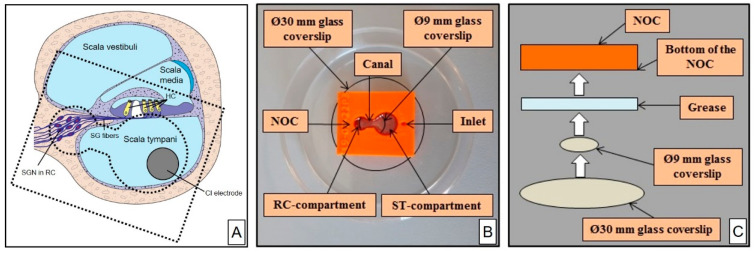
Figure 2.
Illustration of a Neurite Outgrowth Chamber (NOC). (A) Drawn cross-section of the cochlea demonstrating the position of the ST-compartment and RC-compartment of the NOC (dashed lines, down scaled) in reference to the cochlea anatomy to illustrate the concept of the chamber. (B) Photography of the NOC (orange) being attached to the glass coverslips (circled with black lines) and placed within a Petri dish for subsequent cell culture. (C) Schematic drawing of the NOC inside view demonstrating the mount of the Ø9 mm glass coverslip below the canal and of the Ø30 mm glass coverslip to seal the whole NOC bottom mediated by the grease. HC = hair cells; RC = Rosenthal’s canal; SG = spiral ganglion; SGN = spiral ganglion neuron; ST = Scala tympani.

