. 2022 Apr 22;11:e76100. doi: 10.7554/eLife.76100

Figure 4. Docking models of the K48 di-ubiquitin (Ub):UCH37•RPN13DEUBAD complex.

(A–B) HADDOCK docking models show two poses corresponding to the interaction between K48 di-Ub and UCH37 along with their fit to experimental small-angle X-ray scattering data of the K48 di-Ub:UCH37C88A•RPN13DEUBAD complex. The goodness of fit to the experimental intensity is represented by χ2 values. In the first pose (A), the proximal Ub (Ubprox; green) interacts with α5–6 of UCH37. In the second pose (B), the distal Ub (Ubdist; orange) interacts with α5–6 of UCH37. (C) Residues highlighting the interaction between the aromatic-rich region of UCH37 α5–6 and the I44 patch of Ubprox in pose 1 (A). (D) Residues highlighting the interaction between the aromatic-rich region of UCH37 α5–6 and the I44 patch of Ubdist in pose 2 (B). (E) Polar contacts between Ubprox and UCH37 α5–6 in pose 1 (A). (F) Contacts between Ubdist and residues of UCH37 located outside the α5–6 motif in pose 1 (A). (G) Contacts between Ubprox and residues of UCH37 located outside the α5–6 motif in pose 2 (B). (H) The relative location of active site and the scissile, K48 isopeptide bond in molecular docking poses 1 and 2. In pose 1, residues of α6 and the loop leading into the catalytic Cys (C88) form a barrier for the isopeptide bond. In pose 2, only Q82 of UCH37 blocks the K48 isopeptide bond.

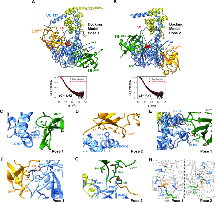
Figure 4.

Figure 4—figure supplement 1. Structural models of the K48 di-ubiquitin (Ub):UCH37•RPN13DEUBAD complex.

Figure 4—figure supplement 1.

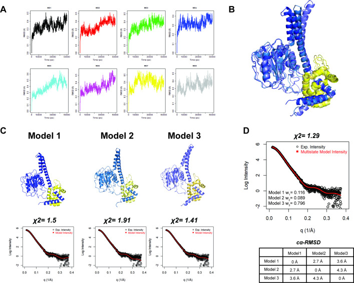
(A) Molecular dynamics (MD) trajectories for eight independent simulations plotted as Cα-RMSD of UCH37 over time. (B) Overlay of MD-derived UCH37•RPN13DEUBAD conformations that afford the best fit to experimental small-angle X-ray scattering (SAXS) data. Flexible N- and C-termini of RPN13DEUBAD are omitted. (C) Individual structures of UCH37•RPN13DEUBAD (models 1–3) with the corresponding fits to the SAXS data. Flexible N- and C-termini of RPN13DEUBAD are omitted. (D) Theoretical and experimental SAXS data for the best-fitting multistate model comprising models 1–3. Pairwise Cα-RMSD is shown between models 1 and 3.
Figure 4—figure supplement 2. Small-angle X-ray scattering (SAXS) of the K48 di-ubiquitin (Ub):UCH37•RPN13DEUBAD complex.

Figure 4—figure supplement 2.

(A–D) Size-exclusion chromatography-SAXS analyses of UCH37C88A•RPN13DEUBAD (blue) and K48 di-Ub:UCH37C88A•RPN13DEUBAD (red) showing distance distribution (E–F), Guinier analyses (G), and Kratky plots (G). (E) HADDOCK docking model of pose 3 showing Ubdist (orange) interacting with RPN13DEUBAD while Ubprox (green) is situated near the C-terminal end of the crossover loop (CL). The fit to the experimental scattering intensity is also shown; the goodness of fit is represented by χ2.
Figure 4—figure supplement 3. Contact frequency analysis of the K48 di-ubiquitin (Ub):UCH37•RPN13DEUBAD complex.

Figure 4—figure supplement 3.

(A) Frequency analysis of pairwise interactions between Ubprox or Ubdist of K48 di-Ub and F117/F121 of UCH37 shown in the form of a bubble plot. (B) Contact frequencies shown for UCH37 residues that are interacting with either Ubprox or Ubdist of K48 di-Ub in poses 1 and 2. Contact frequencies for K48 di-Ub residues interacting with UCH37 in poses 1 and 2.