Clinical Vignette
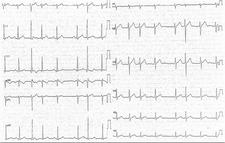
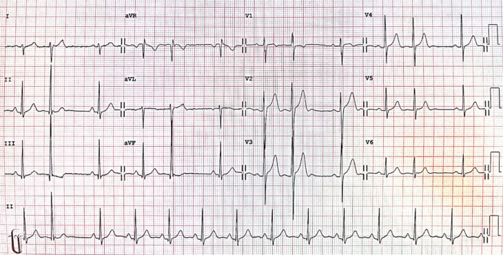
A 19-year-old healthy male presented with frequent palpitations, which interfered with his daily activities. There was no chest pain, reduced effort tolerance, or pre-syncopal episode. No family history of sudden cardiac death nor significant cardiovascular disease was noted. His physical examination was unremarkable. His electrocardiogram on admission is shown in Figure 1. Echocardiogram done showed normal-sized chambers with normal wall motion and preserved ejection fraction. We proceeded to an electrophysiological study (Supplementary material online, Figure S1) which confirmed the presence of a dual node physiology with ventricular ectopics. An atrioventricular (AV) nodal reentrant tachycardia was later induced, for which the patient was symptomatic. The symptoms resolved after successful modification of the slow pathway by radiofrequency ablation. The electrocardiogram post-radiofrequency ablation showed the disappearance of dual node physiology (Figure 2), and there was no complication or recurrence of symptoms post-radiofrequency ablation.
Figure 1.
Electrocardiogram on admission. Note the different QRS morphology (larger amplitude), as depicted by the star. These QRS complexes are narrow, thus are initially presumed to be junctional or arising very near the conduction system. Also, note that these two QRS complexes are different in timings, i.e. the latter occurs earlier (shorter cycle length) than the former. There are also two different PR intervals seen (long left right arrow).
Figure 2.
Electrocardiogram post-ablation. Note the presence of the same ectopic beat without evidence of dual node physiology. As the patient was asymptomatic, we opted for a conservative approach.
Questions
-
1
What is the most likely diagnosis for the different QRS morphology?
Atrial ectopics
Ventricular ectopics
Junctional ectopics
Pre-excitation
One-to-two atrioventricular conduction
The correct answer is B. There is no P wave seen preceding the larger QRS complex; hence, this rules out an atrial ectopic. There are no delta waves to suggest pre-excitation. One-to-two AV conduction may explain the presence of two QRS complexes without a P wave in between but will not explain the prolongation of PR interval after the ectopic beats, as seen in Figure 1. The electrocardiogram (ECG) strip shows two QRS morphologies. Both QRS complexes are narrow and differ only in amplitude. The electrophysiological study (Supplementary material online, Figure S1) is needed to clinch the diagnosis. Since there is no His deflection before the ectopic beat, it points towards an infra-Hisian or ventricular origin. Normally ventricular ectopics have a broad QRS complex; however, the narrow QRS complex, in this case, may be explained by its origin near the conduction system,1 i.e. septal ventricular ectopics. Further evidence to dismiss one-to-two AV conduction can also be derived from the electrophysiological study, as one-to-two AV conduction will show two His signals, one for each pathway utilized.
-
2
Why is the underlying rhythm irregular?
Intermittent complete heart block with occasional ventricular ectopics
Atrial fibrillation
Dual node physiology with ectopics of different coupling intervals
Multifocal atrial rhythm
There is an accessory pathway present
The correct answer is C. This is better explained with the help of a ladder diagram (Supplementary material online, Figure S2). The P–P intervals appear to be somewhat regular on the ECG, except for the last P wave, which coincides with the T wave. There are two septal ventricular ectopics, both of which have different coupling intervals, and the presence of these ectopics seems to prolong the next PR interval as they cause a block in the fast pathway and the subsequent sinus beat travels down the slow pathway (dual node physiology). The presence of an AVNRT in the electrophysiological study confirms the presence of dual node physiology.
-
3
Which of the following conditions are common ECG manifestations of dual node physiology?
AV nodal reentrant tachycardia
AV reentrant tachycardia
Ventricular tachycardia
Atrial flutter
Prolonged QT interval
The correct answer is A. The presence of two pathways in the AV node predisposes the patient to the development of a reentrant circuit within the AV node, as is the case for this patient. The other choices are not commonly seen in a patient with dual node physiology. Other ECG manifestations include irregularly irregular rhythm in atrial fibrillation2,3 and dual AV nodal non-reentrant tachycardia.3
Supplementary material
Supplementary material is available at European Heart Journal – Case Reports online.
Consent: The authors confirm that written consent for submission and publication of this case report, including images and associated text, has been obtained from the patient.
Conflict of interest: None declared.
Funding: None declared.
Supplementary Material
References
- 1. Zheng C, Li J, Lin JX, Wang L-P, Lin J-F. Where is the exact origin of narrow premature ventricular contractions manifesting qR in inferior wall leads? BMC Cardiovasc Disord 2016;16:64. [DOI] [PMC free article] [PubMed] [Google Scholar]
- 2. Peiker C, Pott C, Eckardt L, Kelm M, Shin D-I, Willems S, Meyer C. Dual atrioventricular nodal non-re-entrant tachycardia. Europace 2016;18:332–339. [DOI] [PubMed] [Google Scholar]
- 3. Mani BC, Pavri BB. Dual atrioventricular nodal pathways physiology: a review of relevant anatomy, electrophysiology, and electrocardiographic manifestations. Indian Pacing Electrophysiol J 2014;14:12–25. [DOI] [PMC free article] [PubMed] [Google Scholar]
Associated Data
This section collects any data citations, data availability statements, or supplementary materials included in this article.


