Abstract
Background
Plants have the lifelong ability to generate new organs due to the persistent functioning of stem cells. In seed plants, groups of stem cells are housed in the shoot apical meristem (SAM), root apical meristem (RAM), and vascular cambium (VC). In ferns, a single shoot stem cell, the apical cell, is located in the SAM, whereas each root initiates from a single shoot-derived root initial. WUSCHEL-RELATED HOMEOBOX (WOX) family transcription factors play important roles to maintain stem-cell identity. WOX genes are grouped phylogenetically into three clades. The T3WOX/modern clade has expanded greatly in angiosperms, with members functioning in multiple meristems and complex developmental programs. The model fern Ceratopteris richardii has only one well-supported T3WOX/modern WOX gene, CrWUL. Its orthologs in Arabidopsis, AtWUS, AtWOX5, and AtWOX4, function in the SAM, RAM, and VC, respectively. Identifying the function of CrWUL will provide insights on the progenitor function and the diversification of the modern WOX genes in seed plants.
Results
To investigate the role of CrWUL in the fern, we examined the expression and function of CrWUL and found it expresses during early root development and in vasculature but not in the SAM. Knockdown of CrWUL by RNAi produced plants with fewer roots and fewer phloem cells. When expressed in Arabidopsis cambium, CrWUL was able to complement AtWOX4 function in an atwox4 mutant, suggesting that the WOX function in VC is conserved between ferns and angiosperms. Additionally, the proposed progenitor of T3WOX genes from Selaginella kraussiana is expressed in the vasculature but not in the shoot apical meristem. In contrast to the sporophyte, the expression of CrWUL in the gametophyte exhibits a more general expression pattern and when knocked down, offered little discernable phenotypes.
Conclusions
The results presented here support the occurrence of co-option of the T3WOX/modern clade gene from the gametophyte to function in vasculature and root development in the sporophyte. The function in vasculature is likely to have existed in the progenitor of lycophyte T3WOX/modern clade genes and this function predates its SAM function found in many seed plants.
Supplementary Information
The online version contains supplementary material available at 10.1186/s12870-022-03590-0.
Keywords: WOX, CrWUL, Vascular cambium, Apical meristem, Fern, Ceratopteris, Arabidopsis, Selaginella kraussiana
Background
Meristems of plant bodies house self-renewing stem cells. In seed plants, these centers include the shoot apical meristem (SAM), root apical meristem (RAM), and the vascular cambium (VC). Stem cells within these tissues divide to produce two daughter cells; and once displaced outside the niche actively proliferate to form new organs or differentiated tissues [1–4]. To achieve a balanced population of stem cells and differentiating cells, hormonal and other cellular signals regulate specific families of transcription factors, such as WUSCHEL-Related HOMEOBOX (WOX) transcription factors, SHOOT-MERISTMELESS (STM), and SHORTROOT (SHR) in the apical meristems of Arabidopsis thaliana [5–7].
In the SAM, RAM, and VC of Arabidopsis, WOX genes maintain the size of the stem cell pool in response to auxin, cytokinin and/or CLAVATA signaling [8]. In the SAM, the AtWUS protein is expressed in the organizing center (OC) where it acts non-cell autonomously in the central zone (CZ), a few cell layers above, to maintain the stem cell fate [9]. This SAM maintenance model is likely conserved in the monocot maize [10]. The stem cells of the RAM are also maintained non-cell autonomously by AtWOX5, which moves from the quiescent center (QC) outward to surrounding stem cells [11, 12]. Recent results show that although mobile, movement is not required for AtWOX5 to inhibit stem cells from differentiation [13]. While AtWUS and AtWOX5 are mobile, AtWOX4 maintains VC stem cells in a cell-autonomous fashion [14, 15].
All land plants examined contain WOX genes [16, 17]. In a recently updated phylogeny of WOX proteins in Viridiplantae [17], three ancient superfamilies emerged, the Type 1 (T1WOX), Type 2 (T2WOX), and Type 3 (T3WOX) clades, which represent the previously named ancient, intermediate, and modern clades, respectively [16] with minor differences. These include the assignment of a lycophyte WOX gene as sister to the T2WOX and T3WOX clades and the absence of lycophyte and fern sequences from the T2WOX clade. In addition, four lycophyte WOX genes, three from Isoetes and one from Selaginella moellendorfii, SmWOXII, are now sister to the T3WOX clade. The SmWOXII’s ortholog in Selaginella kraussiana, SkWOX11C (Additional file 1: Fig. S1), has been shown to express in various sporophyte tissues [18]. By this new convention, each clade has members from as early as lycophytes and could allow for a clearer trajectory of WOX gene evolution. Despite the differences, both phylogenetic trees place three fern genes, one from Ceratopteris richardii (CrWUL) and two from Azolla filiculoides (Azfi_s0343.g065738, Azfi_s0051.g031311), firmly within the T3WOX/modern clade. As the sister clade to seed plants, ferns offer a unique opportunity to investigate whether the fern T3WOX/modern gene plays a function in the fern SAM and subsequently uncover the ancestral function of the T3WOX/modern clade progenitor.
Unlike Arabidopsis and other seed plants, most extant ferns have a single stem cell at the apex of the SAM called the shoot apical cell. As in Arabidopsis, the fern sporophyte SAM contains two regions that comprise distinct transcriptional profiles [19], a core region and a shoot apical cell (reviewed by [20]); however, the domains within the core region are not defined. How the shoot apical cell maintains its identity and what the roles, if any, the core region plays in the shoot apical cell maintenance are unknown. What role could a T3WOX/modern gene play in stem cell maintenance of a fern, despite the contrast in meristem organization?
In the fern model Ceratopteris, only one T3WOX/modern gene is well supported, CrWUL, has been found. It would be reasonable to speculate that CrWUL functions in the meristem or even maintains apical cell identity. The CrWUL protein has been shown to move and exhibit stem-cell maintenance functions in Arabidopsis SAM and RAM only after it is truncated and expressed under the control of AtWUS or AtWOX5 promoter, respectively [21]. Those authors proposed that these two Arabidopsis T3WOX/modern genes have evolved through a two-step selection to acquire the stem-cell maintenance function in the SAM and RAM; the first was stem-cell maintenance activity and the second was intercellular mobility. However, in Ceratopteris, previous studies showed that CrWUL is expressed in the leaf vasculature [22], the merophytes of lateral roots [16] and adventitious roots [23] but not in the root apical cell [16]. These results suggest that T3WOX/modern genes were recruited to function in vascular tissues before the divergence of ferns and seed plants from the land plant phylogeny. Here, we show that CrWUL expresses and functions in the vascular tissue but not in the SAM of Ceratopteris. In addition, we provide evidence that the progenitor of T3WOX/modern genes were present in the last common ancestor of lycophyte and fern. Finally, we show that CrWUL can replace AtWOX4 function in Arabidopsis cambium cells. These results support the hypothesis that T3WOX/modern genes acquired apical stem-cell maintenance activity by first being recruited to function in vascular tissues.
Results
Expression of CrWUL during gametophyte and sporophyte development
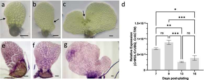
Because ferns have meristems not only in the sporophyte generation, but also in the gametophyte generation, the expression of CrWUL was examined in both generations. Initially, we determined the expression levels of CrWUL in gametophytes at different developmental stages. Hermaphroditic gametophytes establish the notch meristem 7-days post plating (dpp) and reach sexual maturity by 13-dpp (Fig. 1a-c). Expression of CrWUL increased slightly during sexual maturation in 7- to 9-dpp gametophytes and decreased by more than half after sexual maturation in 13-dpp gametophytes and remains low from 13- to 16-dpp (Fig. 1d). Whole mount in situ hybridization was performed to localize CrWUL transcripts during gametophyte development (Fig. 1e-g). At 7- and 9-dpp, CrWUL expression is present throughout the hermaphroditic gametophyte (Fig. 1e, f). After the establishment of the notch meristem, expression decreases in the hermaphrodite thallus (Fig. 1g) and becomes difficult to discern from background (see sense probe images, Additional File 1: Fig. S2), suggesting a drastic decline of CrWUL levels. In the male gametophyte, CrWUL expression was not detected by whole-mount in situ (Additional File 1: Fig. S2). Thus, expression of CrWUL was observed in the hermaphroditic gametophyte prior to sexual maturation and declined subsequently.
Fig. 1.
Expression of CrWUL in the gametophyte generation of C. richardii. (a-c) Representative wild-type hermaphrodite gametophytes at 7-, 9-, and 13-days post plating. (d) Expression of CrWUL during gametophyte development measured by RT-qPCR (mean ± SEM; *, P ≤ 0.05; **, P ≤ 0.01; ***, P ≤ 0.001; N = 3). (e–g) Whole mount in situ hybridization of 7-day (g), 9-day (h), and 13-day (i) old hermaphroditic gametophytes. Black arrowhead in (a-c) indicates notch meristem. Scale Bars = 0.05 mm
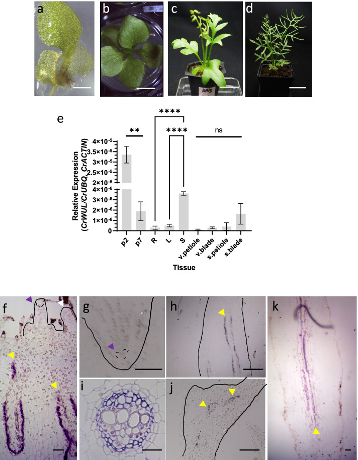
To examine CrWUL expression in different tissues and developmental stages, we took samples from sporophytes ranging from individuals with one expanded leaf to mature sporophytes with sporophylls (Fig. 2a-d) for use in qPCR analysis. Expression was the highest in p2 sporophyte (whole plant including first leaf, leaf primordia, and the first root) (Fig. 2a, e), then decreased dramatically in the p7 sporophyte (whole plant including 6 leaves, leaf primordia, and roots) (Fig. 2b, e). Among isolated roots, leaves, and shoots from p13 plants, expression is highest in the shoot tissues which contain leaf and root primordia (fern root primordia are next to leaf primordia, both are near the SAM) along with the petiole base of more mature leaves (Fig. 2e and Additional File 1: Fig S3). Sporophytes were moved to soil for the remainder of development, where they continue to generate vegetative leaves until sporophyll production (Fig. 2c, d). Expression is low in the last vegetative leaf before the sporophylls but higher in the first fully expanded sporophyll of sexually mature sporophytes (Figs. 2d, e). In situ hybridization was used to localize CrWUL expression. CrWUL expression is absent from the apical cell and core region of the SAM, developing leaf primordia (Fig. 2f), and developing root primordia including the root apical cells (Fig. 2g). In contrast, expression of CrWUL is detected in the longitudinal section of the shoot and leaf petiole as two long bands on either side of xylem tissues (Fig. 2f, h) and in the cross section of the vascular bundle between the pericycle and xylem, containing phloem plus any residual procambium cells (Fig. 2i). Expression continues up the petiole vascular bundles into developing leaf blades (Fig. 2j). In addition, expression of CrWUL is present within root vascular bundles (Fig. 2k). All sense probe control images are provided in Additional File 1: Fig S4. In conclusion, CrWUL is specifically expressed in phloem and is conspicuously absent from the SAM and leaf primordia.
Fig. 2.
Expression of CrWUL in the sporophyte generation of C. richardii. (a-d) Stages of wild-type sporophytes used for expression assays; sporophyte with one leaf (p2) (a), six leaves (p7) (b), 13 expanded leaves (p13) (c), and mature sporophyte with sporophyll (d). (e)Expression of CrWUL in sporophyte tissues measured by RT-qPCR (mean ± SEM;**, P ≤ 0.01; ****, P ≤ 0.0001; N = 3): p2 (the whole plant), p7 (the whole plant), L (oldest fully expanded leaf), R (root), S (shoot) from a liquid grown p13 plant; v.petiole ( vegetative petiole), v.blade (vegetative leaf blade), s.petiole (sporophyll petiole), s.blade (sporophyll blade) from a mature sporophyte shown in (d). (f-k) In situ hybridization of sectioned sporophyte tissues; SAM (f), RAM (g), young petiole (h); cross section of vegetative petiole (i), emerging leaf blade (j), and root vascular tissues (k). White arrowhead points to leaf primordium. Purple arrowheads denote shoot (f) and root (g) apical cells, respectively. Yellow arrowheads indicate phloem tissues. Scale bars = 1 mm (a), 4 mm (b), 25 mm (c), 46 mm (d), 0.05 mm (f-k)
Phenotype of CrWUL RNAi knockdown lines (crwul)
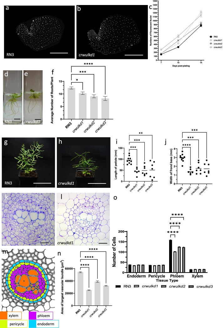
WOX genes are involved in promoting cell division and growth of plant tissues containing meristems. The overall size and morphology of crwul knockdown lines are similar or larger than wild-type gametophytes (Fig. 3a-c). Numbers of cells in the hermaphrodite thallus increases from 7-dpp to 14-dpp and crwul knockdown lines remained at or above wild-type levels during development (Fig. 3c). In addition, Ceratopteris Histone H4 (CrH4) expression was used as marker for cell divisions and when measured, CrH4 expression remained at or above wild-type levels in crwul knockdown gametophytes (Additional File 1: Fig. S5). Knockdown of endogenous CrWUL transcripts in crwul lines was measured in 14-dpp gametophytes (Additional File 1: Fig. S6). Reduction of CrWUL transcripts was highest in line crwul1 at < 7% of wild-type levels. Remaining lines averaged 30%-70% reduction in CrWUL transcripts. Selected gametophytes were selfed and grown in liquid for two weeks until sporophytes reached p7-8. Liquid grown sporophytes were then imaged and adventitious roots quantified (Fig. 3d-f). All Ceratopteris roots are shoot-borne, therefore they are termed either shoot-borne roots or adventitious [24]. crwul lines produced fewer numbers of roots than the wild-type (Fig. 3d, e). On average, crwul lines produced 9.1 roots per plant while wild-type plants produced 12.4 roots per plant, a reduction of 27% (Fig. 3f). crwul knockdown lines are of shorter stature due to shorter leaf petioles (Fig. 3g-j). For quantification, the petiole of the 1st sporophyll of a sporophyte that had produced three sporophylls was measured and overall, petiole length decreased ~ 49% in knockdown lines and width decreased ~ 45% compared with wild-type petioles (Fig. 3i, j). Because expression of CrWUL is absent from the shoot and root meristems but seen in vascular bundles, using anatomical features we counted cells of various tissue types within cross sections of the 1st sporophyll petiole of a sporophyte that had produced three sporophylls (Fig. 3g, h). Ceratopteris produce concentric amphicribral vascular bundles within petioles. During development Ceratopteris petioles increase in width and number of vascular bundles. The 13th vegetative leaf of Ceratopteris contain three vascular bundles, while the 1st sporophyll after three sporophyll produced contain five vascular bundles (data not shown). Ceratopteris sporophyll petioles used for quantification, either from wild-type or crwul lines, each contained five vascular bundles, signifying equivalent developmental time points between lines. A ring of endodermal cells encloses each vascular bundle, in which a central band of xylem is surrounded by parenchyma comprising phloem and any residual procambium, together are referred as phloem from hereon (Fig. 3k-n) [25]. All tissue types were present in the crwul lines but the area of the vascular bundles cross sections decreased (Fig. 3n). Generally, vascular bundles are smaller in younger petioles, therefore we quantified cell number in vascular bundles between equivalent leaves to assess whether the decreased area of crwul vascular bundles was associated with reduced numbers of a specific cell type. Within the crwul lines all tissues except phloem contained similar numbers of cells as wild-type plants (Fig. 3k-o). Phloem cell numbers, on average, were reduced by 27% in crwul lines when compared to wild-type (Fig. 3o). Interestingly, expression of CrH4 decreased in p7-8 crwul whole sporophytes and was also expressed preferentially in the phloem of vascular bundles (Additional File 1: Fig. S5). While the overall stature of the crwul lines were visibly shorter than that of the wildtype, they produced the same number of fronds as the wild-type plants (data not shown). These results showed that knocking down crwul expression caused a decrease of phloem cell numbers, petiole length and width and production of fewer roots but not fewer leaves.
Fig. 3.
Phenotype of crwul knockdown lines. (a, b) Fluorescent images of 10-days post plating (dpp) gametophytes stained with Hoechst dye. (c) Numbers of florescent nuclei 7-, 10-, and 14-dpp of wild-type and crwul knockdown lines (N ≥ 19, statistics in Additional file 3: Table S2). (d, e) Images of wild-type and a representative crwul knockdown line grown for 2 weeks in liquid culture. (f) Numbers of roots per plant (mean ± SEM; *, P ≤ 0.05; **, P ≤ 0.01; ***, P ≤ 0.001; ****, P ≤ 0.0001 N ≥ 16). (g, h) Images of mature wild-type and a crwul knockdown line each has produced three sporophylls. (i) Length of sporophyll petiole from base to first pinna (individual lengths and mean, **, P ≤ 0.01; ***, P ≤ 0.001, N ≥ 6). (j) Width of sporophyll base (individual lengths and mean, **8, P ≤ 0.001; ****, P ≤ 0.0001, N ≥ 6). (k, l) Cross sections of the largest vascular bundles in the first sporophyll from wild-type and a representative crwul line. (m) Diagram of C. richardii vascular bundle with tissue types color coded; en, endoderm; pe, pericycle; ph, phloem; x, xylem. (adapted from [25]). (n) Area of bundles (mean ± SEM, ****,P < 0.0001 N ≥ 9) (o) Numbers of cells per section from each tissue type present in the vascular bundles (mean ± SEM; ****, P ≤ 0.001 N ≥ 10). Scale bars = 0.05 mm (a,b), 10 mm (d, e), 90 mm (g, h) and 0.05 mm (k, l)
CrWUL restores cambiam layer in atwox4 null mutants
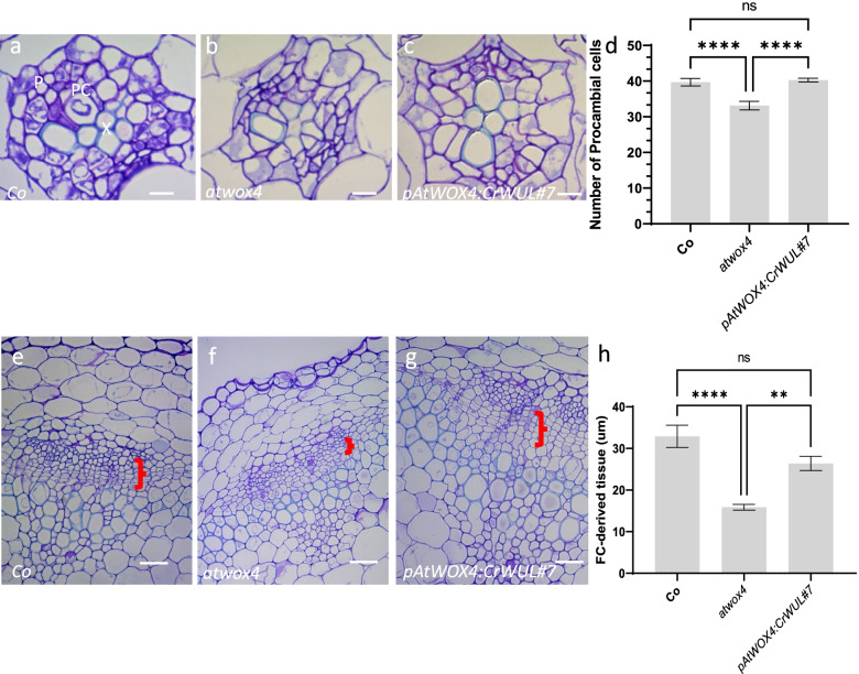
AtWOX4 functions in the cambium of Arabidopsis hypocotyl and stem vascular tissues to maintain the number of cambium cells [14, 15]. Therefore, we asked whether the function of WOX genes in vascular tissues was conserved across ferns and angiosperms by expressing CrWUL under the AtWOX4 endogenous promoter in an atwox4 null mutant (Additional File 1: Fig. S7). This previously characterized T-DNA insertion mutation was chosen because it only affects vascular tissue cell proliferation but not organization [14, 15]. Three transgenic lines were selected and grown for genotyping (Additional File 2: Fig. S8) and phenotyping. atwox4 null mutant plants used in this study have less cambium but no observable phenotype [15]. As expected, atwox4 plants expressing CrWUL are indistiguishable from the wild-type and atwox4 null mutant (Additional File 2: Fig. S9). Arabidopsis hypocotyl cross sections revealed that the organization of the vasculature remain the same in the wild type, atwox4, and pAtWOX4:CrWUL lines, with xylem and phloem separated by cambium (Fig. 4a-c). There were 20% fewer cambium cells produced by atwox4 null mutants than those in the wildtype and pAtWOX4:CrWUL lines. pAtWOX4:CrWUL completely restored the procambium cell numbers in the mutant lines (Fig. 4d). Cross sections of the bottom 1 cm of 15 cm-tall inflorescence stems were examined for the production of vascular tissue (Fig. 4e-h). In wild-type stems, cells of the vascular cambium divided periclinally to produce phloem externally and xylem internally, giving rise to files of cells of each tissue (Fig. 4e). atwox4 plants produced fewer cambial derivatives than Col and pAtWOX4:CrWUL, and therefore the phloem and xylem were separated by a 52% and 40% shorter distance, respectively (Fig. 4f). pAtWOX4:CrWUL plants restored the number of cambial cells and the distance between the phloem and xylem increased to levels measured in wild-type plants (Fig. 4g, h).
Fig. 4.
CrWUL restores cambium layer in atwox4 null mutant lines. a-c Cross section of wild-type Col (a), atwox4 null mutant (b) and pAtWOX4:CrWUL (c) hypocotyl. d Number of procambium cells per section (mean ± SEM; ****, P ≤ 0.0001, N ≥ 10). e–g Cross section of wild-type Col (e), atwox4 (f) and pAtWOX4:CrWUL (g) base of 15 cm primary inflorescence. Red brackets indicate cambium cell layer (e–g). h Length of cambium tissues per section in base of 15 cm primary inflorescences (mean ± SEM; **, P ≤ 0.01; ****, P ≤ 0.0001, N ≥ 10). p: Phloem, x: Xylem, pc: Procambium. Scale Bars = 0.005 mm (a-c), 0.03 mm (e–g)
The S. kraussiana T3WOX/modern gene SkWOX11C is specifically expressed in lycophyte vascular bundles
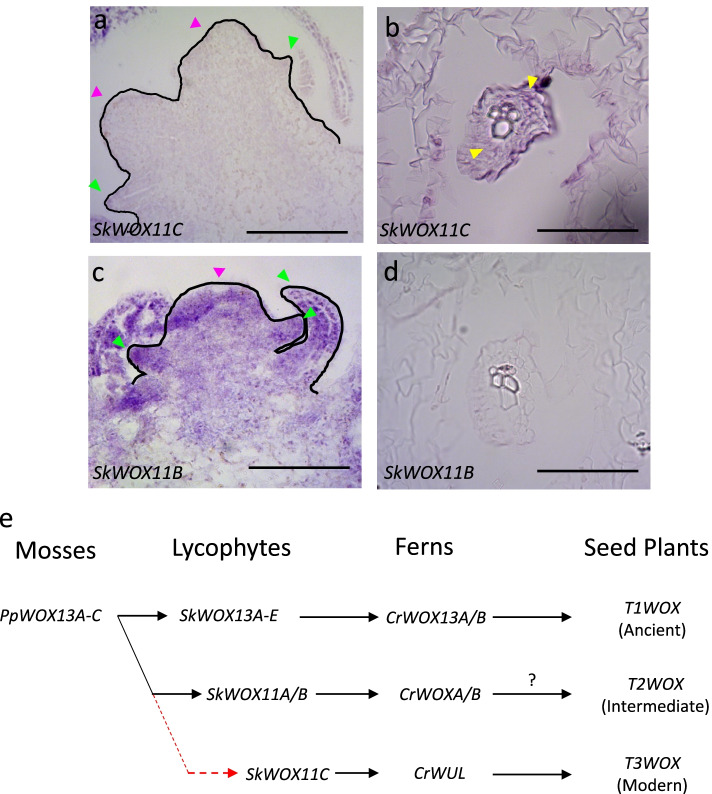
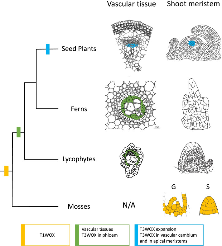
Lycophytes represent the earliest extant vascular plants possessing the T3WOX/modern clade. Therefore, it was of interest to determine whether this gene is expressed in the vascular tissue or in the meristem regions of the lycophyte. Previous expression analysis used RNAseq of RNA samples taken from shoot tip, root tip, microphyll, and stem [26], each likely to include vascular tissue. In order to distinguish whether expression is specific to the SAM apex or to the vasculature present in all tissues, we used in situ hybridization to localize SkWOX11C (Fig. 5a,b). We found that expression of SkWOX11C is absent from the shoot apex and youngest leaf primordia of S. kraussiana (Fig. 5a) but is present in the vascular bundles of the shoot (not shown) and stem, specifically in the phloem, not the xylem (Fig. 5b). Interestingly, SkWOX11B, which is not grouped with T3WOX/modern genes (Additional File 1: Fig. S1), is expressed throughout the S. kraussiana shoot apex including leaf primordia (Fig. 5c) but is absent from vascular tissues (Fig. 5d). Sense probe controls are provided in Additional File 2: Fig. S10. These results support that the AtWOX4-like function may have already existed in the lycophytes. Accordingly, we revised a previously proposed trajectory showing conservation of WOX gene function in land plants [18] by moving SkWOX11C from the T2WOX/intermediate group to the T3WOX/modern clade (Fig. 5e). These results support the existence of the T3WOX/modern clade in the last common ancestor of S. kraussiana and ferns, as well as the recruitment of the T3WOX/modern genes to function in apical meristems after the divergence of ferns and seed plants (Fig. 6).
Fig. 5.
The T3WOX/modern Selaginella kraussiana WOX11C is expressed in lycophyte vascular bundles. a In situ of SkWOX11C in the shoot apex. b In situ of SkWOX11C on cross section of vascular bundle in stem tissues. c Expression of SkWOX11B in the shoot apex. d In situ of SkWOX11B on cross section of stem. e Abbreviated land plant phylogeny, and (f) Revised trajectory of WOX gene function in land plants (adapted from [18]). Pink and green arrows indicate shoot apical meristem and emerging leaf primordia, respectively. Yellow arrows indicate phloem. Scale bars = 0.05 mm
Fig. 6.
Evo/Devo of T3WOX/modern transciption factors. Abbreviated land plant phylogeny depicting the emergence of the T1WOX/ancient clade (yellow box), T3WOX/modern clade (green box), and T3WOX/modern expansion in the seed plant lineage (blue box). Expression of the T1WOX/ancient clade in P. patens is present in both the gametophore apex (G) and the sporophyte apex (S) (yellow cells). Expression of the single T3WOX/modern genes in S. kraussiana and C. richardii are present in the phloem (green cells) but not the SAM. After T3WOX/modern expansion, members of T3WOX/modern genes in seed plants are expressed in vascular tissues and SAM (blue cells)
Discussion
The WOX gene family of homeodomain transcription factors is important in maintaining the identity of stem cells and proliferation of other pluripotent cells in land plants. The T3WOX/modern clade only exists in vascular plants [16, 17]. The earliest known T3WOX/modern clade gene that contains the signature WUS box has been found in Ceratopteris (CrWUL), and the T3WOX/modern clade has subsequently expanded in seed plants. Among the many T3WOX/modern clade members in Arabidopsis, AtWUL, AtWOX5, and AtWOX4 are involved in stem cell maintenance of the SAM, RAM, and VC, respectively [9, 12, 27]. Although ferns are sister to seed plants, they contain a SAM architecture which has only a single apical cell (reviewed by [28, 29]) and shoot-borne roots [30]. Initials for each leaf-root pair arise near each other, in proximity to the apical cell of the SAM. Both leaf and root grow acropetally and all the cells in each leaf and root are descendants of the leaf and root initials, respectively [24, 31]. Here we found the major site of function for CrWUL is the sporophyte vasculature, specifically the phloem. CrWUL expression levels affect the number of roots, but not leaves, produced. The expression of CrWUL was also observed in the gametophytes, where it negatively regulates cell numbers.
CrWUL negatively regulates cell number in the gametophyte
CrWUL is widely expressed in immature hermaphroditic gametophytes, but expression declines once gametophytes reach sexual maturity (Fig. 1). The expression pattern in immature hermaphrodites is different from that of CrWOXB, which is more prominent in the notch region [32]. In males, the expression of CrWUL is not detected but CrWOXB is detected in undifferentiated cells, suggesting a hermaphrodite-specific role for CrWUL. Unlike CrWOXB, which is involved in cell division at the notch region [32], knocking down the expression of CrWUL has a positive effect on the cell number of the gametophytes (Fig. 3). This suggest that CrWUL negatively regulates cell division in the gametophyte, a function opposed to that of CrWOXB. Negative regulation by CrWUL may be attributed to the presence of WUS-box and to a lesser extent, the EAR domain, which is important for AtWUS nuclear export and cytoplasmic stability [33–35], both domains are absent from CrWOXB. In addition, Arabidopsis shoot size is inversely proportional to AtWUS concentration [36]. Perhaps CrWUL dosage and/or negative regulation of certain cell-cycle genes by CrWUL contribute to the larger gametophyte, whereas CrWOXB does not [32]. In Arabidopsis, only one T3WOX/modern clade gene, AtWOX2, is expressed in the female gametophyte in addition to sporophyte, but the function in the gametophyte is unknown [37]. Interestingly, our results show that CrWUL played opposite role in regulating cell division in the two generations of Ceratopteris. This may not be surprising as AtWUS is known as a bifunctional transcription factor [34]. In addition, many transcription factors change their regulatory functions in a tissue specific manner (reviewed in [38]) and/or contain dual roles (positive and negative) in either a “context-dependent” or “signal-dependent” fashion (reviewed in[39]). In sum, CrWUL is expressed in both generations, providing evidence that the T3WOX/modern clade gene may have an ancestral role of negative regulation that was co-opted from the gametophyte to the sporophyte before the divergence of ferns.
A role for CrWUL in fern root and shoot development
Once specified, the Ceratopteris root initial (RI) undergoes four asymmetric cell divisions, resulting in four merophytes that each share a cutting face with the tetrahedral RI [30]. CrWUL is transiently expressed in the three proximal merophytes but never in the RI or the distal merophyte, which is destined for root-cap fate [16]. The observation that the crwul knockdown lines had fewer roots is consistent with its transient expression in the three proximal merophytes where it maintains their pluripotency. With insufficient amount of CrWUL, merophytes arrest, decreasing root numbers (Fig. 3). Fewer roots produced in crwul knockdown lines is also consistent with the report that CrWUL is induced by auxin to promote adventitious root production [23]. Fewer roots in crwul knockdown lines superficially resembled that of the CrWOXB knockdown lines [32] but the temporal and spatial expression patterns of the two genes are very different; CrWOXB is expressed in the root tip region where cells proliferate, whereas CrWUL is not expressed in this region but is expressed in root vasculature and at the very beginning of root initiation (Fig. 2).
In contrast to fewer roots, the number of leaves was similar between crwul knockdown lines and wild-type plants. However, the width and length of the sporophyll petiole were both reduced in crwul knockdown lines. CrWUL is not expressed in the leaf primordia, including the apical cells, and presumably has no role in early leaf development. As such, the decrease in petiole length and width could be due to reduction in plant growth from a combination of fewer roots and decreased phloem. This is different from CrWOXB which is highly expressed in the leaf primordia, including apical cells, and knockdown lines which produce fewer leaves than the wild-type plants [32]. Thus, unlike the root, leaf primordia specification has not recruited the function of a T3WOX/modern clade gene.
CrWUL regulates phloem cell numbers
In the sporophyte, in addition to its expression in the three direct descendants in the RI [16], we showed that CrWUL expresses and functions in the vasculature, specifically for maintaining proper phloem cell numbers (Fig. 3). AtWOX4 is known to regulate cell proliferation of the procambium and cambium of Arabidopsis [14, 15, 26]. Although modern ferns lack secondary growth [40], when CrWUL was expressed under the AtWOX4 promoter, it restored the cambium phenotypes in an Arabidopsis null mutant of AtWOX4 (Fig. 4). Unlike AtWOX4, knocking down CrWUL expression only decreased the phloem cell number but not the xylem or endoderm cell numbers in Ceratopteris, suggesting CrWUL is not involved with specifying these tissues. It also could be due to a redundant function of another CrWOX gene. In Arabidopsis, a T1WOX/ancient clade gene, AtWOX14, acts redundantly with AtWOX4 to regulate vascular proliferation [41]. Ceratopteris has two T1WOX/ancient genes, CrWOX13A and CrWOX13B, both encode proteins which share additional peptide motifs with AtWOX14 outside of the conserved homeodomain ([21], Additional File 2: Fig. S11).
The function of T3WOX/modern clade gene in vasculature predates its SAM function
In contrast to CrWOXB, which is expressed broadly in the SAM and leaf primordia [32], the expression of CrWUL was absent from the SAM and leaf primordia outside of vasculature (Fig. 2). This discovery prompted us to determine if the role of the T3WOX/modern clade in vasculature predates ferns. The T3WOX/modern clade gene in the lycophytes have been suggested to contain the earliest T3WOX genes [17], and expression in the vasculature of S. kraussiana supports this proposed relationship (Fig. 5). However, SkWOX11C does not have a discernable WUS box (Data not shown), which is required for stem cell function [34]. This situation is not unique, as a T3WOX gene from the conifer Araucaria rulei also lacks the WUS-motif [17]. In contrast, SkWOX11B, which is not a T3WOX/modern clade gene, was expressed in SAM apex and young leaf primordia but not in vascular tissues, highlighting the role of T3WOX/modern clade genes in the VC of vascular seed-free plants (Figs. 5, 6). A closely related WOX gene SkWOX11A was not expressed in the lycophyte sporophyte [18] and was not investigated here. In a proposed WOX lineage (Fig. 5e), instead of grouping SkWOX11C with SkWOX11A/B, we place SkWOX11C outside the previous grouping, and in line with the T3WOX/modern clade lineage. This new grouping puts the emergence of T3WOX/modern genes in the last common ancestor of lycophytes and ferns, earlier than previous evidence suggested (Fig. 6). Furthermore, the origination of the T3WOX/modern clade coincides with the emergence of vascular tissues. It is only after the split of seed plants from ferns and subsequent expansion of the T3WOX/modern clade do T3WOX/modern genes function in apical meristems. Therefore, we compared the promoter regions of CrWUL, AtWOX4, and AtWUS. As expected, the upstream regions of CrWUL and AtWOX4 promoters share more predicted motifs than those of CrWUL and AtWUS (Additional File 2: Fig. S12). This lends support for the phloem-specific regulation of the T3WOX/modern genes being the ancestral state. In other duplicated T3WOX/modern genes, such as AtWUS, the upstream regions of the promoter loses the transcription-factor binding sites for phloem-specific expression and gains those for apical meristem expression.
Interestingly, the rice WOX4 gene (OsWOX4), in addition to functioning in the vasculature as does AtWOX4, also maintains stem-cell function in the rice SAM through a possible model different from that proposed for Arabidopsis [42]. The broad expression of OsWOX4 in the SAM overlaps with the expression domain of a CLE-like gene, thus eliminating the requirement of a mobile WOX protein. The dual function of OsWOX4 in the vasculature and the stem-cell maintenance in the SAM further supports that the latter function has evolved more recently. It would be interesting to see whether the full-length CrWUL when expressed in the rice SAM can complement the OsWOX4 function in OsWOX4 knockdown lines [42, 43]. Among Arabidopsis T3WOX/modern clade genes, all but AtWOX4 can complement the AtWUS stem-cell maintenance function in atwus mutants [7], suggesting either divergent transcriptional targets and/or lack of mobility of AtWOX4 protein preventing complementation. CrWUL, when truncated, is mobile and when expressed in the OC and the QC of Arabidopsis, can restore the SAM and RAM functions in atwus and atwox5 mutants, respectively [21], suggesting CrWUL has acquired stem-cell function and the transcriptional targets required for this function. The remaining unanswered question is which, if any, of the CrWOX genes are involved in maintaining apical cell identity in the fern.
In Conclusion, the results presented here support the occurrence of co-option of the T3WOX/modern clade gene from the gametophyte to function in vasculature and root development in the sporophyte. The function in vasculature is likely to have existed in the lycophyte T3WOX/modern clade progenitor and this function predates its SAM function found in many seed plants.
Methods
Plant material and growth conditions
Fern spores used in experiments are of Ceratopteris richardii strain RN3 (Carolina Biological Supply Company, Burlington, NC) as the wildtype. Spores of RN3 and CrWUL RNAi suppression lines (crwul) were surface sterilized with 4% sodium hypochlorite and 0.1% Tween-20 for 4 min, rinsed 4–5 times with sterile water and dark treated for 3–5 days to synchronize germination. Spores were then plated on basal media (1/2 MS salts, pH 6.0, 0.8% agar, 100 µg/mL ampicillin) and grown under humidity domes at 26 °C in 16/8 h light/dark cycle at 100 µM/m−2 s−1 under Bright white (3500 K) and Daylight (6500 K) fluorescent lights (GE Lighting, East Cleveland, OH) for gametophyte development. Basal media with spores were inverted after 10-days post-plating (10-dpp) to prevent water condensation from causing premature fertilization. For selfing, a few drops of sterile water were added to sexually mature hermaphrodite gametophytes in 24-well dishes. Wild-type and crwul sporophytes with 7–8 leaves (p8-p9) were moved to basal liquid media (1/2 MS salts, pH 6.0, 100 µg/mL ampicillin) to facilitate root growth and take samples for root counts. After 2 weeks in basal liquid media, sporophytes were transplanted into Pro-mix PGX (Premier Tech Horticulture, Quakertown, PA) and grown at 23 °C with 16/8 h light/dark cycle at 80 µM/m−2 s−1under fluorescent lights (see above) until sporophylls were harvested.
Arabidopsis atwox4 null-mutant seeds (GK_462G01, N376572) [14, 15, 44] used for complementation were obtained from the Arabidopsis Biological Resource Center (ABRC, Ohio State University, Columbus, OH). Seeds were sown onto Pro-mix PGX soil with one capful of Osmocote Flower and Vegetable (The Scotts Company, Marysville, OH) added to the soil, cold treated for 3-days at 4 °C then grown under the same lighting conditions as Ceratopteris sporophytes (see above). Arabidopsis atwox4 null-mutant plants were genotyped with primer sets 2 and 3 once rosette leaves were formed (See Additional File 3: Table S1 for primer sequences). Col-0 plants were confirmed absent of T-DNA insertions with primer set 1. DNA extraction was carried out as described [45]. S. kraussiana were purchased from miniature-gardening.com (Miniature Gardening, Winter, WI). Plants were grown in Pro-mix PGX soil in a transparent glass trough, with a glass lid to maintain humidity, under natural light at 40 µM/m−2 s−1, 23 °C and 16/8 h light/dark cycle.
In situ hybridization
Antisense and sense RNA in situ probes were synthesized according to [32]. Templates for probe synthesis were cloned into the pENTR vector (Life Technologies, Carlsbad, CA) and sequenced at the Carver Center for Genomics or Iowa Institute of Human Genetics (University of Iowa, Iowa City, IA). From 1 µg of PCR products amplified using primers containing T7 promoter sequences (IDT, Coralville, IA), DIG-labeled RNA probes were synthesized using T7 RNA polymerase (Agilent, Santa Clara, CA) and DIG-RNA labeling mix (Roche Diagnostics, Indianapolis, IN). DIG-labeled RNA was precipitated in 2.25 M LiCl overnight at -20 °C and resuspended in ddH20. RNA probe concentration was estimated with a Nanodrop One (Thermo-Scientific, Waltham, MA) then diluted 1:1 with deionized formamide. Diluted probes were stored at -20 °C until use.
Tissues for whole mount and sectioned in situ hybridization were prepared as described previously [32]. Gametophytes were fixed in FAA (formaldehyde: ethanol: acetic acid, 3.7%:50%:5% v/v respectively) at room temperature for 1–2 h. FAA was replaced with 70% ethanol and tissues were stored at -20 °C. Sporophyte tissues from Arabidopsis, Ceratopteris and S. kraussiana were fixed in 4% paraformaldehyde in 1 × PBS under vacuum for 45 min. Fixative was replaced and tissues were incubated in fix for 1–2 days at 4 °C. Tissue dehydration, embedding, pre-hybridization, hybridization and detection were all described previously, except dehydration post-color development was omitted and slides were mounted in glycerol [32]. Embedded tissues for sectioned in situ hybridization were sectioned at 8–10 µm with a rotary microtome.
Plant transformation
A 737 bp (region 1) or a 368 bp (region 2) (see Additional File 3: Table S1 for primer sequences) of CrWUL coding sequence was cloned into pH7GWIWG2(I) vector using the Gateway technology as described by [46, 47]. Constructs were introduced into Agrobacterium tumefaciens strain GV3101 from E. coli with an E. coli helper strain containing the pRK 2013 plasmid. Stable transformation of young Ceratopteris gametophytes was conducted as described previously [47]. Successfully transformed gametophytes (T0) were selected on media containing 5 µg/mL hygromycin, selfed and grown as described in [47]. From more than 20 independent transgenic lines isolated, 10 were chosen for qPCR analysis and phenotyping.
For complementation experiments, a 2 kb fragment upstream of the ATG start codon of AtWOX4 was amplified with primers as described in [14] adapted with either HindIII or PacI restriction sites for cloning. The amplified fragment was cloned into a pMDC83 vector carrying CrWUL by replacing the 35S promoter with the AtWOX4 2 kb upstream sequences. Constructs were introduced into A. tumefaciens as described above. Transformation of Arabidopsis atwox4 null mutant sporophytes was performed by the floral spraying method [48]. Seeds were collected and positive transformants were selected on ½ MS media with 15 µg/mL hygromycin. Resistant plants were transferred to soil before seed collection.
RNA extraction and RT-qPCR
Tissues harvested for RNA extraction were flash frozen in liquid nitrogen then stored at -75 °C. Total RNA was extracted from frozen tissue with the Quick-RNA MiniPrep (Plus) kit (Zymo Research, Irvine, CA) and 500 ng of total RNA was used for cDNA synthesis with MMLV reverse transcriptase (New England Biolabs, Ipswich, MA) with either N9 random or oligo-dT [16] primers (IDT Coralville, IA).
For RT-qPCR, three biological and two technical replicates were performed for each time point. Total RNA extraction and cDNA synthesis is described as above. Detection of amplification was performed using PerfeCTa SYBR Green FastMix (Quantabio, Beverly, MA) with the Roche LightCycler 480 Real-Time PCR system (Roche Diagnostic, Indianapolis, IN). The PCR protocol was as follows: initial denaturing of 10 min at 95 °C, followed by 45 cycles of denaturing (10 s at 95 °C), annealing (10 s at 59 °C) and extension (20 s at 72 °C), with a single fluorescence read at the end of each extension time. Melting curve analysis was performed to verify absence of primer dimers and non-specific products. Expression was measured relative to CrUBQ alone or CrUBQ and CrActin using the delta Ct method [49].
Phenotyping
The following tissues were used for observation and cell counts of vascular bundles: 1 cm from the base of the 1st fully expanded sporophyll of Ceratopteris sporophytes with three sporophylls, 2-week-old Arabidopsis hypocotyl and 1 cm from the base of 15 cm tall Arabidopsis primary inflorescences. Tissues were embedded in Technovit 8100 (Kulzer, Hanau, Germany) resin according to user instructions with the following modifications: fixation was carried out as described above and dehydration was carried out in a graded acetone series. Resin blocks were sectioned at 1.5–1.75 µm with a glass knife on a Leica EM UC6 ultramicrotome (Leica Microsystems, Buffalo Grove, IL) at the Central Microscopy Research Facility (University of Iowa, Iowa, IA). Sections were dried and stained briefly in 0.025% (w/v) Toluidine blue O, mounted in Permount™ and imaged as described above.
For gametophyte cell counts, 7-,10- and 14- dpp gametophytes were collected and cleared in 100% EtOH overnight at 4 °C, rinsed in water and stained in Hoechst 33,342 (7-, 10-dpp: 40 µg/mL; 14-dpp: 4 µg/mL). For gametophytes aged 7–10 dpp, images were collected using a Leica stereomicroscope with a DAPI filter and a Qicam camera (Qimaging, Surrey, BC, Canada). Gametophytes 14-dpp, were captured with a Sony α35 digital camera through the ocular of a Zeiss compound light microscope with DAPI filter. Images were then processed using ImageJ (National Institute of Health, Bethesda, MD). First, images were mean filtered with a radius of 15 pixels and subtracted from the original image to reduce noise. Gametophytes were selected by outlining the thallus with a Wacom Intuos tablet (Wacom, Saitama, Japan). Processed images were then passed through an object identification pipeline in CellProfiler v3.1.9 (Broad Institute, Cambridge, MA). The pipeline used a global Otsu threshold with a smoothing scale of 1.3488, distinguished clumped objects by shape, and used a propagation method of drawing dividing lines between objects. The typical diameter of objects allowed was adjusted for each gametophyte timepoint, with a Min–Max range of 6–25 pixels and narrowed until the output no longer identified background cell wall fluorescence. The number of accepted objects was collected for each image. Pipeline is available via Github [50].
Ceratopteris petiole length and width measurements were performed on images of the 1st fully expanded sporophyll of sporophytes with three sporophylls. The petiole width was measured 2–3 mm above the site of dissection from the plant. The length was measured from 2–3 mm above the dissection site up the petiole to the first set of pinnae. Width and length were measured using Photoshop CC (Adobe Inc., Mountain View, CA).
Microscopy and cell counting
The images of in situ and Toluidine Blue-O-stained samples were acquired with a Zeiss Axiocam ERC 5 s digital camera on a Zeiss compound light microscope (Carl Zeiss Microscopy LLC, Thornwood, NY). To confirm gene expression patterns, each in situ experiment was repeated at least two times using different biological samples. Photoshop CC (Adobe Inc., Mountain View, CA) was used for counting cells in the vascular bundle and numbers of adventitious roots from images.
Statistical analysis
Statistical analysis of CrWUL transcript abundance in crwul lines, vascular-bundle cell numbers in Arabidopsis and root numbers were conducted with one-way ANOVA, while Ceratopteris vascular bundle cell numbers and gametophyte cell counts were determined with two-way ANOVA with the Greenhouse–Geisser correction. For vascular bundle cell numbers, the one-way ANOVA was followed by Tukey’s multiple comparison test. For the gametophyte cell counts, a Dunnett’s multiple comparisons test was used. All calculations were done in GraphPad Prism version 9.0.0 (GraphPad Software, San Diego, CA).
Bioinformatic analysis
Upstream sequence for promoter comparisons of AtWUS and AtWOX4 were obtained from TAIR [51]. CrWUL upstream sequence were obtained from [23]. Sequences were compared with PlantPAN3.0 [52].
Phylogeny of Selaginella WOX proteins
Multiple sequence alignment of full-length Selaginella WOX proteins was conducted with M-coffee [53] and Maximum-Likelihood trees were generated in MEGA X [54] with 500 bootstrap replicates. Protein sequences for Osctreococcus tauri, Ostreococcus lucimarinus, Selaginella kraussiana and Selaginella moellendorffii SmWOX13 were obtained from Phytozome [55]. Selaginella moellendorffii SmWOXII was obtained from Genbank [56]. Full-length protein sequences are provided in Additional File 4.
Supplementary Information
Acknowledgements
We thank Linh Bui (Translational Genomics Research Institute, Phoenix, AZ) for initiating the project and Angela R. Cordle for critical reading of the manuscript.
Abbreviations
- SAM
Shoot apical meristem
- RAM
Root apical meristem
- VC
Vascular cambium
- WOX
Wuschel-related homeobox
- STM
Shoot-meristemless
- SHR
Shortroot
- OC
Organizing center
- CZ
Central zone
- Dpp
Days-post-plating
- qPCR
Quantitative polymerase chain reaction
- WUS
Wushcel
- RI
Root initials
- QC
Quiescent center
- MS
Murashige and Skoog
- FAA
Formaldehyde ethanol acetic acid
- UBQ
Ubiquitin
- DAPI
4′,6-Diamidino-2-phenylindole
- NCBI
National Center for Biotechnology Information
- TAIR
The Arabidopsis Information Resource
Authors' contributions
CY designed and carried out the experiments, analyzed data and drafted the manuscript. Rachel M. Orpano and KW developed the cell counting pipeline and edited the manuscript. EI edited the manuscript. CLC supervised the research, drafted and edited the manuscript. All authors read and approved the final version of the manuscript.
Funding
NSF (NSF1555487) and the UI Research Council. CY and KW were also supported by the Avis Cone Summer Fellowship and the UI Graduate Summer Fellowship. The funding bodies had no role in the design of the study or analysis, interpretation of data, or writing the manuscript.
Availability of data and materials
The materials, datasets used and/or analyzed during the current study are available from the corresponding author on reasonable request.
Declarations
Ethics approval and consent to participate
All the recombinant DNA procedures were carried out in accordance with NIH guidelines. Ceratopteris richardii strain RN3 was purchased from Carolina Biological Supply Company, (Burlington, NC). CrWUL RNAi suppression lines (crwul) were constructed by CY.
Consent for publication
Not applicable.
Competing interests
The authors declare that they have no competing interests.
Footnotes
Publisher's Note
Springer Nature remains neutral with regard to jurisdictional claims in published maps and institutional affiliations.
References
- 1.Shi D, Lebovka I, López-Salmerón V, Sanchez P, Greb T. Bifacial cambium stem cells generate xylem and phloem during radial plant growth. Development. 2019;146(1):dev171355. doi: 10.1242/dev.171355. [DOI] [PMC free article] [PubMed] [Google Scholar]
- 2.Rahni R, Birnbaum KD. Week-long imaging of cell divisions in the Arabidopsis root meristem. Plant Methods. 2019;15(1):30. doi: 10.1186/s13007-019-0417-9. [DOI] [PMC free article] [PubMed] [Google Scholar]
- 3.Willis L, Refahi Y, Wightman R, Landrein B, Teles J, Huang Kerwyn C, et al. Cell size and growth regulation in the Arabidopsis thaliana apical stem cell niche. Proc Natl Acad Sci. 2016;113(51):E8238–E8246. doi: 10.1073/pnas.1616768113. [DOI] [PMC free article] [PubMed] [Google Scholar]
- 4.Nishihama R, Naramoto S. Apical stem cells sustaining prosperous evolution of land plants. J Plant Res. 2020;133(3):279–282. doi: 10.1007/s10265-020-01198-9. [DOI] [PubMed] [Google Scholar]
- 5.Long JA, Moan EI, Medford JI, Barton MK. A member of the KNOTTED class of homeodomain proteins encoded by the STM gene of Arabidopsis. Nature. 1996;379(6560):66–69. doi: 10.1038/379066a0. [DOI] [PubMed] [Google Scholar]
- 6.Hirsch S, Oldroyd GED. GRAS-domain transcription factors that regulate plant development. Plant Signal Behav. 2009;4(8):698–700. doi: 10.4161/psb.4.8.9176. [DOI] [PMC free article] [PubMed] [Google Scholar]
- 7.Dolzblasz A, Nardmann J, Clerici E, Causier B, van der Graaff E, Chen J, et al. Stem Cell Regulation by Arabidopsis WOX Genes. Mol Plant. 2016;9(7):1028–1039. doi: 10.1016/j.molp.2016.04.007. [DOI] [PubMed] [Google Scholar]
- 8.Su Y-H, Liu Y-B, Zhang X-S. Auxin–Cytokinin Interaction Regulates Meristem Development. Mol Plant. 2011;4(4):616–625. doi: 10.1093/mp/ssr007. [DOI] [PMC free article] [PubMed] [Google Scholar]
- 9.Yadav RK, Perales M, Gruel J, Girke T, Jonsson H, Reddy GV. WUSCHEL protein movement mediates stem cell homeostasis in the Arabidopsis shoot apex. Genes Dev. 2011;25(19):2025–2030. doi: 10.1101/gad.17258511. [DOI] [PMC free article] [PubMed] [Google Scholar]
- 10.Chen Z, Li W, Gaines C, Buck A, Galli M, Gallavotti A. Structural variation at the maize WUSCHEL1 locus alters stem cell organization in inflorescences. Nat Commun. 2021;12(1):2378. doi: 10.1038/s41467-021-22699-8. [DOI] [PMC free article] [PubMed] [Google Scholar]
- 11.Kamiya N, Nagasaki H, Morikami A, Sato Y, Matsuoka M. Isolation and characterization of a rice WUSCHEL-type homeobox gene that is specifically expressed in the central cells of a quiescent center in the root apical meristem. Plant J. 2003;35(4):429–441. doi: 10.1046/j.1365-313X.2003.01816.x. [DOI] [PubMed] [Google Scholar]
- 12.Sarkar AK, Luijten M, Miyashima S, Lenhard M, Hashimoto T, Nakajima K, et al. Conserved factors regulate signalling in Arabidopsis thaliana shoot and root stem cell organizers. Nature. 2007;446(7137):811–814. doi: 10.1038/nature05703. [DOI] [PubMed] [Google Scholar]
- 13.Berckmans B, Kirschner G, Gerlitz N, Stadler R, Simon R. CLE40 Signaling Regulates Root Stem Cell Fate. Plant Physiol. 2020;182(4):1776–1792. doi: 10.1104/pp.19.00914. [DOI] [PMC free article] [PubMed] [Google Scholar]
- 14.Hirakawa Y, Kondo Y, Fukuda H. TDIF peptide signaling regulates vascular stem cell proliferation via the WOX4 homeobox gene in Arabidopsis. Plant Cell. 2010;22(8):2618–2629. doi: 10.1105/tpc.110.076083. [DOI] [PMC free article] [PubMed] [Google Scholar]
- 15.Suer S, Agusti J, Sanchez P, Schwarz M, Greb T. WOX4 imparts auxin responsiveness to cambium cells in Arabidopsis. Plant Cell. 2011;23(9):3247–3259. doi: 10.1105/tpc.111.087874. [DOI] [PMC free article] [PubMed] [Google Scholar]
- 16.Nardmann J, Werr W. The invention of WUS-like stem cell-promoting functions in plants predates leptosporangiate ferns. Plant Mol Biol. 2012;78(1):123–134. doi: 10.1007/s11103-011-9851-4. [DOI] [PubMed] [Google Scholar]
- 17.Wu C-C, Li F-W, Kramer EM. Large-scale phylogenomic analysis suggests three ancient superclades of the WUSCHEL-RELATED HOMEOBOX transcription factor family in plants. PLOS ONE. 2019;14(10):e0223521. doi: 10.1371/journal.pone.0223521. [DOI] [PMC free article] [PubMed] [Google Scholar]
- 18.Ge Y, Liu J, Zeng M, He J, Qin P, Huang H, et al. Identification of WOX Family Genes in Selaginella kraussiana for Studies on Stem Cells and Regeneration in Lycophytes. Front Plant Sci. 2016;7:93. doi: 10.3389/fpls.2016.00093. [DOI] [PMC free article] [PubMed] [Google Scholar]
- 19.Frank MH, Edwards MB, Schultz ER, McKain MR, Fei Z, Sørensen I, et al. Dissecting the molecular signatures of apical cell-type shoot meristems from two ancient land plant lineages. New Phytol. 2015;207(3):893–904. doi: 10.1111/nph.13407. [DOI] [PubMed] [Google Scholar]
- 20.Ambrose BA, Vasco A. Bringing the multicellular fern meristem into focus. New Phytol. 2016;210(3):790–793. doi: 10.1111/nph.13825. [DOI] [PubMed] [Google Scholar]
- 21.Zhang Y, Jiao Y, Jiao H, Zhao H, Zhu Y-X. Two-Step Functional Innovation of the Stem-Cell Factors WUS/WOX5 during Plant Evolution. Mol Biol Evol. 2017;34(3):640–653. doi: 10.1093/molbev/msw263. [DOI] [PMC free article] [PubMed] [Google Scholar]
- 22.Nardmann J, Werr W. Symplesiomorphies in the WUSCHEL clade suggest that the last common ancestor of seed plants contained at least four independent stem cell niches. New Phytol. 2013;199(4):1081–1092. doi: 10.1111/nph.12343. [DOI] [PubMed] [Google Scholar]
- 23.Yu J, Zhang Y, Liu W, Wang H, Wen S, Zhang Y, et al. Molecular Evolution of Auxin-Mediated Root Initiation in Plants. Mol Biol Evol. 2020;37(5):1387–93. [DOI] [PubMed]
- 24.Hou G, Blancaflor EB. Fern Root Development. Annual Plant Reviews Volume 37: Root Development: Wiley-Blackwell; 2009; 37:192–208.
- 25.Eeckhout S, Leroux O, Willats WGT, Popper ZA, Viane RLL. Comparative glycan profiling of Ceratopteris richardii ‘C-Fern’ gametophytes and sporophytes links cell-wall composition to functional specialization. Ann Bot-London. 2014;114(6):1295–1307. doi: 10.1093/aob/mcu039. [DOI] [PMC free article] [PubMed] [Google Scholar]
- 26.Ji J, Strable J, Shimizu R, Koenig D, Sinha N, Scanlon MJ. WOX4 Promotes Procambial Development. Plant Physiol. 2009;152(3):1346–1356. doi: 10.1104/pp.109.149641. [DOI] [PMC free article] [PubMed] [Google Scholar]
- 27.Hirakawa Y, Bowman JL. A Role of TDIF Peptide Signaling in Vascular Cell Differentiation is Conserved Among Euphyllophytes. Frontiers in Plant Science. 2015;6(1048). [DOI] [PMC free article] [PubMed]
- 28.Evert RF. Esau's Plant anatomy : meristems, cells, and tissues of the plant body : their structure, function, and development / Ray F. Evert with the assistance of Susan E. Eichhorn. Third edition.. ed. Esau K, editor. Hoboken, N.J.: Hoboken, N.J. : Wiley-Interscience; 2006. 133-174.
- 29.Imaichi R. Meristem organization and organ diversity. In: Haufler CH, Ranker TA, editors. Biology and Evolution of Ferns and Lycophytes. Cambridge: Cambridge University Press; 2008. pp. 75–104. [Google Scholar]
- 30.Hou G, Hill JP. Heteroblastic Root Development in Ceratopteris richardii (Parkeriaceae) Int J Plant Sci. 2002;163(3):341–351. doi: 10.1086/339156. [DOI] [Google Scholar]
- 31.Hill JP. Meristem Development at the Sporophyll Pinna Apex in Ceratopteris richardii. Int J Plant Sci. 2001;162(2):235–247. doi: 10.1086/319576. [DOI] [Google Scholar]
- 32.Youngstrom CE, Geadelmann LF, Irish EE, Cheng C-L. A fern WUSCHEL-RELATED HOMEOBOX gene functions in both gametophyte and sporophyte generations. BMC Plant Biol. 2019;19(1):416. doi: 10.1186/s12870-019-1991-8. [DOI] [PMC free article] [PubMed] [Google Scholar]
- 33.Rodriguez K, Perales M, Snipes S, Yadav Ram K, Diaz-Mendoza M, Reddy GV. DNA-dependent homodimerization, sub-cellular partitioning, and protein destabilization control WUSCHEL levels and spatial patterning. Proc Natl Acad Sci. 2016;113(41):E6307–E6315. doi: 10.1073/pnas.1607673113. [DOI] [PMC free article] [PubMed] [Google Scholar]
- 34.Ikeda M, Mitsuda N, Ohme-Takagi M. Arabidopsis WUSCHEL is a bifunctional transcription factor that acts as a repressor in stem cell regulation and as an activator in floral patterning. Plant Cell. 2009;21(11):3493–3505. doi: 10.1105/tpc.109.069997. [DOI] [PMC free article] [PubMed] [Google Scholar]
- 35.Plong A, Rodriguez K, Alber M, Chen W, Reddy GV. CLAVATA3 mediated simultaneous control of transcriptional and post-translational processes provides robustness to the WUSCHEL gradient. Nat Commun. 2021;12(1):6361. doi: 10.1038/s41467-021-26586-0. [DOI] [PMC free article] [PubMed] [Google Scholar]
- 36.Perales M, Rodriguez K, Snipes S, Yadav Ram K, Diaz-Mendoza M, Reddy GV. Threshold-dependent transcriptional discrimination underlies stem cell homeostasis. Proc Natl Acad Sci. 2016;113(41):E6298–E6306. doi: 10.1073/pnas.1607669113. [DOI] [PMC free article] [PubMed] [Google Scholar]
- 37.Haecker A, Gross-Hardt R, Geiges B, Sarkar A, Breuninger H, Herrmann M, et al. Expression dynamics of WOX genes mark cell fate decisions during early embryonic patterning in Arabidopsis thaliana. Development. 2004;131(3):657–668. doi: 10.1242/dev.00963. [DOI] [PubMed] [Google Scholar]
- 38.Chang W, Guo Y, Zhang H, Liu X, Guo L. Same Actor in Different Stages: Genes in Shoot Apical Meristem Maintenance and Floral Meristem Determinacy in Arabidopsis. Frontiers in Ecology and Evolution. 2020;8.
- 39.Boyle P, Després C. Dual-function transcription factors and their entourage: unique and unifying themes governing two pathogenesis-related genes. Plant Signal Behav. 2010;5(6):629–634. doi: 10.4161/psb.5.6.11570. [DOI] [PMC free article] [PubMed] [Google Scholar]
- 40.Spicer R, Groover A. Evolution of development of vascular cambia and secondary growth. New Phytol. 2010;186(3):577–592. doi: 10.1111/j.1469-8137.2010.03236.x. [DOI] [PubMed] [Google Scholar]
- 41.Etchells JP, Provost CM, Mishra L, Turner SR. WOX4 and WOX14 act downstream of the PXY receptor kinase to regulate plant vascular proliferation independently of any role in vascular organisation. Development (Cambridge, England) 2013;140(10):2224–2234. doi: 10.1242/dev.091314. [DOI] [PMC free article] [PubMed] [Google Scholar]
- 42.Ohmori Y, Tanaka W, Kojima M, Sakakibara H, Hirano H-Y. WUSCHEL-RELATED HOMEOBOX4 is involved in meristem maintenance and is negatively regulated by the CLE gene FCP1 in rice. Plant Cell. 2013;25(1):229–241. doi: 10.1105/tpc.112.103432. [DOI] [PMC free article] [PubMed] [Google Scholar]
- 43.Yasui Y, Ohmori Y, Takebayashi Y, Sakakibara H, Hirano H-Y. WUSCHEL-RELATED HOMEOBOX4 acts as a key regulator in early leaf development in rice. PLOS Genetics. 2018;14(4):e1007365. doi: 10.1371/journal.pgen.1007365. [DOI] [PMC free article] [PubMed] [Google Scholar]
- 44.Kleinboelting N, Huep G, Kloetgen A, Viehoever P, Weisshaar B. GABI-Kat SimpleSearch: new features of the Arabidopsis thaliana T-DNA mutant database. Nucleic Acids Res. 2012;40(Database issue):D1211–5. [DOI] [PMC free article] [PubMed]
- 45.Springer NM. Isolation of plant DNA for PCR and genotyping using organic extraction and CTAB. Cold Spring Harbor protocols. 2010;2010(11):pdb.prot5515. doi: 10.1101/pdb.prot5515. [DOI] [PubMed] [Google Scholar]
- 46.Curtis MD, Grossniklaus U. A Gateway Cloning Vector Set for High-Throughput Functional Analysis of Genes in Planta. Plant Physiol. 2003;133(2):462–469. doi: 10.1104/pp.103.027979. [DOI] [PMC free article] [PubMed] [Google Scholar]
- 47.Bui LT, Cordle AR, Irish EE, Cheng C-L. Transient and stable transformation of Ceratopteris richardii gametophytes. BMC Res Notes. 2015;8(1):214. doi: 10.1186/s13104-015-1193-x. [DOI] [PMC free article] [PubMed] [Google Scholar]
- 48.Chung MH, Chen MK, Pan SM. Floral spray transformation can efficiently generate Arabidopsis transgenic plants. Transgenic Res. 2000;9(6):471–476. doi: 10.1023/A:1026522104478. [DOI] [PubMed] [Google Scholar]
- 49.Livak KJ, Schmittgen TD. Analysis of relative gene expression data using real-time quantitative PCR and the 2(-Delta Delta C(T)) Method. Methods (San Diego, Calif) 2001;25(4):402–408. doi: 10.1006/meth.2001.1262. [DOI] [PubMed] [Google Scholar]
- 50.Withers K. WUL_Cell_Count 2021 [Available from: https://github.com/Cheng-Fern-Lab/WUL_Cell_Count.
- 51.The Arabidopsis Information Resource (TAIR) [Available from: https://www.arabidopsis.org/servlets/TairObject?id=226904&type=locus.
- 52.Chow CN, Lee TY, Hung YC, Li GZ, Tseng KC, Liu YH, et al. PlantPAN3.0: a new and updated resource for reconstructing transcriptional regulatory networks from ChIP-seq experiments in plants. Nucleic Acids Res. 2019;47(D1):D1155–d63. doi: 10.1093/nar/gky1081. [DOI] [PMC free article] [PubMed] [Google Scholar]
- 53.Notredame C, Higgins DG, Heringa J. T-Coffee: A novel method for fast and accurate multiple sequence alignment. J Mol Biol. 2000;302(1):205–217. doi: 10.1006/jmbi.2000.4042. [DOI] [PubMed] [Google Scholar]
- 54.Kumar S, Stecher G, Li M, Knyaz C, Tamura K. MEGA X: Molecular Evolutionary Genetics Analysis across Computing Platforms. Mol Biol Evol. 2018;35(6):1547–1549. doi: 10.1093/molbev/msy096. [DOI] [PMC free article] [PubMed] [Google Scholar]
- 55.Phytozome. Department of Energy's Joint Genome Institute. 2017 https://phytozome.jgi.doe.gov/pz/portal.html. Accessed 5 Mar 2019.
- 56.Benson DA, Cavanaugh M, Clark K, Karsch-Mizrachi I, Lipman DJ, Ostell J, et al. GenBank. Nucleic Acids Res. 2013;41(D1):D36–D42. [DOI] [PMC free article] [PubMed]
Associated Data
This section collects any data citations, data availability statements, or supplementary materials included in this article.
Supplementary Materials
Data Availability Statement
The materials, datasets used and/or analyzed during the current study are available from the corresponding author on reasonable request.


