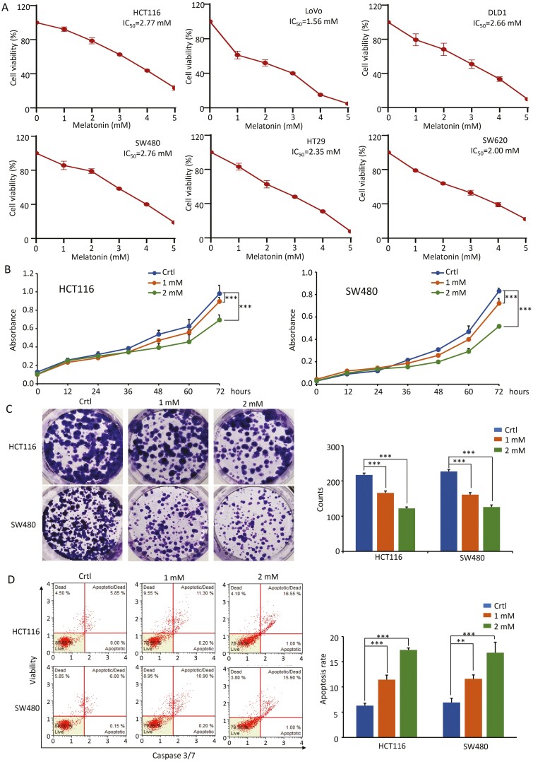
Figure 1.
MLT inhibits the growth of CRC cells in a dose-dependent manner. (A) The cell viability of six CRC cell lines treated with different concentrations of MLT (1–5 mM) for 48 h was determined by CCK-8 assay. (B) Results of CCK-8 assay showing the effect of different MLT concentrations (1 mM, 2 mM) on CRC cell viability. Complete medium containing MLT was added at 24 h and the absorbance was measured at 450 nm after cell culture for 0, 12, 24, 36, 48, 60 and 72 h. (C) Representative results of colony formation assay in CRC cells after exposure to MLT (1 mM, 2 mM) for 7 days. Colony formation count analysis is shown in the right panel. (D) Representative results of FACS analysis using the Muse Caspase-3/7 Kit after treatment on CRC cells by MLT (1 mM, 2 mM) for 48 h. Apoptosis rate analysis is shown in the right panel. Statistical significance: ∗∗P < 0.01, ∗∗∗P < 0.001.

