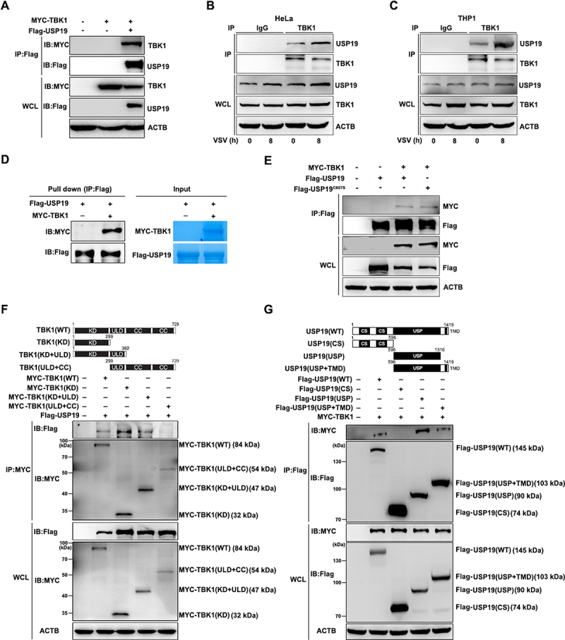
Figure 2.

USP19 interacts with TBK1. (A) HEK293T cells were transfected with MYC-TBK1 and Flag-USP19 plasmids, and then subjected to immunoprecipitation with a Flag antibody before immunoblot analysis. (B and C) HeLa (B) or THP1 cells (C) were infected with VSV for the indicated times, and then subjected to immunoprecipitation with a TBK1 antibody before immunoblot analysis of endogenous TBK1and USP19 protein levels. (D) HEK293T cells were transfected with MYC-TBK1 or Flag-USP19 plasmids, using immunoprecipitation to purify the proteins using Flag or MYC beads. The purified proteins, including Flag-usp19 and MYC-TBK1, were mixed in a Co-IP buffer to perform an in vitro pull-down assay and the results were analyzed via Coomassie Brilliant Blue staining and an immunoblot assay. (E) HEK293T cells were transfected with MYC-TBK1, Flag-USP19, or Flag-USP19C607S plasmids, and then subjected to immunoprecipitation with a Flag antibody before immunoblot analysis. (F) Schematic of TBK1 and the derivatives used (top). HEK293T cells were transfected with MYC-TBK1 (WT), MYC-TBK1 (KD), MYC-TBK1 (KD+ULD), MYC-TBK1 (ULD+CC), and Flag-USP19 plasmids, and then analyzed via immunoprecipitation and immunoblotting with the indicated antibodies (bottom). (G) Schematic of USP19 and the derivatives used (top). HEK293T cells were transfected with Flag-USP19 (WT), Flag-USP19 (CS), Flag-USP19 (USP), Flag-USP19 (USP+TMD), and MYC-TBK1, and then analyzed via immunoprecipitation and immunoblotting with the indicated antibodies (bottom). The data are representative of three independent experiments.
