ABSTRACT
tRNAs purified from non-pathogenic Escherichia coli strains (NPECSs) possess cytotoxic properties on colorectal cancer cells. In the present study, the bioactivity of tRNA halves and tRNA fragments (tRFs) derived from NPECSs are investigated for their anticancer potential. Both the tRNA halves and tRF mimics studied exhibited significant cytotoxicity on colorectal cancer cells, with the latter being more effective, suggesting that tRFs may be important contributors to the bioactivities of tRNAs derived from the gut microbiota. Through high-throughput screening, the EC83 mimic, a double-strand RNA with a 22-nucleotide (nt) 5′-tRF derived from tRNA-Leu(CAA) as an antisense chain, was identified as the one with the highest potency (50% inhibitory concentration [IC50] = 52 nM). Structure-activity investigations revealed that 2′-O-methylation of the ribose of guanosine (Gm) may enhance the cytotoxic effects of the EC83 mimic via increasing the stability of its tertiary structure, which is consistent with the results of in vivo investigations showing that the EC83-M2 mimic (Gm modified) exhibited stronger antitumor activity against both HCT-8 and LoVo xenografts. Consistently, 4-thiouridine modification does not. This provides the first evidence that the bioactivity of tRF mimics would be impacted by chemical modifications. Furthermore, the present study provides the first evidence to suggest that novel tRNA fragments derived from the gut microbiota may possess anticancer properties and have the potential to be potent and selective therapeutic molecules.
IMPORTANCE While the gut microbiota has been increasingly recognized to be of vital importance for human health and disease, the current literature shows that there is a lack of attention given to non-pathogenic Escherichia coli strains. Moreover, the biological activities of tRNA fragments (tRFs) derived from bacteria have rarely been investigated. The findings from this study revealed tRFs as a new class of bioactive constituents derived from gut microorganisms, suggesting that studies on biological functional molecules in the intestinal microbiota should not neglect tRFs. Research on tRFs would play an important role in the biological research of gut microorganisms, including bacterium-bacterium interactions, the gut-brain axis, and the gut-liver axis, etc. Furthermore, the guidance on the rational design of tRF therapeutics provided in this study indicates that further investigations should pay more attention to these therapeutics from probiotics. The innovative drug research of tRFs as potent druggable RNA molecules derived from intestinal microorganisms would open a new area in biomedical sciences.
KEYWORDS: tRF, chemical modifications, gut microbiota, antitumor activity
INTRODUCTION
The microbiome in the human gastrointestinal tract, consisting of trillions of microorganisms, comes into being within days after birth (1). Recent studies revealed that the gut microbiota in general provides beneficial effects to the host by aiding gastrointestinal and immune functions (2). While it can promote health, it may also sometimes cause diseases. For example, non-pathogenic fatty liver, type 2 diabetes, inflammatory bowel disease, obesity, and colorectal cancer (CRC) have been linked to the microbiota (3). Escherichia coli is a Gram-negative gut commensal bacterium found in the colon of mammals and reptiles (4–6). It has become one of the most important model organisms due to its fast growth in chemically defined media in vitro. E. coli can be categorized into four main phylogenetic groups, A, B1, B2, and D. Pathogenic E. coli strains (PECSs) from group B2 have been found to cause gastroenteritis (7), neonatal meningitis (8), hemorrhagic colitis (9), urinary tract infections (10), and Crohn’s disease (11), etc. Advanced studies have revealed that PECSs containing the peptide synthetase-polyketide synthetase (PKS) island could induce DNA double-strand breaks in human cell lines, and thus, PECSs may be able to accelerate the development of colorectal cancer (12–14), which is a leading cause of cancer-related deaths worldwide (15, 16). On the other hand, E. coli strains of groups A and D are not known to cause diseases and can even be beneficial to their hosts, e.g., by producing vitamin K2 (5, 17, 18). Thus, the functions of these non-pathogenic E. coli strains (NPECSs) demand attention from researchers.
In one of our previous studies, tRNA-Val(UAC) and tRNA-Leu(CAG), purified from NPECSs by two-dimensional liquid chromatography, have been shown to exhibit significant cytotoxicity on HCT-8 human colorectal cancer cells (19). As the most abundant class of small RNAs (<200 nucleotides [nt]) (20), tRNAs have been revealed to regulate RNA splicing, RNA translation, and DNA replication (21). It has been reported that endogenous tRNAs may be cleaved by multiple ribonucleases and, under stress conditions, may produce tRNA-derived fragments (tRFs) and tRNA halves (22–25), which have been associated with broad biological functions, such as microRNA (miRNA)-like regulation of protein translation and cellular stress responses (26, 27). In addition, endogenous tRFs can suppress human breast cancer by targeting YBX1, suggesting possible applications as pharmacological agents (28). On the other hand, tRNA halves were found to suppress breast cancer by targeting FZD3 (29). Therefore, it is reasonable to hypothesize that the cytotoxic effects of tRNA-Val(UAC) and tRNA-Leu(CAG) from NPECSs reported in our previous paper (19) may be mediated by the formation of tRFs and tRNA halves. Therefore, we embarked on a study to evaluate and compare tRNA halves and tRF mimics (double-stranded RNA with tRF as an antisense chain) derived from these two RNAs in terms of their antitumor activities against CRC in vitro and in vivo. Moreover, the structure-activity relationship of tRF mimics was investigated using chemically modified tRF mimics.
RESULTS
tRFs may be important contributors to the bioactivities of tRNAs from NPECSs.
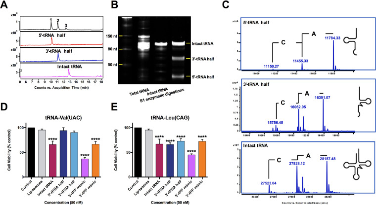
tRNA-Val(UAC) and tRNA-Leu(CAG) were first purified from NPECS total tRNAs (see Fig. S1 in the supplemental material), which were further treated by S1 nuclease to prepare tRNA halves. The results of liquid chromatography-mass spectrometry (LC-MS) analysis and urea-polyacrylamide gel electrophoresis (PAGE) analysis showed that intact tRNAs were successfully cleaved into two half fragments at their anticodon loop (Fig. 1A to C). Cytotoxicity studies, using HCT-8 cells with liposomal transfection at a concentration of 50 nM, showed that intact tRNA-Val(UAC) and tRNA-Leu(CAG) and their tRNA halves and tRF mimics (double-strand RNAs with 22-nt-long 5′- and 3′-tRFs as the antisense chains) significantly decreased the cell viability of HCT-8 cells to various extents, ranging from approximately 36 to 80%, with the exception of the 5′-tRNA half of tRNA-Val(UAC) (Fig. 1D and E). However, the 5′-tRF mimics of both tRNAs are the most effective RNAs.
FIG 1.
The cytotoxicity of tRF mimics is significantly stronger than that of tRNA halves on HCT-8 cells. (A) Typical UHPLC chromatogram of 5′- and 3′-tRNA halves of tRNA-Leu(CAG) under UV at 260 nm. (B) Urea-polyacrylamide gel electrophoresis of 5′- and 3′-tRNA halves of tRNA-Leu(CAG) confirming that the purified tRNA was successfully digested into two products. (C) UHPLC-MS analysis of 5′- and 3′-tRNA halves of tRNA-Leu(CAG) showing that the molecular weights of the digestion products were in accordance with the 5′- and 3′-tRNA halves of E. coli tRNA. (D) Cytotoxic comparison of tRNA halves, tRF mimics, and individual tRNAs of tRNA-Val(UAC) on HCT-8 cells. (E) Cytotoxic comparison of tRNA halves, tRF mimics, and individual tRNAs of tRNA-Leu(CAG) on HCT-8 cells. Data are shown as the means ± SD from three independent experiments. ****, P < 0.0001 (by one-way ANOVA followed by post hoc analysis).
Workflow of individual tRNA purification from NPECSs. Download FIG S1, TIF file, 0.9 MB (911.8KB, tif) .
Copyright © 2022 Cao et al.
This content is distributed under the terms of the Creative Commons Attribution 4.0 International license.
5′-tRF-Leu(CAA) with a length of 22 nt exhibits the strongest inhibitory effects on CRC cells.
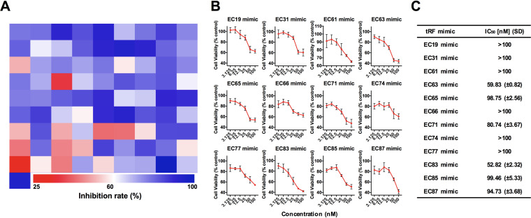
Double-stranded RNA has been widely employed in investigations on the molecular functions of miRNA or tRF since the double-stranded format is more stable than the single-stranded one (30, 31). A total of 82 tRFs, including 5′-tRFs and 3′-tRFs 22 nt in length derived from 41 tRNAs of NPECSs reported in the MODOMICS database and tRNA db (25) (Table S1), were selected as the antisense strands of tRF mimics in the small interfering RNA (siRNA) form (Table S2) since they are the most abundant types of tRFs (32). Their cytotoxic effects were screened using HCT-8 cells (Fig. 2A). Following an initial screen at 50 nM, a total of 12 tRF mimics that exhibited the greatest effects were selected for detailed analysis. Figure 2B shows the dose-dependent cytotoxic effects of these 12 tRF mimics. The most effective tRF mimic is the EC83 mimic [a double-strand RNA with a 22-nt 5′-tRF derived from tRNA-Leu(CAA) as an antisense chain], with a 50% inhibitory concentration (IC50) value of 52.8 nM (Fig. 2C).
FIG 2.
High-throughput screening hits of EC83 mimics with the strongest cytotoxicity compared to other NPECS tRF mimics on HCT-8 cells. (A) Heatmap of high-throughput screening of 82 tRF mimics with hits of 12 tRF mimics with potency in reducing the cell viability of HCT-8 cells. (B) Dose-dependent investigations of the top 12 hits of tRF mimics. (C) IC50 values with standard deviations for the top 12 hits of tRF mimics showing that the EC83 mimic has the strongest cytotoxicity against HCT-8 cells.
Sequence information of E. coli tRNAs. Download Table S1, DOCX file, 0.02 MB (18.3KB, docx) .
Copyright © 2022 Cao et al.
This content is distributed under the terms of the Creative Commons Attribution 4.0 International license.
Designed tRF mimics based on the sequence information of E. coli tRNAs. Download Table S2, DOCX file, 0.02 MB (24.9KB, docx) .
Copyright © 2022 Cao et al.
This content is distributed under the terms of the Creative Commons Attribution 4.0 International license.
Gm modification increases the cytotoxicity of EC83 mimics toward CRC cells.
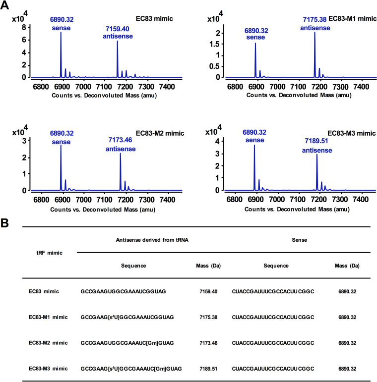
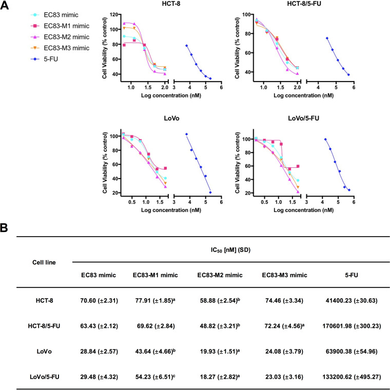
It has been demonstrated that chemical modifications of tRNA can alter the biological functions of tRFs (33). According to the reported modifications of 2′-O-methylguanosine (Gm) and 4-thiourdine (s4U) in tRNA-Leu(CAA), three modified derivatives of EC83 were designed and synthesized, namely, the EC83-M1 mimic (s4U modified), the EC83-M2 mimic (Gm modified), and the EC83-M3 mimic (both s4U and Gm modified). Figure 3A shows the results of the LC-MS analysis, which confirmed the good agreement of their molecular weights with the theoretical values (Fig. 3B) (34). Figure 4A shows that the EC83 mimic and its derivatives decreased the cell viability of colorectal cancer cells (HCT-8 and its fluorouracil [5-FU]-resistant strain and LoVo and its 5-FU-resistant strain). Notably, the EC83-M2 mimic exhibited the strongest cytotoxicity with the lowest IC50 value among the four tRF mimics in the four cancer cell lines (Fig. 4B). These results indicated that Gm modification may increase the cytotoxicity of the EC83 mimic, while s4U modification does not. In comparison, 5-FU has IC50 values in the micromolar concentration range, which is more than 700 times of those of the EC83-M2 mimic, indicating this tRF mimic’s extraordinary cytotoxicity toward cancer cells. It is noteworthy that all tRF mimics showed the same potency in the 5-FU-resistant cells as that in the nonresistant cells. Furthermore, three cancer cell lines (the human ovarian cancer cell line A2780, the liver cancer cell line HepG2, and the breast cancer cell line MDA-MB-231) together with the human colon epithelial cell line HCoEpiC were supplemented in the 3-(4,5-dimethylthiazol-2-yl)-2,5-diphenyltetrazolium bromide (MTT) assay as control groups. As shown in Fig. S2A, the results demonstrated that no significant cytotoxicity of the EC83 mimic and its modified derivatives was observed on A2780 cells, but they exhibited strong cytotoxicity on HepG2 and MDA-MB-231 cells, in which the EC83-M2 mimic had a much lower IC50 value than those of the other RNAs (Fig. S2B). Meanwhile, no significant cytotoxicity of these tRF mimics was observed on HCoEpiC cells. The above-described results demonstrated that the cytotoxicity of tRF mimics derived from non-pathogenic Escherichia coli has cell specificity.
FIG 3.
UHPLC-MS analysis confirmed the accurate molecular weights of the EC83 mimic and its chemically modified derivatives. (A) UHPLC-MS analysis of the EC83 mimic and its modified derivatives confirmed the agreement of their molecular weights with the theoretical values. (B) Sequence information and deconvolution MS of the EC83 mimic and its modified derivatives.
FIG 4.
Gm modification increases the cytotoxicity of EC83 mimics on CRC cells and their 5-FU-resistant strains, while s4U does not. (A) Dose-dependent investigation of EC83 mimics and 5-FU on HCT-8, 5-FU-resistant HCT-8, LoVo, and 5-FU-resistant LoVo cells. (B) IC50 values with SD of EC83 mimics and 5-FU demonstrating that the EC83-M2 mimic exhibits stronger cytotoxicity than the EC83 mimic, while the EC83-M1 mimic has weaker cytotoxicity. a, P < 0.1; b, P < 0.05; c, P < 0.01 (by one-way ANOVA followed by post hoc analysis).
MTT assay of EC83 mimic and its modified mimics on selected cell lines. Download FIG S2, TIF file, 0.9 MB (898.7KB, tif) .
Copyright © 2022 Cao et al.
This content is distributed under the terms of the Creative Commons Attribution 4.0 International license.
Gm modification increases the inhibitory effects of EC83 mimics on colony formation and migration of CRC cells.
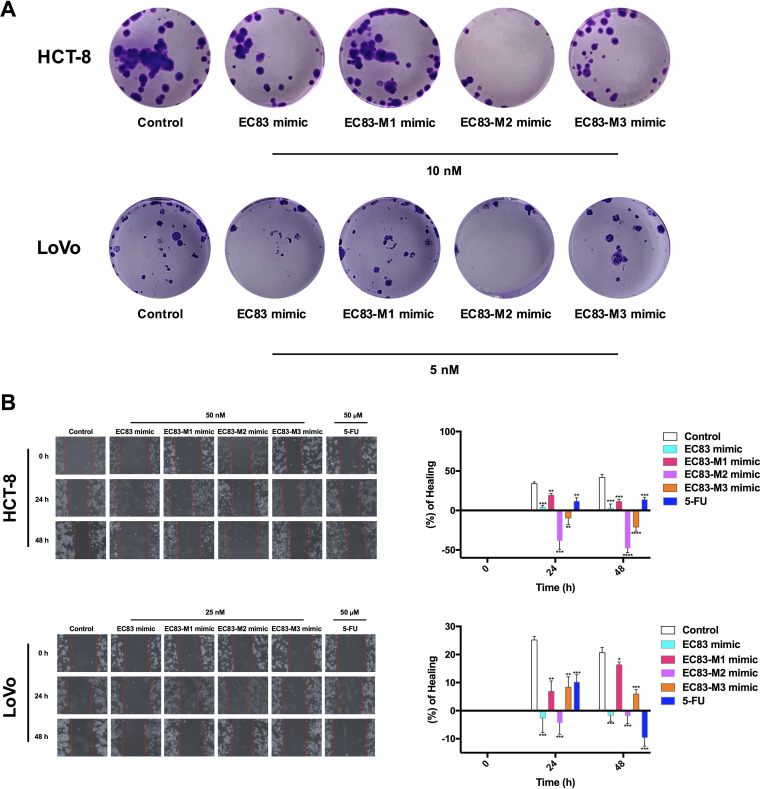
The effectiveness of the EC83 mimic and its modified derivatives in preventing colony formation in a clonogenic assay was determined on HCT-8 and LoVo cells. The results indicated that all tRF mimics together with 5-FU are very effective in suppressing clonogenic ability in both cell lines. The EC83-M2 mimic (Gm modified) exhibited significantly stronger inhibitory effects on the colony formation of both HCT-8 and LoVo cells than the EC83 mimic, while the EC83-M1 mimic and the EC83-M3 mimic did not exhibit such effect (Fig. 5A).
FIG 5.
Gm modification increases the inhibitory effects of EC83 mimics on colony formation and migration of CRC cells, while s4U does not. (A) Clonogenic assay of the EC83 mimic, the EC83-M1 mimic, the EC83-M2 mimic, and the EC83-M3 mimic on HCT-8 and LoVo cells. (B) Wound-healing assay of the EC83 mimic, the EC83-M1 mimic, the EC83-M2 mimic, and the EC83-M3 mimic on HCT-8 and LoVo cells. Data are shown as the means ± SD from three independent experiments. *, P < 0.05; **, P < 0.01; ***, P < 0.001; ****, P < 0.0001 (by two-tailed Student’s t test).
The migration of cancer cells is important for cancer development. As shown in Fig. 5B, at 24 and 48 h, EC83-M2 mimic-treated HCT-8 cells at 50 nM (−38.6% ± 10.6% and −48.0% ± 5.3%, respectively) exhibited a significantly lower wound-healing rate (WHR) than EC83 mimic-treated cells (4.0% ± 2.4% and 2.8% ± 5.3%), while the EC83-M1 mimic-treated ones had a higher WHR at the same concentration (19.4% ± 2.1% and 11.4% ± 2.8%). For LoVo cells, EC83-M2 mimic-treated cells at 25 nM (−4.3% ± 4.0% and −1.9% ± 2.7%) exhibited a WHR comparable to that with the EC83 mimic (−2.6% ± 5.1% and −1.6% ± 2.1%) at 24 and 48 h, while the EC83-M1 mimic-treated ones had a significantly higher WHR at the same concentration (6.8% ± 3.7% and 16.3% ± 2.9%). Meanwhile, EC83-M3 mimic-treated cells had lower WHRs than those of EC83-M2 mimic-treated cells. As a positive control, 5-FU-treated HCT-8 cells (11.7% ± 4.2% and 13.8% ± 2.9%) and LoVo cells (10.1% ± 2.6% and −9.5% ± 3.0%) at 50 μM exhibited a low WHR at 24 and 48 h. These results, together with those of the clonogenic assay, demonstrated that Gm modification may increase the cytotoxic effectiveness of the EC83 mimic toward CRC cells, while s4U modification does not.
Gm modification increases the inhibitory effects of EC83 mimic toward CRC tumor growth in vivo.
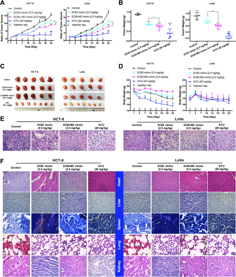
To further confirm the antitumor activity of EC83 mimic and EC83-M2 mimic and their structure-activity relationship, a CRC cell (HCT-8 and LoVo) xenograft-bearing nude mouse model was developed for in vivo experiments. The results showed that at day 22, compared to the control group, both EC83 mimic and EC83-M2 mimic could significantly suppress the tumor growth rate (Fig. 6A), tumor weight (Fig. 6B), and tumor size (Fig. 6C), while 5-FU was used as a positive control. Interestingly, the EC83-M2 mimic exhibited significantly stronger inhibitory effects than those of EC83 mimic, suggesting that Gm modification might enhance the anticancer activity of EC83 mimics. This is consistent with the in vitro results. However, 5-FU was found to significantly reduce the body weight of nude mice at the end of the experiment, while no significant change was observed in the RNA-treated group (Fig. 6D). Meanwhile, hematoxylin and eosin (H&E) staining showed that EC83 mimic and EC83-M2 mimic-treated tumors have less eosinophilic cytoplasm than the control group, indicating their efficacy as tumor suppressants (Fig. 6E). To determine the side effects of the two RNAs, morphological imaging and H&E staining of major organs, including heart, liver, spleen, lung, and kidney, from HCT-8 and LoVo xenograft-bearing nude mice were performed (Fig. S3 and Fig. 6F). The results demonstrated that there are no significant differences in the major organs between the RNA-treated group and the control group, suggesting that RNA treatment is potentially safe.
FIG 6.
Gm modification increases the inhibitory effects of EC83 mimics toward CRC tumor growth in vivo. (A) Growth rates of tumors with the encapsulated EC83 mimic and the EC83-M2 mimic, 5-FU, or HKP alone as a control. (B) Weights of tumors removed from the mice after day 22. (C) Images of each tumor in all groups. (D) Body weights of mice in all groups. (E and F) Representative images of hematoxylin and eosin-stained tumors (E) and major organs (F) of CRC xenograft-bearing nude mice in all groups. Data are shown as the means ± standard errors of the means (SEM). *, P < 0.05; ***, P < 0.001; ****, P < 0.0001 (by two-tailed Student’s t test).
Morphological images of major organs from CRC xenograft-bearing nude mice. Download FIG S3, TIF file, 2.1 MB (2.1MB, tif) .
Copyright © 2022 Cao et al.
This content is distributed under the terms of the Creative Commons Attribution 4.0 International license.
Gm modification increases the stability of the EC83 mimic’s tertiary structure.
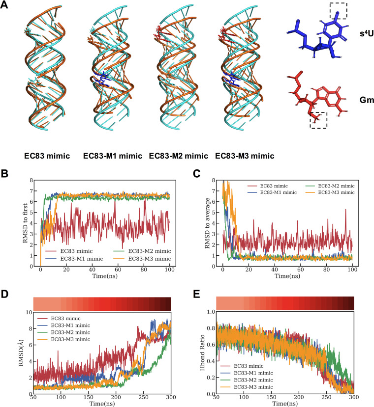
To evaluate the stability of the EC83 mimic and its derived modified mimics at the molecular level, their structures were initially assumed to be the standard A-form double helix from molecular dynamics (MD) simulations. Starting from each of these initial structures, a 100-ns simulation at 300 K in isothermal‐isobaric (NPT) ensemble revealed that the EC83 mimic remained as an A-form double helix, while the three modified derivatives transformed into more compact helical conformations rapidly (Fig. 7A). Moreover, as shown in Fig. 7B and C, the fluctuation of the root mean square deviation (RMSD) of the EC83 mimic is significantly larger than those of the modified mimics, indicating that the transformed helical structures (EC83-M1, -M2, and -M3 mimics) are more stable than the A-form standard helix (EC83 mimic).
FIG 7.
Gm modification increases the tertiary structural stability of EC83 mimics. (A) Simulated three-dimensional (3D) initial structure (cyan) and transformed structure (gold) of the EC83 mimic and its modified derivatives. (B and C) RMSD for the initial A-form structure (B) and stable structures for four RNA mimics changing over time (C). (D and E) With the increasing temperature, the RMSD, using the stable structure as a reference (D), and the hydrogen bond (Hbond) ratios (E) of four RNA mimics change over time. The temperature at each time point is shown in the color bars at the top.
To further evaluate the stability of the modified mimics, 200-ns simulations were carried out by increasing the temperature gradually from 300 K to 500 K (intervals of 10 K). The results showed that mimics would lose their stable structures in the order of EC83 mimic, EC83-M1 mimic or EC83-M3 mimic, and EC83-M2 mimic (Fig. 7D). Here, the unfolding structures were defined by their RMSD values above 5 Å relative to the stable structure. As further shown in Fig. 7E, the hydrogen bond (Hbond) ratio of the EC83-M2 mimic remained above 40% at 260 ns, while those of the other 3 were under 20%. These results demonstrated that the EC83-M2 mimic is structurally the most stable of the derivatives, which are also more stable than the unmodified EC83 mimic, suggesting that Gm modification enhances the tertiary structural stability of EC83 mimics. On the other hand, s4U modification appears to not exhibit such a strong effect.
DISCUSSION
CRC has become the third most frequently diagnosed malignancy worldwide (10.0%), accounting for ∼576,858 deaths in 2020 (35). Risk factors of CRC include family history, a diet high in red and processed meat, heavy alcohol intake, smoking, being overweight or obese, age (being older), and inflammatory bowel disease (36). Currently, cytotoxic chemotherapy is widely used as a monotherapy or a component of combined therapy for CRC treatment (37). However, the development of resistance to treatment is a great challenge. RNAs are able to regulate gene expression and therefore may have the potential to be a new class of cancer therapeutics that are nontoxic and have desirable selectivity for a specific target gene (38). The present study describes, for the first time, the ability of tRNA halves and tRFs derived from NPECS that exhibit anticancer activity against colorectal cancer. They are very effective in vitro, with IC50 values at the 10−8 M concentration range, compared to the 10−4 M range for 5-FU. Interestingly, tRFs exhibited higher potency than tRNA halves, suggesting that tRFs might play an important role in the cytotoxic effects of tRNAs derived from the gut microbiota. Importantly, the more potent EC83 mimic and its modified derivatives were observed to be just as effective on 5-FU-resistant CRC cells as on non-pathogenic cells, indicating that microbiota-derived tRFs may be an effective treatment even in CRC resistant to conventional chemotherapy. This observation supports the idea that the cytotoxic actions of tRFs differ from that of 5-FU. The exact mechanism of action remains to be investigated. It has been revealed that endogenous tRFs derived from human breast cancer cells could suppress breast cancer in vitro and in vivo by targeting YBX1 mRNA, suggesting that tRFs could target signaling pathways related to cell proliferation, thus enabling an effective intervention with the treatment of cancers (28). This, together with our previous study on a tRF derived from Chinese yew, which suppresses ovarian cancer by targeting the oncogene TRPA1 (31), raises the hypothesis that tRFs derived from non-pathogenic E. coli might also function via an RNA interference (RNAi) pathway. Further investigations on the molecular target of EC83 mimics should be performed via mRNA sequencing and experimental validations.
As a class of highly modified nucleic acids (39–44), tRFs have been proven to possess miRNA-like regulatory function in relation to RNA interference (33). Functional improvements have been reported following chemical modifications of tRFs (45). The loss of cytosine-5 RNA methylation was found to enhance the angiogenin-mediated endonucleolytic cleavage of tRNA, leading to an accumulation of 5′-tRFs and, subsequently, to a reduced cell size and increased apoptosis (46). Consistently, the present results revealed that Gm modification enhanced the cytotoxic effects of EC83 mimics perhaps via the observed increased stability of the tertiary structure. This was supported by the observation that s4U modification does not exhibit such a strong effect. Indeed, different RNAs have different potentials for encapsulation within lipid vesicles, which might be greatly impacted by their negative charges. However, the retention time values of the EC83 mimic and its derived modified mimics in our ion-pair chromatography analysis are very close (see Fig. S4 in the supplemental material). This indicated that the EC83 mimic and its derived modified mimics have similar negative charges, demonstrating that these RNAs have similar encapsulation rates within lipid vesicles. Overall, this first structure-activity investigation provided evidence that the bioactivity of tRF mimics can be impacted by chemical modifications. Further structural analysis is required to decipher the critical structural differences between these modified tRF mimics.
Typical ion-pair chromatograms of EC83 mimic and its derived modified mimics. Download FIG S4, TIF file, 0.5 MB (535.5KB, tif) .
Copyright © 2022 Cao et al.
This content is distributed under the terms of the Creative Commons Attribution 4.0 International license.
Recently, it was reported that colonic bacteria, especially PECSs, bear a close relationship with CRC development (47, 48). Thus, current interests in clinical studies focus on PECSs, such as enterohemorrhagic E. coli (EHEC) O157:H7, which produces metabolites or toxins that can cause DNA damage in the colon and CRC development (49–51). As the genome size and gene number of NPECSs (3.98 Mb and 3,696, respectively) differ from those of PECSs (5.86 Mb and 5,919) (52), NPECSs and PECSs may produce completely different profiles of tRNA fragments through different chemical modifications. This may raise the possibility of identifying CRC biomarkers from tRNA molecules derived from PECSs.
In summary, these findings revealed tRNA halves and especially tRFs as a new class of bioactive constituents derived from gut microorganisms themselves and provided new insights into their therapeutic effects on human diseases, which broadened our knowledge of the beneficial effects of gut microbiota. Also, the present study demonstrates that studies on biological functional molecules in the intestinal microbiota should not neglect tRFs since they are bioactive constituents (28). Research on tRFs will play an important role in biological research on gut microorganisms, including bacterium-bacterium interactions, the gut-brain axis, and the gut-liver axis, etc. Furthermore, with the increasing interest in the identification of tRFs in bacteria (53, 54), the guidance on the rational design of tRF therapeutics provided in this study suggests that further investigations should pay more attention to these therapeutics from probiotics. The innovative drug research on tRFs as potent druggable RNA molecules derived from intestinal microorganisms will open a new area in biomedical sciences.
MATERIALS AND METHODS
Chemicals and reagents.
Escherichia coli MRE600 total tRNA was purchased from Roche (Switzerland). Biotin-labeled single-stranded DNA oligonucleotides were obtained from BGI, China. A low-range single-stranded RNA (ssRNA) ladder was purchased from New England BioLabs (USA). Diethylpyrocarbonate (DEPC)-treated water, S1 nuclease, and polyacrylamide containing a ratio of acrylamide/bis of 19:1 (wt/wt) were purchased from Thermo (USA). Triethylammonium acetate, hexafluoro-2-propanol, and fluorouracil (5-FU) were purchased from Sigma (USA). Deionized water was prepared using a Millipore MilliQ Plus system. All reagents used were of analytical grade.
Affinity purification of tRNAs from an NPECS.
Affinity purification was performed according to methods described previously by Tsurui et al. (55), with minor modifications. Briefly, biotin-labeled single-strand DNA (antisense) oligonucleotides complementary to tRNA-Val(UAC) (5′-GCCGACCCCCTCCTTGTAAGGGAGGTGCTC-3′) or tRNA-Leu(CAG) (5′-ACGTCCGTAAGGACACTAACACCTGAAGCT-3′) were mixed with total tRNA from E. coli and then denatured at 95°C for 5 min. After incubation at a temperature 5°C lower than the melting temperature (Tm) of each DNA probe for 1.5 h, streptavidin magnetic beads (Beaverbio, China) were added, and the mixture was incubated for another 30 min. Subsequently, the biotinylated DNA/tRNA-coated beads were separated with a magnet and washed at 40°C. Finally, the magnetic beads were incubated in RNase-free water at 65°C for 5 min to release the immobilized tRNA with a probe and then centrifuged at 10,000 × g for 1 min. The supernatants were analyzed immediately by ultrahigh-performance liquid chromatography (UHPLC).
S1 nuclease hydrolysis.
S1 nuclease was added to purified tRNA samples (8 U enzyme/500 ng tRNA) with reaction buffer (2 μL), the final volume was adjusted to 20 μL, and the mixture was incubated at 25°C for 40 min. The reaction was stopped by the addition of an EDTA solution (0.5 M; 0.5 μL). The supernatant was collected by centrifugation at 10,000 × g for 1 min for UHPLC-MS analysis.
Urea-polyacrylamide gel electrophoresis.
All samples were separated by vertical slab gel electrophoresis (Mini-Protean Tetra system; Bio-Rad, USA) using a 15% urea-polyacrylamide gel. Samples were electrophoresed at 250 V for 1 h at room temperature (25°C) and stained with 1× SYBR gold nucleic acid gel stain (Thermo) in MilliQ Plus water for 30 min, followed by imaging using a Bio-Rad imaging system under UV light.
UHPLC-MS.
UHPLC (Agilent 1290 system) was performed using a C18 column (Acquity UPLC [ultraperformance liquid chromatography] OST, 2.1 × 100 mm, 1.7-μm internal diameter [i.d.]; Waters, USA) at 60°C with a diode array detector. UHPLC-MS was performed using an Agilent 1290 system (Agilent Technologies, USA), equipped with a vacuum degasser, a quaternary pump, an autosampler, a diode array detector, and an Agilent ultrahigh-definition 6545 quadrupole time of flight (Q-TOF) mass spectrometer. Separation was carried out on an Acquity UPLC OST C18 column (2.1 × 100 mm, 1.7-μm i.d.; Waters, USA) at 60°C. tRNAs were separated by eluting the column at a flow rate of 0.2 mL/min with a mobile phase of 100 mM 1,1,1,3,3,3-Hexafluoro-2-propanol (HFIP) plus 15 mM triethylammonium acetate (TEAA) containing methanol (MeOH) at the following concentrations: 1% (vol/vol) for 1.5 min, 1 to 14% over 1.5 to 8.3 min, and finally 14 to 17% over 8.3 to 16.5 min. Electrospray ionization (ESI) conditions were as follows: gas temperature of 320°C, spray voltage of 3.5 kV, and sheath gas flow and temperature set at 12 L/min and 350°C, respectively. Fractions corresponding to each chromatographic peak were collected and freeze-dried. For MS experiments, samples were analyzed in negative mode over an m/z range of 500 to 3,200.
Cell culture.
The human ovarian cancer cell line A2780 was purchased from KeyGen Biotech (China). The human liver cancer cell line HepG2, the human breast cancer cell line MDA-MB-231, the human colorectal cancer cell lines HCT-8 and LoVo along with their 5-FU-resistant strains (HCT-8/5-FU and LoVo/5-FU), and the human colon epithelial cell line HCoEpiC were purchased from the American Type Culture Collection (ATCC). A2780, LoVo/5-FU, HCT-8, HCT-8/5-FU, and HCoEpiC cells were cultured in RPMI 1640 medium (Gibco, New Zealand). LoVo cells were cultured in F-12K medium (Thermo). HepG2 cells were cultured in minimal essential medium (MEM) (Thermo). MDA-MB-231 cells were cultured in Leibovitz’s L-15 medium (Thermo). All culture media contained 10% fetal bovine serum (FBS) and 1% penicillin/streptomycin (P/S). All cell lines were cultured in a humidified 5% CO2 atmosphere at 37°C, except for MDA-MB-231 cells, which were cultured in a humidified 100% air atmosphere at 37°C. All tested RNA samples were dissolved in nuclease-free water and stored at −80°C before use. 5-FU was dissolved in dimethyl sulfoxide (DMSO) and used as a positive control.
Cytotoxicity determination.
Cells (5 × 10³ in 100 μL culture medium) were seeded onto 96-well plates. After 20 h, cells were treated with various concentrations of RNA sample solutions with Lipofectamine RNAiMAX transfection reagent in Opti-MEM medium (Thermo) according to the manufacturer’s instructions. Cells without any treatment were used as controls, and cells treated with liposomes were used as treatment controls. Cell viability was determined after 48 h. An MTT [3-(4,5-dimethylthiazol-2-yl)-2,5-diphenyltetrazolium bromide] solution (50 μL per well; 1 mg/mL) (Thermo) was added to each well; the mixture was incubated for 4 h at 37°C, followed by DMSO (200 μL); and the A570 was measured using a SpectraMax 190 microplate reader (Molecular Devices, USA). Dose-response curves were constructed and the IC50 values were calculated using GraphPad Prism 5.0 (GraphPad, USA). Each experiment was carried out three times. IC50 results were expressed as means ± standard deviations (SD).
Clonogenic assay.
The clonogenic assay was performed according to methods described previously by Franken et al. (56), with minor modifications. Briefly, cells were plated at a density of 1,000 cells/well with culture medium in 6-well plates. After 20 h, the medium was changed to medium containing RNA samples (10 nM for HCT-8 and 5 nM for LoVo cells) with liposomal transfection or blank Opti-MEM for a further 48 h. Cells were maintained in normal culture medium for the following 14 days. After fixation for 20 min with a 4% paraformaldehyde (PFA) fix solution (Beyotime, China), the cells were stained with crystal violet (Beyotime, China) for 10 min. Finally, the numbers of colonies with more than 50 individual cells were counted using ImageJ software.
Wound-healing assay.
Cells (5 × 10⁵ in 100 μL culture medium) were grown in 6-well plates for 20 h until confluent. A scratch was made by using a sterile 1-mL pipette tip, and the medium was changed to medium containing RNA samples (50 nM for HCT-8 and 25 nM for LoVo cells) with liposomal transfection or 5-FU at 50 μM. The cells were viewed with a 10× objective and photographed using a phase-contrast microscope (Leica Microsystems, Germany) at various time points (0, 24, and 48 h). ImageJ software was applied to quantify the area of the wound created. The wound-healing rate was calculated using the following formula: wound-healing rate = [(wound area at 0 h − wound area at 24 or 48 h)/wound area at 0 h] × 100.
Animal study.
Animal care and use protocols were approved by the Ethics Committee of the Macau University of Science and Technology. HCT-8 cells (2.0 × 10⁶) and LoVo cells (1.0 × 10⁷) were injected subcutaneously under the armpits of 8-week-old BALB/c female nude mice (Shanghai SLAC Laboratory Animal Co., Ltd., China). When the tumors reached 100 mm3, the EC83 mimic and EC83-M2 mimic encapsulated in HKP nanoparticles were administered by intratumoral injection (2.5 mg/kg of body weight) once every 3 days. 5-FU (20 mg/kg) and histidine-lysine polymer (HKP) were used as positive and negative controls, respectively. The animals were sacrificed on day 22. Tumor diameters were measured at the points of maximum length and maximum width with digital calipers. Tumor volumes were calculated by the following formula: volume = (width)2 × length/2. Data were statistically analyzed using GraphPad Prism 5.0.
Histological analysis.
The tissues from nude mice were fixed in 4% PFA and embedded in paraffin. Three-micrometer to 5-μm paraffin sections were cut and stained with hematoxylin and eosin (H&E) to determine the efficacy and side effects of the EC83 mimic and the EC83-M2 mimic.
Computational simulations of tertiary structures.
The initial standard A-form double-helix structures for the EC83 mimic and its modified derivatives were built by NAB (57) in the Amber18 package (58). The force field used was OL3 (59) (leaprc.RNA.OL3 in Amber18), and the parameters for the modified nucleotides (s4U and Gm) were described previously by Aduri et al. (60). Each system was solvated in a 12-Å transferable interatomic potential with three points model (TIP3P) cubic water box (∼6,000 water molecules), and 42 Na+ were then randomly added to neutralize the whole system. To minimize the systems before simulation, RNA molecules were first restricted by 20 kcal/mol Å harmonic potential with the energy of the solvent minimized for 8,000 steps, where the first 4,000 were steepest-descent steps and the last 4,000 were conjugated-gradient steps. Furthermore, another 8,000 minimization steps were performed in the same way except that the constraints exerted on the solvate were removed.
After energy optimization, the system was heated from 0 to 300 K, and temperature equilibration was allowed at 300 K for 200 ps. Subsequently, another 200-ps equilibration simulation was performed. The solvate was restricted as described above. Besides, the time step was set to 1 fs and run at NPT ensemble using the parallel version of pmemd (61), with the primary molecular dynamics engine within Amber. A 300-ns production simulation was performed for each RNA molecule. The temperature was equilibrated at 300 K by Langevin dynamics in the first 100 ns and then gradually increased to 500 K in the last 200 ns with 10 K intervals. The time step was increased from 1 to 2 fs due to the application of the SHAKE algorithm. The nonbond cutoff was set to 10 Å, and the long-range electrostatic interactions were evaluated by the pattern method extension (PME) method. The pressure was kept at around 105 Pa (0.987 atm), and all simulations were performed on Nvidia graphic processing unit (GPU) cards by the GPU version of pmemd.
The stabilities of the RNA molecules were compared in terms of backbone (P, O3′, O5′, C3′, C4′, and C5′) RMSDs based on reference to the stable structures and the extent of hydrogen bonding in 22 bp. As the fluctuation of the RMSD for each RNA molecule was small, their stable structures were the average structures in 50 to 100 ns. Furthermore, the two criteria were calculated by cpptraj (62) in Amber, and the following data analysis was based on numpy and matplotlib, which are two popular python packages. The tertiary structures were visualized using PyMOL (63).
Statistical and data analyses.
All experimental results were expressed as means ± SD. Statistical significance was analyzed using two-tailed Student’s t test (GraphPad Prism) or one-way analysis of variance (ANOVA) followed by post hoc analysis.
Supplementary Material
ACKNOWLEDGMENTS
This work was financially funded by the Science and Technology Development Fund, Macau SAR (file no. 0023/2019/AKP, 015/2017/AFJ), and the State Key Laboratory of Chemical Oncogenomics.
We declare that there are no conflicts of interest.
Contributor Information
Zhi-Hong Jiang, Email: zhjiang@must.edu.mo.
Zackery Bulman, University of Illinois at Chicago.
REFERENCES
- 1.Ley RE, Peterson DA, Gordon JI. 2006. Ecological and evolutionary forces shaping microbial diversity in the human intestine. Cell 124:837–848. doi: 10.1016/j.cell.2006.02.017. [DOI] [PubMed] [Google Scholar]
- 2.Neish AS. 2009. Microbes in gastrointestinal health and disease. Gastroenterology 136:65–80. doi: 10.1053/j.gastro.2008.10.080. [DOI] [PMC free article] [PubMed] [Google Scholar]
- 3.Marchesi JR, Adams DH, Fava F, Hermes GD, Hirschfield GM, Hold G, Quraishi MN, Kinross J, Smidt H, Tuohy KM, Thomas LV, Zoetendal EG, Hart A. 2016. The gut microbiota and host health: a new clinical frontier. Gut 65:330–339. doi: 10.1136/gutjnl-2015-309990. [DOI] [PMC free article] [PubMed] [Google Scholar]
- 4.Gordon DM, Cowling A. 2003. The distribution and genetic structure of Escherichia coli in Australian vertebrates: host and geographic effects. Microbiology (Reading) 149:3575–3586. doi: 10.1099/mic.0.26486-0. [DOI] [PubMed] [Google Scholar]
- 5.Bentley R, Meganathan R. 1982. Biosynthesis of vitamin K (menaquinone) in bacteria. Microbiol Rev 46:241–280. doi: 10.1128/mr.46.3.241-280.1982. [DOI] [PMC free article] [PubMed] [Google Scholar]
- 6.Kosek M, Bern C, Guerrant RL. 2003. The global burden of diarrhoeal disease, as estimated from studies published between 1992 and 2000. Bull World Health Organ 81:197–204. [PMC free article] [PubMed] [Google Scholar]
- 7.Eltai NO, Al Thani AA, Al Hadidi SH, Al Ansari K, Yassine HM. 2020. Antibiotic resistance and virulence patterns of pathogenic Escherichia coli strains associated with acute gastroenteritis among children in Qatar. BMC Microbiol 20:54. doi: 10.1186/s12866-020-01732-8. [DOI] [PMC free article] [PubMed] [Google Scholar]
- 8.Liu Y, Zhu M, Fu X, Cai J, Chen S, Lin Y, Jiang N, Chen S, Lin Z. 2021. Escherichia coli causing neonatal meningitis during 2001–2020: a study in eastern China. Int J Gen Med 14:3007–3016. doi: 10.2147/IJGM.S317299. [DOI] [PMC free article] [PubMed] [Google Scholar]
- 9.Quinto EJ, Marín JM, Caro I, Mateo J, Schaffner DW. 2020. Modelling growth and decline in a two-species model system: pathogenic Escherichia coli O157:H7 and psychrotrophic spoilage bacteria in milk. Foods 9:331. doi: 10.3390/foods9030331. [DOI] [PMC free article] [PubMed] [Google Scholar]
- 10.Li D, Reid CJ, Kudinha T, Jarocki VM, Djordjevic SP. 2020. Genomic analysis of trimethoprim-resistant extraintestinal pathogenic Escherichia coli and recurrent urinary tract infections. Microb Genom 6:mgen000475. doi: 10.1099/mgen.0.000475. [DOI] [PMC free article] [PubMed] [Google Scholar]
- 11.Elhenawy W, Hordienko S, Gould S, Oberc AM, Tsai CN, Hubbard TP, Waldor MK, Coombes BK. 2021. High-throughput fitness screening and transcriptomics identify a role for a type IV secretion system in the pathogenesis of Crohn’s disease-associated Escherichia coli. Nat Commun 12:2032. doi: 10.1038/s41467-021-22306-w. [DOI] [PMC free article] [PubMed] [Google Scholar]
- 12.Arthur JC, Gharaibeh RZ, Muhlbauer M, Perez-Chanona E, Uronis JM, McCafferty J, Fodor AA, Jobin C. 2014. Microbial genomic analysis reveals the essential role of inflammation in bacteria-induced colorectal cancer. Nat Commun 5:4724. doi: 10.1038/ncomms5724. [DOI] [PMC free article] [PubMed] [Google Scholar]
- 13.Dejea CM, Fathi P, Craig JM, Boleij A, Taddese R, Geis AL, Wu X, DeStefano Shields CE, Hechenbleikner EM, Huso DL, Anders RA, Giardiello FM, Wick EC, Wang H, Wu S, Pardoll DM, Housseau F, Sears CL. 2018. Patients with familial adenomatous polyposis harbor colonic biofilms containing tumorigenic bacteria. Science 359:592–597. doi: 10.1126/science.aah3648. [DOI] [PMC free article] [PubMed] [Google Scholar]
- 14.Nougayrede JP, Homburg S, Taieb F, Boury M, Brzuszkiewicz E, Gottschalk G, Buchrieser C, Hacker J, Dobrindt U, Oswald E. 2006. Escherichia coli induces DNA double-strand breaks in eukaryotic cells. Science 313:848–851. doi: 10.1126/science.1127059. [DOI] [PubMed] [Google Scholar]
- 15.Ferlay J, Steliarova-Foucher E, Lortet-Tieulent J, Rosso S, Coebergh JW, Comber H, Forman D, Bray F. 2013. Cancer incidence and mortality patterns in Europe: estimates for 40 countries in 2012. Eur J Cancer 49:1374–1403. doi: 10.1016/j.ejca.2012.12.027. [DOI] [PubMed] [Google Scholar]
- 16.Siegel R, Desantis C, Jemal A. 2014. Colorectal cancer statistics. CA Cancer J Clin 64:104–117. doi: 10.3322/caac.21220. [DOI] [PubMed] [Google Scholar]
- 17.Gao Q, Chen H, Wang G, Yang W, Zhong X, Liu J, Huo X, Liu W, Huang J, Tao Y, Lin B. 2021. Highly efficient production of menaquinone-7 from glucose by metabolically engineered Escherichia coli. ACS Synth Biol 10:756–765. doi: 10.1021/acssynbio.0c00568. [DOI] [PubMed] [Google Scholar]
- 18.Mladěnka P, Macáková K, Kujovská Krčmová L, Javorská L, Mrštná K, Carazo A, Protti M, Remião F, Nováková L, on behalf of the OEMONOM Researchers and Collaborators . 2021. Vitamin K—sources, physiological role, kinetics, deficiency, detection, therapeutic use, and toxicity. Nutr Rev 80:677–698. doi: 10.1093/nutrit/nuab061. [DOI] [PMC free article] [PubMed] [Google Scholar]
- 19.Cao KY, Pan Y, Yan TM, Jiang ZH. 2020. Purification, characterization and cytotoxic activities of individual tRNAs from Escherichia coli. Int J Biol Macromol 142:355–365. doi: 10.1016/j.ijbiomac.2019.09.106. [DOI] [PubMed] [Google Scholar]
- 20.Storz G. 2002. An expanding universe of noncoding RNAs. Science 296:1260–1263. doi: 10.1126/science.1072249. [DOI] [PubMed] [Google Scholar]
- 21.Barciszewska MZ, Perrigue PM, Barciszewski J. 2016. tRNA—the golden standard in molecular biology. Mol Biosyst 12:12–17. doi: 10.1039/c5mb00557d. [DOI] [PubMed] [Google Scholar]
- 22.Fu H, Feng J, Liu Q, Sun F, Tie Y, Zhu J, Xing R, Sun Z, Zheng X. 2009. Stress induces tRNA cleavage by angiogenin in mammalian cells. FEBS Lett 583:437–442. doi: 10.1016/j.febslet.2008.12.043. [DOI] [PubMed] [Google Scholar]
- 23.Yamasaki S, Ivanov P, Hu GF, Anderson P. 2009. Angiogenin cleaves tRNA and promotes stress-induced translational repression. J Cell Biol 185:35–42. doi: 10.1083/jcb.200811106. [DOI] [PMC free article] [PubMed] [Google Scholar]
- 24.Thompson DM, Parker R. 2009. The RNase Rny1p cleaves tRNAs and promotes cell death during oxidative stress in Saccharomyces cerevisiae. J Cell Biol 185:43–50. doi: 10.1083/jcb.200811119. [DOI] [PMC free article] [PubMed] [Google Scholar]
- 25.Gebetsberger J, Polacek N. 2013. Slicing tRNAs to boost functional ncRNA diversity. RNA Biol 10:1798–1806. doi: 10.4161/rna.27177. [DOI] [PMC free article] [PubMed] [Google Scholar]
- 26.Shen Y, Yu X, Zhu L, Li T, Yan Z, Guo J. 2018. Transfer RNA-derived fragments and tRNA halves: biogenesis, biological functions and their roles in diseases. J Mol Med (Berl) 96:1167–1176. doi: 10.1007/s00109-018-1693-y. [DOI] [PubMed] [Google Scholar]
- 27.Keam SP, Hutvagner G. 2015. tRNA-derived fragments (tRFs): emerging new roles for an ancient RNA in the regulation of gene expression. Life (Basel) 5:1638–1651. doi: 10.3390/life5041638. [DOI] [PMC free article] [PubMed] [Google Scholar]
- 28.Goodarzi H, Liu X, Nguyen HCB, Zhang S, Fish L, Tavazoie SF. 2015. Endogenous tRNA-derived fragments suppress breast cancer progression via YBX1 displacement. Cell 161:790–802. doi: 10.1016/j.cell.2015.02.053. [DOI] [PMC free article] [PubMed] [Google Scholar]
- 29.Mo D, Jiang P, Yang Y, Mao X, Tan X, Tang X, Wei D, Li B, Wang X, Tang L, Yan F. 2019. A tRNA fragment, 5′-tiRNAVal, suppresses the Wnt/β-catenin signaling pathway by targeting FZD3 in breast cancer. Cancer Lett 457:60–73. doi: 10.1016/j.canlet.2019.05.007. [DOI] [PubMed] [Google Scholar]
- 30.Zhang L, Hou D, Chen X, Li D, Zhu L, Zhang Y, Li J, Bian Z, Liang X, Cai X, Yin Y, Wang C, Zhang T, Zhu D, Zhang D, Xu J, Chen Q, Ba Y, Liu J, Wang Q, Chen J, Wang J, Wang M, Zhang Q, Zhang J, Zen K, Zhang C-Y. 2012. Exogenous plant MIR168a specifically targets mammalian LDLRAP1: evidence of cross-kingdom regulation by microRNA. Cell Res 22:107–126. doi: 10.1038/cr.2011.158. [DOI] [PMC free article] [PubMed] [Google Scholar]
- 31.Cao K-Y, Yan T-M, Zhang J-Z, Chan T-F, Li J, Li C, Leung EL-H, Gao J, Zhang B-X, Jiang Z-H. 2022. A tRNA-derived fragment from Chinese yew suppresses ovarian cancer growth via targeting TRPA1. Mol Ther Nucleic Acids 27:718–732. doi: 10.1016/j.omtn.2021.12.037. [DOI] [PMC free article] [PubMed] [Google Scholar]
- 32.Martinez G, Choudury SG, Slotkin RK. 2017. tRNA-derived small RNAs target transposable element transcripts. Nucleic Acids Res 45:5142–5152. doi: 10.1093/nar/gkx103. [DOI] [PMC free article] [PubMed] [Google Scholar]
- 33.Diebel KW, Zhou K, Clarke AB, Bemis LT. 2016. Beyond the ribosome: extra-translational functions of tRNA fragments. Biomark Insights 11:1–8. doi: 10.4137/BMI.S35904. [DOI] [PMC free article] [PubMed] [Google Scholar]
- 34.Boccaletto P, Machnicka MA, Purta E, Piatkowski P, Baginski B, Wirecki TK, de Crecy-Lagard V, Ross R, Limbach PA, Kotter A, Helm M, Bujnicki JM. 2018. MODOMICS: a database of RNA modification pathways. 2017 update. Nucleic Acids Res 46:D303–D307. doi: 10.1093/nar/gkx1030. [DOI] [PMC free article] [PubMed] [Google Scholar]
- 35.Sung H, Ferlay J, Siegel RL, Laversanne M, Soerjomataram I, Jemal A, Bray F. 2021. Global cancer statistics 2020: GLOBOCAN estimates of incidence and mortality worldwide for 36 cancers in 185 countries. CA Cancer J Clin 71:209–249. doi: 10.3322/caac.21660. [DOI] [PubMed] [Google Scholar]
- 36.Keum N, Giovannucci E. 2019. Global burden of colorectal cancer: emerging trends, risk factors and prevention strategies. Nat Rev Gastroenterol Hepatol 16:713–732. doi: 10.1038/s41575-019-0189-8. [DOI] [PubMed] [Google Scholar]
- 37.Rejhová A, Opattová A, Čumová A, Slíva D, Vodička P. 2018. Natural compounds and combination therapy in colorectal cancer treatment. Eur J Med Chem 144:582–594. doi: 10.1016/j.ejmech.2017.12.039. [DOI] [PubMed] [Google Scholar]
- 38.Setten RL, Rossi JJ, Han SP. 2019. The current state and future directions of RNAi-based therapeutics. Nat Rev Drug Discov 18:421–446. doi: 10.1038/s41573-019-0017-4. [DOI] [PubMed] [Google Scholar]
- 39.Song J, Yi C. 2017. Chemical modifications to RNA: a new layer of gene expression regulation. ACS Chem Biol 12:316–325. doi: 10.1021/acschembio.6b00960. [DOI] [PubMed] [Google Scholar]
- 40.Nachtergaele S, He C. 2018. Chemical modifications in the life of an mRNA transcript. Annu Rev Genet 52:349–372. doi: 10.1146/annurev-genet-120417-031522. [DOI] [PMC free article] [PubMed] [Google Scholar]
- 41.Roundtree IA, Evans ME, Pan T, He C. 2017. Dynamic RNA modifications in gene expression regulation. Cell 169:1187–1200. doi: 10.1016/j.cell.2017.05.045. [DOI] [PMC free article] [PubMed] [Google Scholar]
- 42.Harcourt EM, Kietrys AM, Kool ET. 2017. Chemical and structural effects of base modifications in messenger RNA. Nature 541:339–346. doi: 10.1038/nature21351. [DOI] [PMC free article] [PubMed] [Google Scholar]
- 43.Durdevic Z, Schaefer M. 2013. tRNA modifications: necessary for correct tRNA-derived fragments during the recovery from stress? Bioessays 35:323–327. doi: 10.1002/bies.201200158. [DOI] [PubMed] [Google Scholar]
- 44.Molla-Herman A, Angelova MT, Ginestet M, Carré C, Antoniewski C, Huynh JR. 2020. tRNA fragments populations analysis in mutants affecting tRNAs processing and tRNA methylation. Front Genet 11:518949. doi: 10.3389/fgene.2020.518949. [DOI] [PMC free article] [PubMed] [Google Scholar]
- 45.Chen Q, Zhang X, Shi J, Yan M, Zhou T. 2021. Origins and evolving functionalities of tRNA-derived small RNAs. Trends Biochem Sci 46:790–804. doi: 10.1016/j.tibs.2021.05.001. [DOI] [PMC free article] [PubMed] [Google Scholar]
- 46.Blanco S, Dietmann S, Flores JV, Hussain S, Kutter C, Humphreys P, Lukk M, Lombard P, Treps L, Popis M, Kellner S, Hölter SM, Garrett L, Wurst W, Becker L, Klopstock T, Fuchs H, Gailus-Durner V, Hrabĕ de Angelis M, Káradóttir RT, Helm M, Ule J, Gleeson JG, Odom DT, Frye M. 2014. Aberrant methylation of tRNAs links cellular stress to neuro-developmental disorders. EMBO J 33:2020–2039. doi: 10.15252/embj.201489282. [DOI] [PMC free article] [PubMed] [Google Scholar]
- 47.Eckburg PB, Bik EM, Bernstein CN, Purdom E, Dethlefsen L, Sargent M, Gill SR, Nelson KE, Relman DA. 2005. Diversity of the human intestinal microbial flora. Science 308:1635–1638. doi: 10.1126/science.1110591. [DOI] [PMC free article] [PubMed] [Google Scholar]
- 48.Arthur JC, Perez-Chanona E, Mühlbauer M, Tomkovich S, Uronis JM, Fan T-J, Campbell BJ, Abujamel T, Dogan B, Rogers AB, Rhodes JM, Stintzi A, Simpson KW, Hansen JJ, Keku TO, Fodor AA, Jobin C. 2012. Intestinal inflammation targets cancer-inducing activity of the microbiota. Science 338:120–123. doi: 10.1126/science.1224820. [DOI] [PMC free article] [PubMed] [Google Scholar]
- 49.Gatsios A, Kim CS, Crawford JM. 2021. Escherichia coli small molecule metabolism at the host-microorganism interface. Nat Chem Biol 17:1016–1026. doi: 10.1038/s41589-021-00807-5. [DOI] [PMC free article] [PubMed] [Google Scholar]
- 50.Khan AA, Khan Z, Malik A, Kalam MA, Cash P, Ashraf MT, Alshamsan A. 2017. Colorectal cancer-inflammatory bowel disease nexus and felony of Escherichia coli. Life Sci 180:60–67. doi: 10.1016/j.lfs.2017.05.016. [DOI] [PubMed] [Google Scholar]
- 51.Lucas C, Barnich N, Nguyen HTT. 2017. Microbiota, inflammation and colorectal cancer. Int J Mol Sci 18:1310. doi: 10.3390/ijms18061310. [DOI] [PMC free article] [PubMed] [Google Scholar]
- 52.Markowitz VM, Chen IM, Palaniappan K, Chu K, Szeto E, Grechkin Y, Ratner A, Jacob B, Huang J, Williams P, Huntemann M, Anderson I, Mavromatis K, Ivanova NN, Kyrpides NC. 2012. IMG: the Integrated Microbial Genomes database and comparative analysis system. Nucleic Acids Res 40:D115–D122. doi: 10.1093/nar/gkr1044. [DOI] [PMC free article] [PubMed] [Google Scholar]
- 53.He X, Li F, Bor B, Koyano K, Cen L, Xiao X, Shi W, Wong DTW. 2018. Human tRNA-derived small RNAs modulate host-oral microbial interactions. J Dent Res 97:1236–1243. doi: 10.1177/0022034518770605. [DOI] [PMC free article] [PubMed] [Google Scholar]
- 54.Coskun FS, Płociński P, van Oers NSC. 2021. Small RNAs asserting big roles in mycobacteria. Noncoding RNA 7:69. doi: 10.3390/ncrna7040069. [DOI] [PMC free article] [PubMed] [Google Scholar]
- 55.Tsurui H, Kumazawa Y, Sanokawa R, Watanabe Y, Kuroda T, Wada A, Watanabe K, Shirai T. 1994. Batchwise purification of specific tRNAs by a solid-phase DNA probe. Anal Biochem 221:166–172. doi: 10.1006/abio.1994.1393. [DOI] [PubMed] [Google Scholar]
- 56.Franken NAP, Rodermond HM, Stap J, Haveman J, van Bree C. 2006. Clonogenic assay of cells in vitro. Nat Protoc 1:2315–2319. doi: 10.1038/nprot.2006.339. [DOI] [PubMed] [Google Scholar]
- 57.Macke TJ, Case DA. 1998. Modeling unusual nucleic acid structures. ACS Symp Ser Am Chem Soc 682:379–393. [Google Scholar]
- 58.Case DA, Cheatham TE, III, Darden T, Gohlke H, Luo R, Merz KM, Jr, Onufriev A, Simmerling C, Wang B, Woods RJ. 2005. The Amber biomolecular simulation programs. J Comput Chem 26:1668–1688. doi: 10.1002/jcc.20290. [DOI] [PMC free article] [PubMed] [Google Scholar]
- 59.Zgarbová M, Otyepka M, Sponer J, Mládek A, Banáš P, Cheatham TE, III, Jurečka P. 2011. Refinement of the Cornell et al. nucleic acids force field based on reference quantum chemical calculations of glycosidic torsion profiles. J Chem Theory Comput 7:2886–2902. doi: 10.1021/ct200162x. [DOI] [PMC free article] [PubMed] [Google Scholar]
- 60.Aduri R, Psciuk BT, Saro P, Taniga H, Schlegel HB, SantaLucia J. 2007. Amber force field parameters for the naturally occurring modified nucleosides in RNA. J Chem Theory Comput 3:1464–1475. doi: 10.1021/ct600329w. [DOI] [PubMed] [Google Scholar]
- 61.Salomon-Ferrer R, Gotz AW, Poole D, Le Grand S, Walker RC. 2013. Routine microsecond molecular dynamics simulations with Amber on GPUs. 2. Explicit solvent particle mesh Ewald. J Chem Theory Comput 9:3878–3888. doi: 10.1021/ct400314y. [DOI] [PubMed] [Google Scholar]
- 62.Roe DR, Cheatham TE, III.. 2013. Ptraj and Cpptraj: software for processing and analysis of molecular dynamics trajectory data. J Chem Theory Comput 9:3084–3095. doi: 10.1021/ct400341p. [DOI] [PubMed] [Google Scholar]
- 63.DeLano WL. 2002. Pymol: an open-source molecular graphics tool. CCP4 Newsl Protein Crystallogr 40:82–92. [Google Scholar]
Associated Data
This section collects any data citations, data availability statements, or supplementary materials included in this article.
Supplementary Materials
Workflow of individual tRNA purification from NPECSs. Download FIG S1, TIF file, 0.9 MB (911.8KB, tif) .
Copyright © 2022 Cao et al.
This content is distributed under the terms of the Creative Commons Attribution 4.0 International license.
Sequence information of E. coli tRNAs. Download Table S1, DOCX file, 0.02 MB (18.3KB, docx) .
Copyright © 2022 Cao et al.
This content is distributed under the terms of the Creative Commons Attribution 4.0 International license.
Designed tRF mimics based on the sequence information of E. coli tRNAs. Download Table S2, DOCX file, 0.02 MB (24.9KB, docx) .
Copyright © 2022 Cao et al.
This content is distributed under the terms of the Creative Commons Attribution 4.0 International license.
MTT assay of EC83 mimic and its modified mimics on selected cell lines. Download FIG S2, TIF file, 0.9 MB (898.7KB, tif) .
Copyright © 2022 Cao et al.
This content is distributed under the terms of the Creative Commons Attribution 4.0 International license.
Morphological images of major organs from CRC xenograft-bearing nude mice. Download FIG S3, TIF file, 2.1 MB (2.1MB, tif) .
Copyright © 2022 Cao et al.
This content is distributed under the terms of the Creative Commons Attribution 4.0 International license.
Typical ion-pair chromatograms of EC83 mimic and its derived modified mimics. Download FIG S4, TIF file, 0.5 MB (535.5KB, tif) .
Copyright © 2022 Cao et al.
This content is distributed under the terms of the Creative Commons Attribution 4.0 International license.


