Abstract
GM237354 is a novel sordarin derivative with a broad spectrum of potent activity against a wide range of fungi. The members of this new class of antifungal agents act as potent inhibitors of fungal protein synthesis. In this study, the therapeutic effects of GM237354 were investigated in a novel experimental oral Candida albicans infection model in immunosuppressed rats. The animals were immunosuppressed with dexamethasone in their drinking water and infected on three alternate days. GM237354 was given three times per day for seven consecutive days at 1.25, 2.5, 5, or 10 mg/kg of body weight per dose. In addition, to provide a preliminary idea of the correlation between regimen administration and therapeutic efficacy, GM237354 was administered to two additional groups of rats at 5 mg/kg once or twice a day for 7 days. The drug efficacy was assessed microbiologically, histologically, and by a morphometric study of lesions. Evident agreement was observed among results obtained by the different methods in all of the animals studied. Microbiologically, the efficacy of GM237354 was determined by measuring the number of C. albicans organisms in the oral cavities of rats in the middle (day 4) and at the end (day 7) of the treatment. GM237354 administered at 5, 7.5, 10, 15, or 30 mg/kg/day for 7 days significantly reduced the number of CFU in the oral cavities of treated rats compared with the number of CFU in the oral cavities of the untreated controls. A significant reduction was also observed when GM237354 was administered at 7.5, 10, 15, or 30 mg/kg/day for 4 days. Furthermore, C. albicans was not detected in oral swabs from any infected rats after 1 week of treatment when GM237354 was administered at 15 or 30 mg/kg/day or after 4 days of treatment at 30 mg/kg/day. Histologically, untreated control animals showed extensive colonization of the epithelium of the dorsal tongue by numerous hyphae. Animals treated with GM237354 at 7.5 mg/kg/day showed small areas with superficial hyphal penetration into the epithelium that produced intraepithelial microabscesses. However, animals treated with GM237354 at 15 mg/kg/day showed multiple regenerative areas of the covering epithelium, and only focalized zones of the tongue surface were occupied by hyphae. No hyphal colonization of the epithelium was seen in rats treated with GM237354 at 30 mg/kg/day and which showed extensive areas of epithelial regeneration of the tongue. The histopathology findings were confirmed by morphometry studies, and the percentage of epithelium occupied by C. albicans hyphae decreased from 17.5% in the control group to 4.8 and 0.1% in animals treated with GM237354 at 7.5 and 15 mg/kg/day, respectively. These results demonstrated that the sordarin derivative GM237354 was effective against experimental oral candidiasis in immunosuppressed rats, and further studies are needed to determine the potential of GM237354 for use in the treatment of this infection in humans.
Results of epidemiological surveys indicate that Candida organisms are present as commensals in the oral cavities of approximately 40% of healthy subjects (4) and that Candida albicans specifically is carried as a commensal organism in the mouths of approximately one-third of the population (20). As a consequence of this, the opportunistic fungus C. albicans is a major cause of oral and esophageal infections in immunocompromised patients (8, 9) and affects up to 90% of patients with human immunodeficiency virus infection or AIDS (17). The expression of C. albicans virulence in the oral cavity is strongly correlated with impairment of the immune system (1, 20). Other conditions predisposing individuals to oral C. albicans infection include hyposalivation (16, 19), diabetes mellitus, prolonged use of antibiotics or immunosuppressive drugs, and poor oral hygiene (1, 22).
In recent years, fluconazole has become one of the drugs of choice for treating this fungal infection (21) because of its excellent pharmacokinetic characteristics and low toxicity (10). Resistance of Candida spp. to azole agents has been considered an infrequent event, though recent studies have indicated the possibility of treatment failures associated with Candida resistance to fluconazole (6, 17). In addition, the development of resistance in patients with extensive prior azole use is frequent (24). Therefore, new and effective drugs are needed to treat this fungal infection.
The sordarins are a new class of antifungal compounds that act by inhibiting the protein synthesis elongation cycle (7). Sordarin derivatives have demonstrated a potent and relatively broad-spectrum antifungal activity in several in vitro (14) and in vivo (18) studies. The novel mode of action and potent antifungal activity of sordarins have led to the design of several new compounds for potential clinical development, including GM237354.
The need for an animal model of oral candidiasis arises from the fact that human beings are notoriously variable, and several animal models have been developed to study the pathogenesis of C. albicans oral infections (1). We have developed an experimental model in rats with impaired immune function and a stable yeast population in the oral cavity. The efficacy of GM237354 against experimental oral C. albicans infection in immunosuppressed rats was demonstrated by microbiological and histopathological studies.
(This work was presented in part at the 39th Interscience Conference on Antimicrobial Agents and Chemotherapy, San Francisco, California, 26 to 29 September 1999 [A. Martinez, S. Ferrer, E. Jimenez, J. Sparrowe, F. Gomez de las Heras, and D. Gargallo-Viola, Abstr. 39th Intersci. Conf. Antimicrob. Agents Chemother., abstr. J-1999, 1999].)
MATERIALS AND METHODS
Antifungal agents.
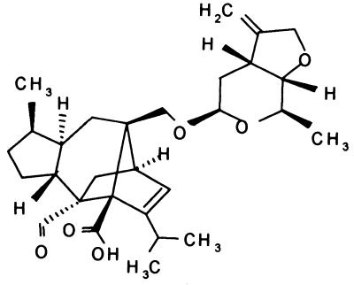
The sordarin derivative GM237354 (Fig. 1) was synthesized at the Glaxo Wellcome Research Centre in Madrid, Spain, and was supplied as a sodium salt powder. Immediately before each experiment, the compound was dissolved in sterile deionized water, and dilutions were made to the desired concentrations.
FIG. 1.
Chemical structure of GM237354.
Organism.
Therapeutic efficacy studies were performed against C. albicans 4711E, a clinical isolate obtained from the Glaxo Wellcome Laboratories (Greenford, United Kingdom) culture collection. This strain was stored at −80°C in Sabouraud dextrose broth (Difco Laboratories, Madrid, Spain) containing 15% glycerol in our laboratory until the experiment was performed. C. albicans 4711E was grown on Sabouraud dextrose agar (Difco Laboratories) plates at 30°C for 24 to 48 h. After incubation, the cells were harvested, washed three times in sterile saline, and resuspended in sterile saline to a final concentration of 3 × 10⁸ per ml. The inoculum size was verified by quantitative cultures of serial 10-fold dilutions on Sabouraud dextrose agar plates. All counts are expressed as CFU of viable organisms. The MIC for this isolate was determined by Herreros et al. (14) according to the methods of the National Committee for Clinical Laboratory Standards.
Animals.
Male Sprague-Dawley rats (age, 6 weeks; weight, approximately 200 g; Iffa-Credo France Inc., Lyon, France) were used in this study. The rats were housed in groups of five in 480- by 270- by 200-mm Apec cages (Techniplast; Letica Scientific Instruments, Madrid, Spain) on corncob granules (Panlab, Barcelona, Spain). The photoperiods were adjusted to 12 h of light and 12 h of darkness daily, and the environmental temperature was constantly maintained at 21°C. The rats were given ad libitum access to food and water. The research complied with European legislation and with company policy on the care and use of animals and with related codes of practice.
Oral candidiasis in rats.
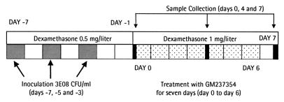
An animal model of oral candidiasis was induced basically as reported by Jones et al. (15) with some modifications. The rats were immunosuppressed, starting 1 week before experimental infection and continuing throughout the experiment, by administration of dexamethasone (Fortecortin; Merck Laboratories, Madrid, Spain) in the drinking water at a dose of 0.5 mg/liter. Also, a 0.1% aqueous solution of tetracycline hydrochloride (Terramicine; Pfizer Laboratories, Alcobendas, Spain) was given to the rats, beginning 7 days before infection. The concentration of tetracycline hydrochloride was reduced to 0.01% on the day of infection and maintained throughout the experiment. The rats were orally infected three times at 48-h intervals (days −7, −5, and −3) with 0.1 ml of a saline suspension containing 3 × 10⁸ viable cells of C. albicans 4711E. Oral infection was performed by means of a cotton swab rolled twice over all parts of the mouth, following a standarized protocol. A schematic diagram of the oral candidiasis model is presented in Fig. 2.
FIG. 2.
Schematic diagram of the oral candidiasis model in immunosuppressed rats.
Antifungal treatment.
Just before treatment, the animals were sampled to confirm the presence of C. albicans and to quantify the number of CFU in the oral cavity. Then, the animals were randomized and assigned to groups of five. Treatment was administered for seven consecutive days (from day 0 to day 6). GM237354 was administered every 8 h (TID) by subcutaneous injection (0.5 ml) at doses of 1.25, 2.5, 5, and 10 mg/kg of body weight. Two additional groups treated at 5 mg/kg once or twice a day were added to the experiment to study the impact of the regimen administration on the therapeutic efficacy. The control group (n = 13) received sterile saline by the subcutaneous route.
Microbiology.
The drug efficacy was assessed microbiologically by measuring the number of C. albicans organisms in oral swabs obtained in the middle (day 4) and 24 h after the end (day 7) of treatment. Oral samples were collected by rolling a sterile cotton swab over the oral cavity and suspending it in 1.0 ml of sterile saline. The oral samples were cultured in duplicate onto Bengal Rose chloramphenicol agar (Microkit Iberica, S. A., Barcelona, Spain) by using an autoplate (Spiral Biotech, Aplicaciones Analíticas, Barcelona, Spain). The plates were incubated at 37°C for 24 to 48 h, the CFU were counted, and the totals per swab were calculated. Plates with less than two colonies were considered negatives cultures (the detection threshold was 40 CFU/swab).
Pathology.
Gross observations, histological findings, and morphometry studies were performed in untreated animals and animals treated with GM237354 at 1.25, 2.5, 5, and 10 mg/kg TID for 7 days.
(i) Gross observations.
At the end of the experiment (24 h after the last administration of GM237354), the animals were sacrificed by an overdose of pentobarbital (Eutalender, Normon, Spain). The tongues were removed by an incision at the base, and gross observations and photographs were made immediately, following gentle rinsing of the tongue.
(ii) Histological findings.
Tongues were routinely processed for light microscopy. Briefly, the tongues were fixed in toto by immersion in neutral buffered formalin solution for 48 h, and serial transverse sections were made of the whole tongue. These sections were again fixed in formalin for 12 h, followed by embedding in paraffin. Finally, 5-μm- thick sections were obtained from the paraffin blocks and stained with hematoxylin and eosin (HE), as well as with periodic acid-Schiff (PAS) stain, for histological findings and fungal visualization.
(iii) Morphometric study.
In order to quantify the extent of the oral candidiasis and the evolution of the infection with and without antifungal treatment, several histological sections of each tongue were selected. In these sections, the length of the surface of the epithelium occupied by hyphae (Lh) was measured; this value, as a percentage, represents the proportion of epithelium occupied by hyphae with respect to the total epithelial surface on the dorsum of the tongue. In those areas occupied by hyphae, the following volume density (VD) parameters were also determined. (i) VDEh, expressed as a percentage, represents the VD of the epithelium with hyphae, considering all epithelial layers (from basal to superficial layers). This parameter represents the extension and range of C. albicans mycelial penetration into the epithelium of the tongue. (ii) VDh corresponds to the number and size of the hyphae. This parameter is expressed as a percentage with respect to the total surface of the epithelium occupied by hyphae. Lh and VDEh were measured at ×10 magnification, while VDh was determined under ×40 magnification. Lh was directly determined from the slides by using an eyepiece with a millimeter scale. In order to quantify the VD measures (Eh and h), a stereologic procedure based on the counting of points was used, as previously described by Gundersen et al. (12, 13). Basically, the microscopic image of the area under study was captured and digitized by means of a Videoplan Kontron image analyzer, and a frame was superimposed over the image on a monitor. This frame included a set of points (176) and a set of test lines, and the estimation of the VD measures was made by counting the points that intercepted the quantified structure (hyphae or epithelium).
Statistical analysis.
An analysis of variance on ranks was used to statistically compare the CFU of C. albicans isolated from the mouths of the experimental groups. Multiple comparisons of treatment groups versus the control group were performed by Dunn's method. Data from the morphometric studies were statistically analyzed using a Student t test. All statistical evaluations were performed using the analysis of variance program of the Sigmastat statistical package (Jandel Scientific, Erkrath, Germany). P values of ≤0.05 were considered statistically significant. All mean values given in the text and tables include the standard deviations of the means.
RESULTS
The therapeutic efficacy of GM237354 against experimental oral candidiasis was studied in immunosuppressed rats. The efficacy of GM237354 was determined microbiologically, histologically, and by a morphometric study of the lesions in the epithelium of the dorsal tongue.
Microbiology.
Oral cavity cultures of each rat were performed prior to initiation of the study, and no Candida organisms were found in any case. Oral candidiasis was induced with C. albicans 4711E; the MIC of GM237354 against this strain was 0.001 μg/ml. Infected animals were sampled postinfection, just prior to the start of the treatment, and the oral swabs were all positive for the presence of C. albicans, with a mean log CFU/swab of 5.0 ± 0.7 (n = 43 rats). Then, the animals were randomized in groups of five. On day 4 of the experiment, oral samples were collected and cultured to quantify the CFU in the oral cavities of infected and treated or untreated control animals. The mean log CFU/swab of control animals was 4.0 ± 0.7. Groups treated with 7.5, 10, 15, and 30 mg/kg/day showed a significant reduction (P < 0.05) of the number of CFU/swab compared with controls (Table 1). A good dose-dependent therapeutic effect relationship was observed. In addition, 60% of the animals treated with GM237354 at 15 mg/kg/day and 0% of the rats treated at 30 mg/kg/day showed cultures positive for the presence of C. albicans after 4 days of treatment. At the end of the experiment, (24 h after the last treatment), the mean log CFU/swab for untreated control animals was 3.9 ± 0.6. All groups treated with GM237354 showed significant reductions of C. albicans, with the exception of animals treated at 3.75 mg/kg/day. However, in this group, 20% of the rats showed C. albicans-negative cultures. The percentages of animals with positive cultures from the groups treated with GM237354 at 5, 7.5, and 10 mg/kg/day were 80, 60, and 60%, respectively. Moreover, C. albicans organisms were not detected in the oral cavities of any animals treated with GM237354 at 15 or 30 mg/kg/day after 7 days of treatment (Table 1).
TABLE 1.
| Total dose (mg/kg/day) | Regimen (mg/kg) | Day 4
|
Day 7
|
||
|---|---|---|---|---|---|
| No. of infected animals/total (%) | Log CFU/swab (mean ± SD) | No. of infected animals/total (%) | Log CFU/swab (mean ± SD) | ||
| None | 13/13 (100) | 4.0 ± 0.7 | 13/13 (100) | 3.9 ± 0.6 | |
| 3.75 | 1.25 TID | 5/5 (100) | 3.6 ± 0.9 | 4/5 (80) | 2.7 ± 0.8 |
| 5 | 5 OD | 5/5 (100) | 3.7 ± 0.8 | 4/5 (80) | 2.4 ± 0.4∗ |
| 7.5 | 2.5 TID | 5/5 (100) | 2.8 ± 0.3∗ | 3/5 (60) | 2.5 ± 0.3∗ |
| 10 | 5 BID | 5/5 (100) | 3.3 ± 0.5∗ | 3/5 (60) | 2.6 ± 0.4∗ |
| 15 | 5 TID | 2/5 (40) | 2.7 ± 0.3∗ | 0/5 (0) | <1.6∗∗ |
| 30 | 10 TID | 0/5 (0) | <1.6∗∗ | 0/5 (0) | <1.6∗∗ |
Rats were orally infected three times at 24-h intervals with 3 × 10⁸ C. albicans 4711E cells/ml. GM237354 was administered subcutaneously once a day (OD), twice a day (BID), or TID for seven consecutive days (day 0 to day 6). The mean log CFU/swab just before treatment was 5.0 ± 0.7 (n = 43). ∗, P < 0.05; ∗∗, P < 0.01 (versus the control treatment). The limit of detection was 40 CFU/swab.
Pathology.
Gross observations, histological findings, and morphometry studies were performed in untreated and treated animals administered GM237354 at 1.25, 2.5, 5, and 10 mg/kg TID for 7 days.
(i) Gross observations.
All infected and untreated animals had clinically manifest lesions of the lingual mucosa, consisting of patchy areas of smooth mucosa and well-delimited atrophic areas on the dorsum of the tongue. These lesions were mainly distributed surrounding the giant conical papillae of the tongue. Animals treated with GM237354 at 3.75 or 7.5 mg/kg/day showed lesions similar to those observed in the controls. However, animals given 15 or 30 mg/kg/day showed grossly normal dorsal tongue surfaces.
(ii) Histological findings.
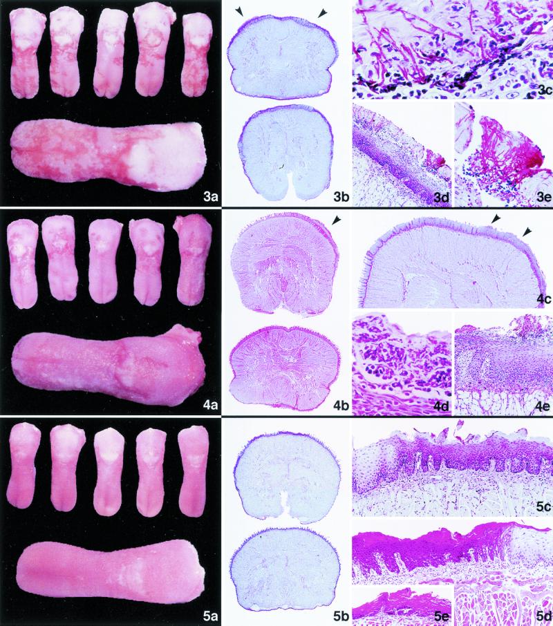
Infected and untreated control animals showed extensive colonization of the epithelium of the dorsal surface of the tongue by numerous hyphae, and in many areas, colonization extended deep through the superficial layers of the horny and squamous layers. Furthermore, in some animals, the hyphae focally penetrated even deeper, reaching the parabasal cells of the epithelium. In these cases, the phenomenon was associated with an increased presence of inflammatory cells in the lamina propria. Multifocal leukodiapedesis into the epithelium was observed in association with these inflammatory infiltrates in the lamina propria. In multiple areas, the intraepithelial hyphae caused keratinocyte destruction and the formation of intraepithelial microabscesses (Fig. 3). All animals treated with GM237354 at 3.75 or 7.5 mg/kg/day showed hyphae on the tongue surface, with focal hyphal penetration that produced intraepithelial microabscesses (Fig. 4). Animals treated with GM237354 at 15 mg/kg/day showed multiple regenerative areas of the covering epithelium, characterized by basal keratinocyte hyperplasia and superficial hyperkeratosis. Only focalized zones of the tongue surface were occupied by hyphae, but in these zones the numbers of hyphae were minimal. No histological evidence of C. albicans within the epithelium of the tongue was seen in rats treated with GM237354 at a dose of 30 mg/kg/day. In addition, these animals showed extensive areas of epithelial regeneration (Fig. 5).
(iii) Morphometry.
The results of the morphometric study (Table 2) confirmed the histological findings described above. A significant increase in the height of the epithelium was observed. This increase was due to epithelial hyperplasia and was evident in the tongues of control rats. Eighteen percent of the Lh of the tongue was filled by a variable amount of C. albicans hyphae in the control group (17.5 versus 19.4% in the group treated at 3.75 mg/kg/day). This value decreased significantly (P < 0.05) to 4.8 and 0.1% in the groups treated with GM237354 at 7.5 or 15 mg/kg/day, respectively. Only two to five animals treated with GM237354 at 15 mg/kg/day showed C. albicans hyphae in the epithelium of the dorsum of the tongue. Animals treated with GM237354 at a dose of 30 mg/kg/day showed no areas of hyphal colonization, and the morphometric study was consequently not performed in such cases. The VDEh values in animals treated with GM237354 at 3.75 mg/kg/day were similar to those of the controls (30.9 versus 25.6% in the controls). The VDEh values were in turn significantly reduced (P < 0.05) to 13.5 and 0.9% in the groups treated at 7.5 and 15 mg/kg/day, respectively. Statistically significant differences (P < 0.05) in VDh values were also observed between the group treated at 15 mg/kg/day and the control group.
TABLE 2.
| dose (mg/kg/day) | No. of infected animals/total (%) | Lh (%) | VDEh (%) | VDh (%) |
|---|---|---|---|---|
| None | 5/5 (100) | 17.5 ± 10.1 | 30.9 ± 5.2 | 6.5 ± 3.6 |
| 3.75 | 5/5 (100) | 19.4 ± 4.9 | 25.6 ± 5.4 | 10.2 ± 2.2 |
| 7.5 | 5/5 (100) | 4.8 ± 2.4* | 13.5 ± 11.8* | 3.2 ± 2.2 |
| 15 | 2/5 (40) | 0.1 ± 0.2* | 0.9 ± 1.3* | 0.7 ± 1.0* |
| 30 | 0/5 (0) | 0 | 0 | 0 |
The morphometric study was performed in several sections of the tongue. The animals were treated TID for 7 consecutive days. Animals treated at 30 mg/kg/day showed no tongue colonization by C. albicans. Infected animals represents the number of animals that were histologically positive for the presence of C. albicans in the epithelium. *, P < 0.05 versus the control treatment.
DISCUSSION
The growing population of immunocompromised patients receiving immunosuppressive or anticancer therapy has led to an increased incidence of opportunistic mycoses. Although oral candidiasis is not a life-threatening disease, the sustained immunosuppression in these patients facilitates the recurrence of infection. Children and adolescents with a compromised or suppressed immune status are particularly susceptible to the development of oral candidiasis (8). We have therefore developed a standardized experimental model of oral C. albicans infection in immunocompromised rats. Systemic corticosteroid dosing in drinking water 1 week before the challenge significantly decreased the white blood cell count. This decrease remained constant throughout the experiment (data not shown). The present immunosuppressed-animal model appears to more closely mimic the situation seen in clinical settings. In addition, the administration of systemic antimicrobials, particularly tetracycline, is widely used to facilitate the development of candidiasis in the rat oral cavity (15, 23). On the other hand, Allen and Beck (2) have described strain-related differences in C. albicans pathogenicity in the rat oral mucosa; consequently, we used our well-characterized strain of C. albicans, which has been widely used for therapeutic studies in rodents with systemic candidiasis, demonstrating its pathogenic properties (3, 18).
Sordarin derivatives are a novel class of antifungals with potent broad-spectrum antifungal activity in several in vitro studies (14), and earlier research indicates that sordarin derivatives possess promising activity in several animal models of infection (18). Clemons and Stevens have recently demonstrated that sordarins (GM193663, GM211676, and GM237354) are effective in the treatment of experimental systemic coccidioidomycosis in mice (5), and Graybill et al. demonstrated that sordarins were effective in a murine model of histoplasmosis (11).
This study has shown that the sordarin derivative GM237354 administered therapeutically to immunosuppressed rats with oral candidiasis effectively reduces organism-mediated oral cavity injury, as measured by colony counts, gross pathology, and histological examination. Microbiologically, GM237354 has shown a good dose-dependent therapeutic effect in the oral-candidiasis model. Thus, the therapeutic efficacy of GM237354 against oral candidiasis was observed when at least 5 mg of the compound per kg per day was administered to rats for 7 days, significantly decreasing the load of C. albicans in the mouths of infected animals compared with that in the controls. However, when the compound was administered every 24, 12, or 8 h, the eradication of microorganisms after 7 days of treatment was observed in 20, 40, or 100% of infected animals, respectively.
Evident agreement between cultures from the oral cavity and the clinical and histological evidence of infection was also observed. Animals treated with lower doses of GM237354 (3.75 mg/kg/day) showed persistent C. albicans culture positivity, as well as abundant mycelial penetration into the epithelium of the tongue. However, in animals treated with GM237354 at 7.5 mg/kg/day, the number CFU of C. albicans obtained from the mouths of infected animals decreased significantly. In these animals, the histological study demonstrated Candida to have disappeared from the surface of the tongue; however, some hyphae remained within the most superficial keratinocyte layers. The viability of these few hyphae within the epithelium is not known. Although the pathogenicity of the mycelia cannot be defined, this small population of hyphae apparently did not have the same morphological features as those in the infected and untreated animals. At a dose of 15 mg/kg/day, the presence of immunocompetent cells with exocytosis and the complete absence of C. albicans on the dorsal surface of the tongue suggest that the sordarin derivative was extremely effective at this dose. These results are completely consistent with the 100% microbiological eradication of C. albicans previously observed in animals treated at 15 mg/kg/day. As expected, the histological study of the group, treated at 30 mg/kg/day showed no hyphae in either the surface or epithelium of the tongue. In this group, the morphology of the lingual epithelium was normal, though in some areas regenerative transformation was frequently seen, reflected by a proliferation of basal cells and an altered maturation of the keratinocytes. These regenerative changes may be directly related to the lesion induced by Candida or to the action of the exudation of immunocompetent cells into the epithelium. It is well established that these changes are completely regressive. The sordarin derivatives eliminate candidal organisms, and the inflammatory response consequently decreases; as a result, the regenerative process is delayed, giving rise to normal proliferation. Therefore, GM237354 eradicated the fungal load from the mouths of infected animals and produced no important or irreversible lesions of the oral mucosa.
The morphometric study clearly demonstrated the therapeutic effect of sordarins, indicating an excellent correlation among the microbiological, histological, and morphometric findings. This kind of study can thus be a useful tool for the in vivo evaluation of new antifungal agents.
In conclusion, the results of the present study are encouraging, although further investigations and comparative toxicity profile studies are needed to confirm sordarin derivatives as very promising and effective antifungal agents in human candidal infections.
FIG. 3.
Fig. 3. Oral candidiasis in immunosuppressed rats; untreated control group. (a and b) Macroscopic (a) and panoramic (b) histological lingual candidiasis; the epithelium of the tongue shows irregular thickness and hyperkeratotic areas with plentiful hyphae (arrowheads). PAS; magnification, ×5. (c) Diffuse, abundant Candida mycelial elements affecting the superficial layers of the epithelium. PAS; magnification, ×600. (d) Large focal group of hyphae penetrating into the deeper layers of the epithelium. PAS; magnification, ×125. (e) Magnification of panel d. Note the abundant hyphae that have destroyed the keratinocytes, associated with small leukocyte infiltrates. PAS; magnification, ×600. Fig. 4. Oral candidiasis in immunosuppressed rats; animals treated with GM237354 at 1.25 mg/kg TID for 7 days. (a and b) Macroscopic (a) and panoramic (b) histological lingual candidiasis; the transverse sections show important focal thickening of the epithelium (arrowhead). HE; magnification, ×5. (c) Multifocal hyperplasia of the epithelium, associated with irregular accumulations of hyphae (arrowheads). PAS; magnification, ×15. (d) Intraepithelial microabscess containing numerous leukocytes, neutrophils, and partially degenerated hyphae. PAS; magnification, ×600. (e) Destruction of several epithelial layers, with extensive neutrophil infiltration in relation to the presence of hyphae in the epithelium. PAS; magnification, ×125. Fig. 5. Oral candidiasis in immunosuppressed rats; animals treated with GM237354 at 10 mg/kg TID for 7 days. (a and b) Macroscopic (a) and panoramic (b) histological lingual candidiasis; note the different coloration of the dorsal surface of the tongue, due to the alternation of hyperplastic and normal epithelial areas. PAS; magnification, ×5. (c) Basal cell hyperplasia, with scant differentiation of squamous cells and horny layer dyskeratosis. There are no C. albicans hyphae in the epithelial surface layer. PAS; magnification, ×125. (d) Focal alteration of the epithelial maturation of the tongue, reflected by intense dyskeratosis alternation associated with normal epithelium. A small increase of lymphocytes in the lamina propria was observed. HE; magnification, ×125. (e) Atrophy of the epithelium associated with intense regenerative changes. HE; magnification, ×125.
ACKNOWLEDGMENTS
We thank Esperanza Herreros for providing all the in vitro data, the members of Centro de Investigacion Farmacologica for their excellent technical assistance, and the members of the Organic Chemistry Group for the synthesis of compounds.
REFERENCES
- 1.Allen C M. Animal models of oral candidiasis. Oral Surg Oral Med Oral Pathol. 1994;78:216–221. doi: 10.1016/0030-4220(94)90150-3. [DOI] [PubMed] [Google Scholar]
- 2.Allen C M, Beck F. Strain-related differences in pathogenicity of Candida albicans for oral mucosa. J Infect Dis. 1983;147:1036–1040. doi: 10.1093/infdis/147.6.1036. [DOI] [PubMed] [Google Scholar]
- 3.Aviles P, Falcoz C, San Roman R, Gargallo-Viola D. Pharmacokinetics-pharmacodynamics of a sordarin derivative ( GM237354) in a murine model of lethal candidiasis. Antimicrob Agents Chemother. 2000;44:2333–2340. doi: 10.1128/aac.44.9.2333-2340.2000. [DOI] [PMC free article] [PubMed] [Google Scholar]
- 4.Challacombe S J. Immunologic aspects of oral candidiasis. Oral Surg Oral Med Oral Pathol. 1994;78:202–210. doi: 10.1016/0030-4220(94)90148-1. [DOI] [PubMed] [Google Scholar]
- 5.Clemons K V, Stevens D A. Efficacies of sordarin derivatives GM193663, GM211676, and GM237354 in a murine model of systemic coccidioidomycosis. Antimicrob Agents Chemother. 2000;44:1874–1877. doi: 10.1128/aac.44.7.1874-1877.2000. [DOI] [PMC free article] [PubMed] [Google Scholar]
- 6.Diaz-Guerra T, Martinez-Suarez J, Laguna F, Rodriguez-Tudela J. Comparison of four molecular typing methods for evaluating genetic diversity among Candida albicans isolates from human immunodeficiency virus-positive patients with oral candidiasis. J Clin Microbiol. 1997;35:856–861. doi: 10.1128/jcm.35.4.856-861.1997. [DOI] [PMC free article] [PubMed] [Google Scholar]
- 7.Domínguez J M, Kelly V A, Kinsman O S, Marriott M S, Gómez de las Heras F, Martín J J. Sordarins: a new class of antifungal with selective inhibition of the protein synthesis elongation cycle in yeast. Antimicrob Agents Chemother. 1998;42:2274–2278. doi: 10.1128/aac.42.9.2274. [DOI] [PMC free article] [PubMed] [Google Scholar]
- 8.Flaitz C M, Hicks M J. Oral candidiasis in children with immune suppression: clinical appearance and therapeutic considerations. ASDC J Dent Child. 1999;66:161–166. [PubMed] [Google Scholar]
- 9.Freire-Garabal M, Nunez M J, Balboa J, Rodriguez-Cobo A, Lopez-Paz J M, Rey-Mendez M, Suarez-Quintanilla J A, Millan J C, Mayan J M. Effects of amphetamine on development of oral candidiasis in rats. Clin Diagn Lab Immunol. 1999;6:530–533. doi: 10.1128/cdli.6.4.530-533.1999. [DOI] [PMC free article] [PubMed] [Google Scholar]
- 10.Graninger W, Presteril E, Schneeweiss B, Teleky B, Georgopoulos A. Treatment of Candida albicans fungaemia with fluconazole. J Infect. 1993;26:133–146. doi: 10.1016/0163-4453(93)92761-k. [DOI] [PubMed] [Google Scholar]
- 11.Graybill J R, Najvar L, Fothergill A, Bocanegra R, Gomez de las Heras F. Activities of sordarins in murine histoplasmosis. Antimicrob Agents Chemother. 1999;43:1716–1718. doi: 10.1128/aac.43.7.1716. [DOI] [PMC free article] [PubMed] [Google Scholar]
- 12.Gundersen H J, Bagger P, Bendtsen T F, Evans S M, Korbo L, Marcussen N, Moller A, Nielsen K, Nyengaard J R, Pakkenberg B. The new stereological tools: dissector, fractionator, nucleator and point sampled intercepts and their use in pathological research and diagnosis. APMIS. 1988;96:857–881. doi: 10.1111/j.1699-0463.1988.tb00954.x. [DOI] [PubMed] [Google Scholar]
- 13.Gundersen H J, Bendtsen T F, Korbo L, Marcussen N, Moller A, Nielsen K, Nyengaard J R, Pakkenberg B, Sorensen F B, Vesterby A. Some new, simple and efficient stereological methods and their use in pathological research and diagnosis. APMIS. 1988;96:379–394. doi: 10.1111/j.1699-0463.1988.tb05320.x. [DOI] [PubMed] [Google Scholar]
- 14.Herreros E, Martinez C M, Almela M J, Marriot M S, Gomez de las Heras F, Gargallo-Viola D. Sordarins: in vitro activities of new antifungal derivatives against pathogenic yeast, Pneumocystis carinii, and filamentous fungi. Antimicrob Agents Chemother. 1998;42:2863–2869. doi: 10.1128/aac.42.11.2863. [DOI] [PMC free article] [PubMed] [Google Scholar]
- 15.Jones J, Russell C, Young C, Owen D. Tetracycline and the colonization and infection of the mouths of germ-free and conventionalized rats with Candida albicans. J Antimicrob Chemother. 1976;2:247–253. doi: 10.1093/jac/2.3.247. [DOI] [PubMed] [Google Scholar]
- 16.Jorge A, Totti M, Almeida O, Scully C. Effect of sialoadenectomy on the carriage of Candida albicans in the mouths of rats. J Oral Pathol Med. 1993;22:138–140. doi: 10.1111/j.1600-0714.1993.tb01045.x. [DOI] [PubMed] [Google Scholar]
- 17.Lopez-Ribot J, McAtee R, Perea S, Kirkpatrick W, Rinaldi M G, Patterson T. Multiple resistant phenotypes of Candida albicans coexist during episodes of oropharyngeal candidiasis in human immunodeficiency virus-infected patients. Antimicrob Agents Chemother. 1999;43:1621–1630. doi: 10.1128/aac.43.7.1621. [DOI] [PMC free article] [PubMed] [Google Scholar]
- 18.Martinez A, Aviles P, Jimenez E, Caballero J, Gargallo-Viola D. Activities of sordarins in experimental models of candidiasis, aspergillosis, and pneumocystosis. Antimicrob Agents Chemother. 2000;44:3389–3394. doi: 10.1128/aac.44.12.3389-3394.2000. [DOI] [PMC free article] [PubMed] [Google Scholar]
- 19.Meitner S, Bowen W, Haidaris C. Oral and esophageal Candida albicans infection in hyposalivatory rats. Infect Immun. 1990;58:2228–2236. doi: 10.1128/iai.58.7.2228-2236.1990. [DOI] [PMC free article] [PubMed] [Google Scholar]
- 20.Odds F. Candidiasis of the oropharynx. In: Odds F, editor. Candida and candidosis: a review and bibliography. 2nd ed. London, United Kingdom: W. B. Saunders; 1988. pp. 117–123. [Google Scholar]
- 21.Quereda C, Polanco A, Giner C, Sánchez-Sousa A, Pereira E, Navas E, Fortún J, Guerrero A, Baquero F. Correlation between in vitro resistance to fluconazole and clinical outcome of oropahryngeal candidiasis in HIV-infected patients. Eur J Clin Microbiol Infect Dis. 1996;15:30–37. doi: 10.1007/BF01586182. [DOI] [PubMed] [Google Scholar]
- 22.Rossie K, Guggenheimer J. Oral candidiasis: clinical manifestations, diagnosis, and treatment. Pract Periodontics Aesthet Dent. 1997;9:635–641. [PubMed] [Google Scholar]
- 23.Russell C, Jones J. Effects of oral inoculation of Candida albicans in tetracycline-treated rats. J Med Microbiol. 1973;6:275–279. doi: 10.1099/00222615-6-3-275. [DOI] [PubMed] [Google Scholar]
- 24.Tumbarello M, Tacconelli E, Caldarola G, Morace G, Cauda R, Ortona L. Fluconazole resistant oral candidiasis in HIV-infected patients. Oral Dis. 1997;3:S110–S112. doi: 10.1111/j.1601-0825.1997.tb00337.x. [DOI] [PubMed] [Google Scholar]


