Abstract
A map in stereotaxic coordinates of the tentorium cerebelli is presented. Its utility in positioning brain stem and cerebellar electrodes is briefly discussed.
Keywords: Cat, Stereotaxic, Tentorium
THE bony tentorium cerebelli of the cat lies between the cerebellum and the cerebral hemispheres. Since its exact location and size are not indicated in the major atlases [1, 2, 3, 4, 5], its discovery often comes as an unpleasant surprise to investigators attempting to explore brain stem or cerebellar areas with depth electrodes or cannulae. A direct hit may destroy both the electrode being positioned and the surrounding brain tissue. Contact with the edge of the tentorium can produce an electrode deflection that goes unnoticed until the brain is subjected to histological analysis.
Therefore, as part of a larger program of brain stem exploration in our laboratory, it became desirable to map the exact location of the bony tentorium. This map allows us to determine when orthogonal coordinates can be used for brain stem exploration and what the optimal angles of approach are when non-orthogonal coordinates must be used.
METHOD
Animals
Five cats, 2 male and 3 female, weighing between 2.7 and 3.2 kg were used. The cats had been killed previously in connection with other investigations or because of infectious disease.
Procedure
The cat’s head was positioned in a Kopf stereotaxic head holder. The cranium was opened and the cerebral hemispheres overlying the tentorium were removed. The cerebellum and brain stem underneath the tentorium were removed through an enlargement of the foramen magnum. A high intensity light projected through the foramen enabled visualization of the junction of the tentorium with the base of the skull.
All mapping was done on the dorsal surface of the tentorium. A calibrated insect pin electrode was lowered to the margins of the tentorium. A-P and H coordinates were recorded as the electrode was moved laterally in 1 mm steps. Sagittal maps, made by moving the electrode over the surface in 1 mm A-P increments, were drawn for 0, 2, 4, and 6 mm lateral to the midline.
RESULTS
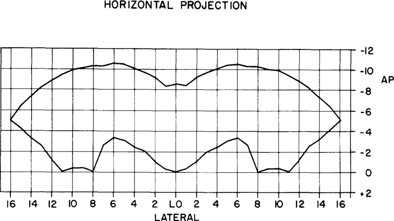
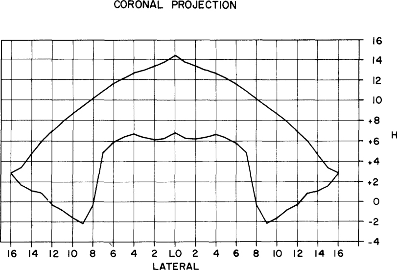
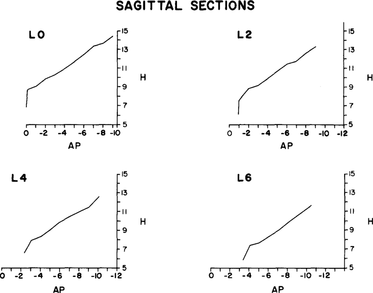
Horizontal and coronal projections of the surface of the tentorium are presented in Figs. 1 and 2. Sagittal views at 0, 2, 4, and 6 mm lateral to the midline are presented in Fig. 3. The point 10 mm dorsal to the ear bars was considered stereotaxic zero for the horizontal plane.
FIG. 1.
Horizontal projection of the surface of the tentorium.
FIG. 2.
Coronal projection of the surface of the tentorium.
FIG. 3.
Sagittal views of the tentorium at 0, 2, 4 and 6 mm lateral to the midline.
The standard deviation of the points on the horizontal and coronal projections averaged 1.25 mm. The standard deviation of points in the sagittal views averaged 1.38 mm.
The thickness of the tentorium ranged from 0.6 to 1.7 mm. The margins and the midline tended to have the greater thicknesses, while the lateral wings were of a uniform thickness less than 1 mm.
DISCUSSION
The predictable location of the tentorium makes it possible to use orthogonal penetrations for some brain stem locations without fear of hitting the bone. It is also possible to pass electrodes under the tentorium from an anterior, posterior, or lateral approach, should such an electrode path be desirable. The optimal angles of approach for brain stem structures can be determined by use of the sagittal views of the tentorium. It should be emphasized that stiff, sharp electrodes, advisable for all stereotaxic work, are especially necessary when working around the tentorium because there are meningeal layers near both of its surfaces.
Acknowledgments
Supported in part by NIH postdoctoral fellowship 1FOGM55512-01.
REFERENCES
- 1.Berman AL The Brain Stem of the Cat. Madison: The University of Wisconsin Press, 1968. [Google Scholar]
- 2.Bleier R The Hypothalamus of the Cat. Baltimore: The Johns Hopkins Press, 1961. [Google Scholar]
- 3.Jasper HH and Ajmone-Marsan C. A Stereotaxic Atlas of the Diencephalon of the Cat. Ottawa: National Research Council of Canada, 1954. [Google Scholar]
- 4.Snider RS and Niemer WT. A Stereotaxic Atlas of the Cat Brain. Chicago: University of Chicago Press, 1961. [Google Scholar]
- 5.Reinoso-Suarez F Topographischer Hirnatlas der Katze fur Experimetal-Physiologische Untersuchungen. Damstadt: Herausgegeben von E. Merck Ag, 1961. [Google Scholar]


