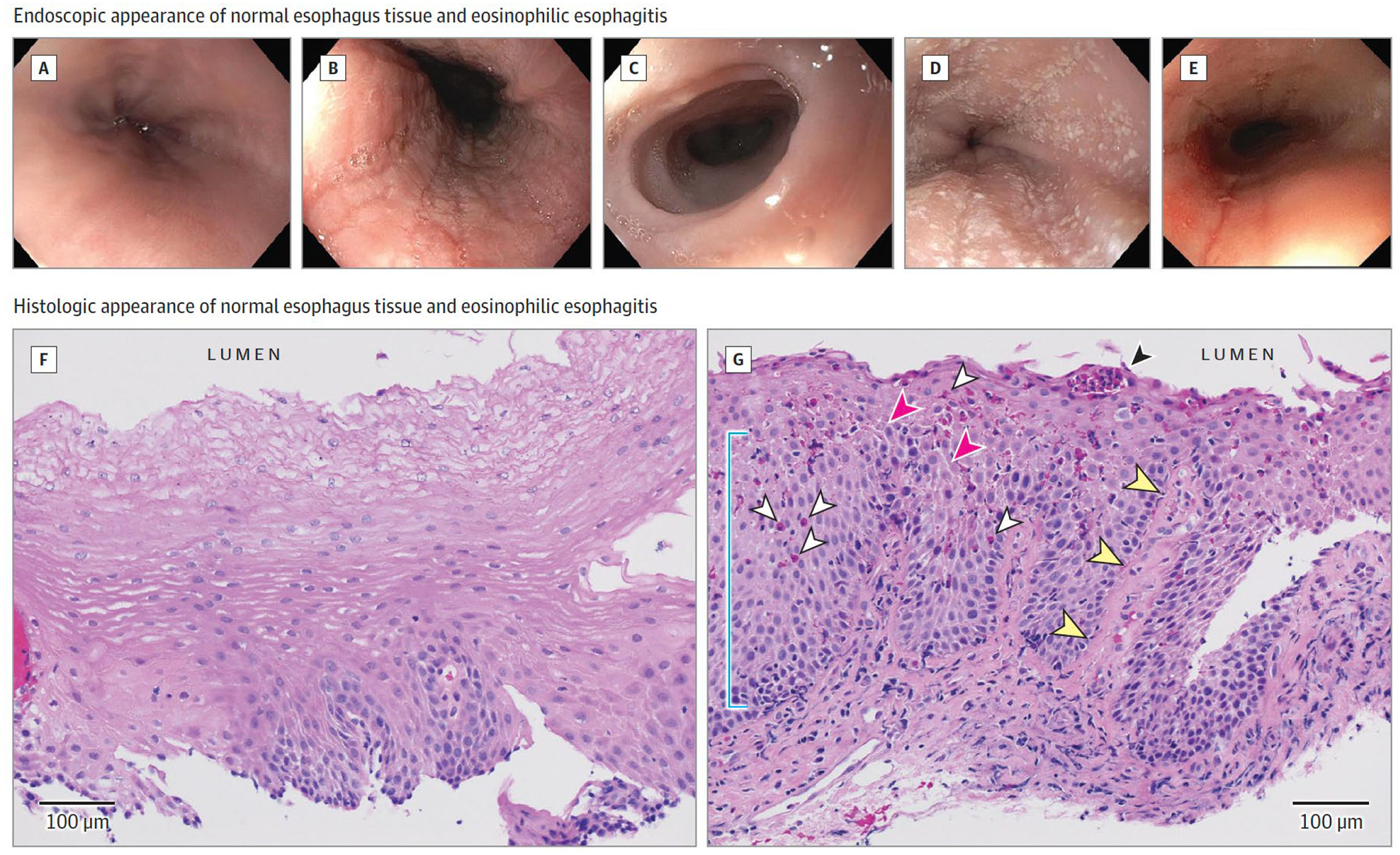
Figure 2.

Endoscopic and Histologic Appearance of the Eosinophilic Esophagitis (EoE) Esophagus
Endoscopy of EoE: normal esophagus (A); linear furrows (B); mucosal pallor representing edema, decreased vascular pattern, and concentric rings or trachealization (C); small white plaques (D); and esophageal narrowing and rent due to endoscope passage (E). Histology (hematoxylin and eosin) of EoE: F, Normal esophageal squamous epithelium with inconspicuous basal layer, luminal squamous differentiation, and absence of inflammation. G, EoE mucosa demonstrating elongated papilla (yellow arrowheads), basal cell hyperplasia (blue line), infiltrating eosinophils (white arrowheads), eosinophil microabscess (black arrowhead), and epithelial spongiosis (pink arrowheads). Images courtesy of Benjamin Wilkins, MD, PhD, Children’s Hospital of Philadelphia.
