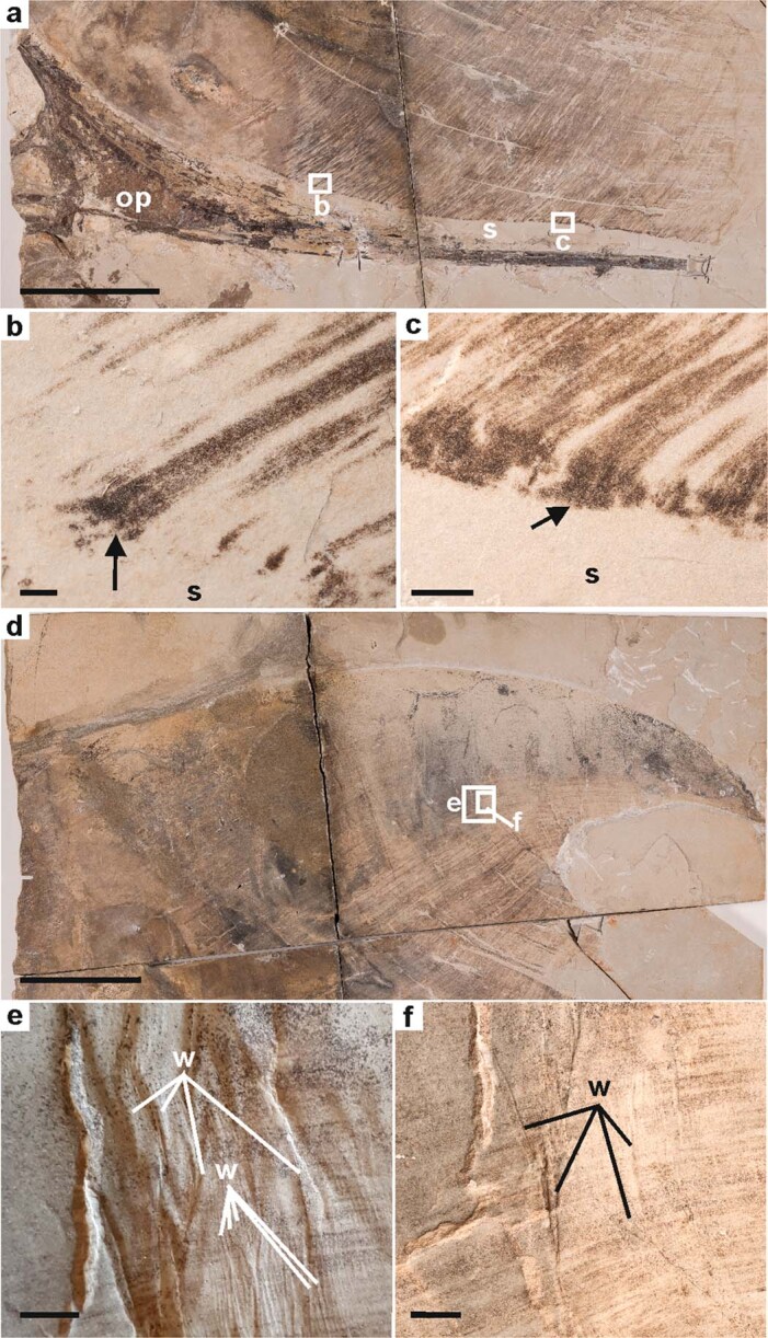
Extended Data Fig. 4. Integumentary structures of the cranial crest of MCT.R.1884.

a, Ventral part of the soft tissue crest separated from the occipital process (op) by a zone lacking soft tissue and showing only sediment (s). b, c, Detail of the basal part of the cranial crest showing dark brown structures at the base of the fibres (see arrows). d, Posterodorsal part of the cranial crest. e, f, Details of regions indicated in (d). The brown fibres of the soft tissue crest are oriented perpendicular to prominent wrinkles, expressed as variation in the topography of the specimen. Scale bars, 10 mm (a, d); 2 mm (b, c); 5 mm (e, f). op, occipital process; s, sediment; w, wrinkle.
