Abstract
Objective
To investigate not only differential patterns of functional connectivity of core brain regions between implicit and explicit verbal memory tasks underlying negatively evoked emotional condition, but also correlations of functional connectivity (FC) strength with clinical symptom severity in patients with generalized anxiety disorder (GAD).
Methods
Thirteen patients with GAD and 13 healthy controls underwent functional magnetic resonance imaging for memory tasks with negative emotion words.
Results
Clinical symptom and its severities of GAD were potentially associated with abnormalities of task-based FC with core brain regions and distinct FC patterns between implicit vs. explicit memory processing in GAD were potentially well discriminated. Outstanding FC in implicit memory task includes positive connections of precentral gyus (PrG) to inferior frontal gyrus and inferior parietal gyrus (IPG), respectively, in encoding period; a positive connection of amygdala (Amg) to globus pallidus as well as a negative connection of Amg to cerebellum in retrieval period. Meanwhile, distinct FC in explicit memory included a positive connection of PrG to inferior temporal gyrus (ITG) in encoding period; a positive connection of the anterior cingulate gyrus to superior frontal gyrus in retrieval period. Especially, there were positive correlation between GAD-7 scores and FC of PrG-IPG (r2 = 0.324, p = 0.042) in implicit memory encoding, and FC of PrG-ITG (r2 = 0.378, p = 0.025) in explicit memory encoding.
Conclusion
This study clarified differential patterns of brain activation and relevant FC between implicit and explicit verbal memory tasks underlying negative emotional feelings in GAD. These findings will be helpful for an understanding of distinct brain functional mechanisms associated with clinical symptom severities in GAD.
Keywords: Explicit memory, Functional connectivity, General anxiety disorder, Implicit memory, Negative emotion
INTRODUCTION
Symptom of generalized anxiety disorder (GAD) is characterized by persistent patterns of worrying, tension and anxiety in daily life. Constant GAD symptom leads to emotional dysregulation [1], executive dysfunction [2], and cognitive and memory impairments [3], resulting in difficulties in social coordination at work and school [4]. Once GAD is developed, it becomes most often chronic and the symptom severity worsens during times of stress. Specifically, abnormalities in the implicit and explicit memory processing of anxiety-related information were commonly addressed in patients with GAD [5-7]. The patients with GAD recalled more negative feeling words than positive or neutral during implicit and explicit memory. Memory bias was observed, especially in tacit memory [8-10]. Therefore, it is important to clarify the neuro-functional abnormalities in connection with the relevant core brain regions and their functional connectivity (FC) in the implicit and explicit memory tasks underlying negatively evoked emotional conditions and these findings are useful to understand the neurofunctional mechanism on the relevant clinical symptom severities in GAD.
Until now, numerous neuroimaging studies [11-15] have investigated the functional abnormalities in response to the emotional expression in various memory tasks in patients with GAD. The key brain centers associated with brain dysfunction in the emotional expression and regulation in GAD include the amygdala (Amg), medial/ventral/dorsal prefrontal cortices and anterior cingulate gyrus (ACG) [11-13]. Although a few works [14,15] have demonstrated the differential brain activity patterns between implicit or explicit memory processing in GAD, those studies were only focused on the identification of the distributed brain areas in either implicit or explicit memory task. A task-based brain functional connection is associated with the complex neuro-functional network linked with the correspondent task performance [16,17]. Therefore, the functional connectivity between the core brain areas is critical for further understanding of the neural network because of its ability to provide the temporal dependency of neural activation patterns of anatomical brain regions [17,18]. These advantages will be expanded to the development of the standardized magnetic resonance (MR) imaging-based neurological biomarker for accurate diagnosis and prognosis of GAD.
To the best of our knowledge, this study is the first attempt to compare the differential patterns of key brain regions and their FC observed in the processing of negatively evoked emotional expressions during the implicit vs. explicit verbal memory tasks in GAD.
METHODS
Subjects
Thirteen patients with GAD (mean age, 35.92 ± 8.61 years) and 13 healthy controls (mean age, 36.54 ± 10.12 years) participated in this study. All patients were diagnosed by the Diagnostic and Statistical Manual of Mental Disorders 5th edition criteria [19] (Table 1) and the duration of illness of the patients was 4.17 ± 4.07 years. Out of 13 GAD patients, 2 patients received a single psychotropic medication including escitalopram (n = 1) and bupropion (n = 1), and the other patients received multiple psychotropic medications (Table 2). We set participant exclusion criteria as follows: 1) age, second, 2) sex, 3) education-level, 4) handedness. This retrospective study has been approved by Institutional Review Board of Jeonbuk National University Hospital (IRB-CBNU No. CBIRB0907- 65). Also, all the analytical procedure and method were performed in accordance with the relevant guidelines and regulations approved by IRB-CBNU.
Table 1.
Comparison of the demographic and clinical characteristics between patients with GAD and healthy controls
| Variable | GAD patients (n = 13) | Healthy controls (n = 13) | p value |
|---|---|---|---|
| Mean age (yr) | 35.92 ± 8.61 | 36.54 ± 10.12 | 0.898* |
| Sex (male/female) | 9/4 | 7/6 | 0.420** |
| Handedness (right:left:mixed) | 13:0:0 | 13:0:0 | 1.000** |
| Education (yr) | 14.54 ± 1.76 | 14.62 ± 1.89 | 0.885* |
| Duration of illness (yr) | 4.17 ± 4.07 | - | - |
| Perceived negative scores for unpleasant words | 6.46 ± 2.37 | 6.46 ± 1.33 | 0.182* |
| Psychiatric rating scales | |||
| HAMA | 19.15 ± 4.00 | 0.46 ± 0.97 | < 0.001* |
| HAMD-17 | 11.77 ± 3.42 | 0.31 ± 0.48 | < 0.001* |
| GAD-7 | 12.62 ± 3.62 | 0.23 ± 0.60 | < 0.001* |
Values are presented as mean ± standard deviation or number only.
HAMA, Hamilton Anxiety Scale; HAMD-17, Hamilton Depression Scale-17; GAD-7, Generalized Anxiety Disorder Scale-7 (cut-off > 4).
*Independent sample ttest. **Chi-square test.
Table 2.
Psychotropic medicine in patients with GAD
| Medicine | Dose |
|---|---|
| Alprazolam (n = 5) | 0.25−1.5 mg |
| Benzodiazepine (n = 2) | 50 mg |
| Bupropion (n = 2) | 150 mg |
| Buspirone (n = 3) | 15−30 mg |
| Duloxetine (n = 2) | 50−60 mg |
| Escitalopram (n = 7) | 5−10 mg |
| Lorazepam (n = 1) | 0.5 mg |
| Paroxetine (n = 2) | 25 mg |
| Zolpidem (n = 1) | 5 mg |
GAD, generalized anxiety disorder.
Clinical Interviews
The patients were diagnosed by using Generalized Anxiety Disorder Scale 7 (GAD-7; 7 items with 4 level scale, cutoff score > 4). In addition, all participants underwent the following clinical interviews: Hamilton Anxiety Scale (HAMA; 14 items with a 5 level scale, cutoff score > 14), Hamilton Depression Scale 17 (HAMD-17; 8 items with five-level scale and nine items with three level scale, cutoff score > 7) and GAD-7.
Paradigm for Brain Activation
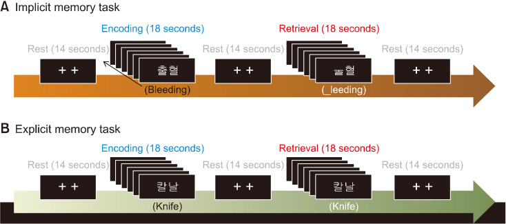
Two different implicit and explicit memory tasks were performed using the two-syllable words arousing negative emotional feelings. Brain activation paradigm consists of the following cycle: rest (14 seconds), first encoding (18 seconds), rest (14 seconds), first retrieval (18 seconds), rest (14 seconds), second encoding (18 seconds), rest (14 seconds), second retrieval (18 seconds), and rest (14 seconds) (Fig. 1). Two fixation crosses were displayed in the rest period. All of the words were displayed on a computer monitor using Superlab Pro software (Cedrus Co., Phoenix, AZ, USA). During the implicit memory task, six different two-syllable words whose first consonant was removed were displayed for 3second per each slide during the encoding period. Then, the two-syllable words given in the encoding period, but without first consonance, were presented during the retrieval period. In the explicit memory task, six different two-syllable words were presented during the encoding period. Then, either new words or the old words given in the “encoding” period were presented in the retrieval period. Participants were instructed to press the button if the presenting word is old one.
Fig. 1.
fMRI paradigms for the brain activation in implicit (A) and explicit (B) memory tasks with unpleasant words. The activation paradigm con-sists of “rest-encoding-rest-implicit or explicit retrieval.”
Data Acquisition
All the participants underwent a 3 Tesla Magnetom Verio MR Scanner (Siemens Medical Solutions, Erlangen, Germany) with an 8-channel birdcage type of head coil. The high resolution T1 weighted images were acquired using a three dimensional magnetization prepared rapid acquisition gradient echo (3D MP-RAGE) sequence with repetition time (TR)/echo time (TE) = 1,900/2.35 ms, field-of-view (FOV) = 22 × 22 cm2, matrix size = 256 × 256, number of excitation (NEX) = 1 and slice thickness = 1 mm. Functional images were acquired using a gradient-echo echo planner image (GRE-EPI) with the follow parameters: TR/TE = 2,000/30 ms, flip angle = 90°, FOV = 22 × 22 cm2, matrix size = 64 × 64, NEX = 1, number of slice = 25 and slice thickness = 5 mm without a slice gap.
Data Processing and Statistical Analysis
Functional brain mapping
Functional imaging data were analyzed using the SPM12 software (Wellcome Department of Cognitive Neurology, London, UK). The data preprocessing was conducted by the following steps: realignment of the functional data for the correction of motion artifacts; co-registration of the structural and functional images; normalization of the co-registered functional data to Montreal Neurological Institute (MNI) space; and spatial smoothing with an 8 mm full-width-at-half-maximum (FWHM) Gaussian kernel. To analyze the group difference of brain activation, two sample t test was used (p < 0.001, uncorrected, cluster size ≥ 15 voxels).
Task-based functional connectivity
Functional connectivity analyses were performed using CONN-fMRI FC toolbox (ver. 17e) with SPM12. The data processing was initiated by slice timing correction, realignment and co-registration. The co-registered images were segmented using standard SPM tissue probability maps, and then the images were smoothed with a 8 mm3 FWHM Gaussian kernel. A component based method (CompCor) [17,20] was performed for the reduction of noise in both blood oxygenation level dependent (BOLD). The band-pass filtering ware performed with a frequency window of 0.01−0.1 Hz.
For the functional connectivity analysis (FCA) of functional magnetic resonance imaging (fMRI) data, we conducted a region of interest (ROI)-to-voxel FCA using the specific ten spherical clusters where demonstrated as core regions in fMRI tasks [21] (four in implicit memory task; six in explicit memory task) with 5 mm diameters and peak coordinates. The seed ROIs for FCA included the precentral gyrus (PrG: x = −44, y = 4, z = 40), thalamus (Th: −2, −22, 16), middle occipital gyrus (MOG: −26, −92, 18) and amygdala (Amg: 24, 0, −12) in implicit memory task, and median cingulate gyrus (MCG: −18, −46, 40), PrG (16, −22, 64), inferior temporal gyrus (ITG: 60, −24, −18), supplementary motor area (MOs: 10, 8, 54), anterior cingulate gyrus (ACG: 4, 38, 2) and MOG (30, −68, 26) in the explicit memory task. General linear model (GLM) was used for the correlation of BOLD time series between the seed area and each voxels. Significant connections were identified by calculating the false discovery rate (FDR), p value < 0.05 (voxel threshold p < 0.001, uncorrected).
Statistical analysis
The difference of symptom severity between healthy controls vs. GAD group was analyzed by independent two-sample t test.
To analyze the correlation between the FCs with seed ROIs and psychiatric symptom severities, a multiple linear regression analysis was used to multiple comparison using the SPSS statistical software package (version 20.0; IBM Co., Armonk, NY, USA).
RESULTS
Demographic Characteristics and Psychiatric Rating Scales
There were no differences between healthy control and GAD groups in terms of age, sex, and education period (Table 1). Average duration of illness in patients with GAD was 4.17 ± 4.07 year. Compared to healthy controls, GAD patients showed significantly higher scores in HAMA, HAMD-17, and GAD-7 (p < 0.001).
Differential Brain Activation Patterns between Implicit vs. Explicit Memory Tasks
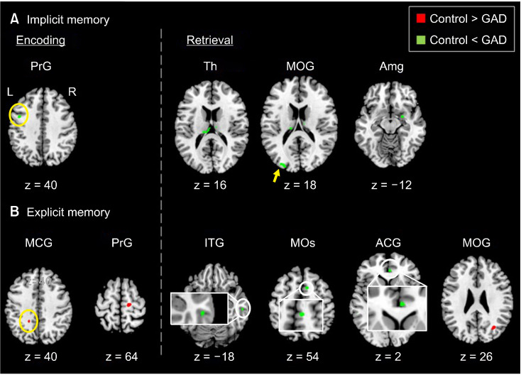
There were predominantly different brain activation patterns between patients with GAD and healthy controls in memory tasks. In the implicit memory tasks, GAD patients showed higher brain activities in PrG during the encoding period; and Th, MOG and Amg during the retrieval period (Fig. 2A and Table 3). In the explicit memory tasks, GAD patients showed lower brain activities in the MCG and PrG during the encoding period; and lower brain activities in the MOG as well as higher activities in the ITG, MOs, and ACG during the retrieval period (Fig. 2B and Table 3).
Fig. 2.
Brain areas demonstrating the neural activity reduction (red) and increase (green) in patients with GAD relative to healthy controls du-ring implicit memory (A) and ex-plicit me-mory (B) tasks.
Table 3.
Comparative brain areas with distinct activity between implicit and explicit memory tasks in patients with GAD vs. healthy controls
| Brain areas | Healthy control < GAD | Healthy control > GAD | |||||||||
|---|---|---|---|---|---|---|---|---|---|---|---|
|
|
|
||||||||||
| MNI coordinates | Cluster size | Maximum t value | MNI coordinates | Cluster size | Maximum t value | ||||||
|
|
|
||||||||||
| x | y | z | x | y | z | ||||||
| Implicit memory | |||||||||||
| Encoding | |||||||||||
| PrG | −44 | 4 | 40 | 15 | 4.21 | ||||||
| Retrieval | |||||||||||
| Th | −2 | −22 | 16 | 58 | 5.60 | ||||||
| MOG | −26 | −92 | 18 | 28 | 5.24 | ||||||
| Amg | 24 | 0 | −12 | 16 | 4.03 | ||||||
| Explicit memory | |||||||||||
| Encoding | |||||||||||
| MCG | −18 | −46 | 40 | 15 | 4.49 | ||||||
| PrG | 16 | −22 | 64 | 16 | 4.39 | ||||||
| Retrieval | |||||||||||
| ITG | 60 | −24 | −18 | 15 | 4.69 | ||||||
| MOs | 10 | 8 | 54 | 17 | 4.44 | ||||||
| ACG | 4 | 38 | 2 | 22 | 3.89 | ||||||
| MOG | 30 | −68 | 26 | 28 | 4.11 | ||||||
GAD, generalized anxiety disorder; MNI, Montreal Neurological Institute; PrG, precental gyrus; Th,thalamus; MOG, middle occipital gyrus; Amg, amygdala; MCG, median cingulate gyrus; PrG, precental gyrus; ITG, inferior temporal gyrus; MOs, supplementary motor area; ACG, anterior cingulate gyrus; MOG, middle occipital gyrus.
Functional Connectivity
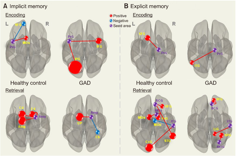
The task-based FC in GAD patients was compared with those in healthy controls. During the implicit memory encoding period, healthy controls showed a positive FC of PrG-MOs as well as a negative FCof PrG-superior frontal gyrus (SFG), while GAD patients showed positive FC of PrG-inferior frontal gyrus (IFG) and PrG-inferior parietal gyrus (IPG). During the implicit memory retrieval period, healthy controls showed positive FC of Amg-its contralateral Amg and Amg-right/left caudate (Rt/Lt Cd), while GAD patients showed a positive FC of Amg-globus pallidus (GP) as well as a negative FC of Amg-cerebellum (Cb) (Fig. 3A and Table 4).
Fig. 3.
Differential brain functional connectivity patterns in patients with GAD and healthy controls during implicit memory (A) and explicit memory (B) tasks. Red lines represent positive connectivity and blue lines represent negative connectivity. Details are shown at Tables 4 (implicit memory) and 5 (explicit memory).
Table 4.
Seed area-based functional connectivity networks during the encoding and retrieval steps in implicit verbal memory task
| Brain area | MNI coordinates | Cluster size |
b-value | Maximum tvalue |
p-FWE | p-FDR | ||
|---|---|---|---|---|---|---|---|---|
|
| ||||||||
| x | y | z | ||||||
| Encoding | ||||||||
| Seed: Precentral gyrus | −44 | 4 | 40 | |||||
| GAD < Healthy control | ||||||||
| SFG | −12 | 56 | 34 | 30 | −0.49 | −6.77 | 0.099 | 0.025 |
| MOs | −2 | 14 | 48 | 31 | 0.41 | 7.06 | 0.085 | 0.025 |
| GAD > Healthy control | ||||||||
| IFG | 42 | 6 | 24 | 50 | 0.49 | 7.66 | 0.004 | 0.000 |
| IPG | −28 | −58 | 40 | 277 | 0.55 | 10.08 | 0.000 | 0.000 |
| Retrieval | ||||||||
| Seed: Amygdala | 24 | 0 | −12 | |||||
| GAD < Healthy control | ||||||||
| Rt. Cd | 18 | 2 | 22 | 59 | 0.50 | 7.33 | 0.001 | 0.000 |
| Lt. Cd | −16 | 2 | 10 | 89 | 0.44 | 10.45 | 0.000 | 0.000 |
| Lt. Amg | −24 | 0 | −10 | 86 | 0.56 | 7.48 | 0.000 | 0.000 |
| GAD > Healthy control | ||||||||
| GP | −16 | 4 | −4 | 90 | 0.50 | 8.75 | 0.000 | 0.000 |
| Cb | 42 | −46 | −26 | 34 | −0.45 | −7.16 | 0.026 | 0.005 |
MNI, Montreal Neurological Institute; p-FWE, family wise error; p-FDR, false discovery rate; GAD, generalized anxiety disorder; SFG, superior frontal gyrus; MOs, supplementary motor area; IFG, inferior frontal gyrus; IPG, inferior parietal gyrus; Rt. Cd, right caudate; Lt. Cd, left caudate; Lt. Amg, left amygdala; GP, globus pallidus; Cb, cerebellum.
During the explicit memory encoding period, healthy controls showed a positive FC of PrG-SFG, while GAD patients showed a positive FC of PrG-ITG. During the explicit memory retrieval period, both groups showed more complex FC compared with the encoding step. Healthy controls showed positive FC of ACC-superior temporal gyrus (STG) and ACC-orbitofrontal gyrus (OFG), while GAD patients showed a positive FC of ACC-SFG. Also, healthy controls showed a positive FC of ITG-calcarine gyrus (CcG) as well as a negative FC of ITG-MOs, while GAD patients showed a positive FC of ITG-lingual gyrus (LiG) as well as another positive FC of MOG-superior parietal gyrus (SPG). In addition, healthy controls showed positive FC of MOs-its contralateral MOs and MOs-postcentral gyrus (PoG) (Fig. 3B and Table 5).
Table 5.
Seed area-based functional connectivity networks during the retrieval steps in explicit verbal memory task
| Brain area | MNI coordinates | Cluster size |
b-value | Maximum tvalue |
p-FWE | p-FDR | ||
|---|---|---|---|---|---|---|---|---|
|
| ||||||||
| x | y | z | ||||||
| Encoding | ||||||||
| Seed: Precentral gyrus | 16 | −22 | 64 | |||||
| GAD < Healthy control | ||||||||
| Superior frontal gyrus | −12 | 16 | 56 | 52 | 0.45 | 7.39 | 0.005 | 0.002 |
| GAD > Healthy control | ||||||||
| Inferior temporal gyrus | −54 | −60 | −6 | 30 | 0.41 | 8.37 | 0.060 | 0.026 |
| Retrieval | ||||||||
| Seed: Inferior temporal gyrus | 60 | −24 | −18 | |||||
| GAD < Healthy control | ||||||||
| Calcarine gyrus | −2 | −94 | −12 | 117 | 0.83 | 15.48 | 0.000 | 0.000 |
| Supplementary motor area | 4 | −8 | 68 | 31 | −0.53 | −6.60 | 0.070 | 0.017 |
| GAD > Healthy control | ||||||||
| Lingual gyrus | 16 | −46 | −10 | 32 | 0.50 | 7.93 | 0.046 | 0.015 |
| Seed: Supplementary motor area | 10 | 8 | 54 | |||||
| GAD < Healthy control | ||||||||
| Left supplementary motor area | −4 | 2 | 54 | 44 | 0.61 | 7.11 | 0.008 | 0.003 |
| Postcentral gyrus | 14 | 8 | 0 | 42 | 0.53 | 8.18 | 0.011 | 0.000 |
| Seed: Aenterior cingulate gyrus | 4 | 38 | 2 | |||||
| GAD < Healthy control | ||||||||
| Superior temporal gyrus | 66 | −52 | 22 | 81 | 0.46 | 7.48 | 0.000 | 0.000 |
| Orbitofrontal gyrus | 30 | 32 | −16 | 38 | 0.56 | 7.37 | 0.024 | 0.008 |
| GAD > Healthy control | ||||||||
| Superior frontal gyrus | 12 | 48 | 50 | 41 | 0.53 | 7.71 | 0.012 | 0.004 |
| Seed: Middle occipital gyrus | 30 | −68 | 26 | |||||
| GAD > Healthy control | ||||||||
| Superior parietal gyrus | 32 | −52 | 58 | 44 | 0.51 | 6.84 | 0.005 | 0.002 |
MNI, Montreal Neurological Institute; p-FWE, family wise error; p-FDR, false discovery rate; SFG, superior frontal gyrus; ITG, inferior temporal gyrus; CcG, calcarine gyrus; MOs, supplementary motor area; LiG, lingual gyrus; Lt. MOs, left supplementary motor area; PoG, postsentral gyrus; STG, superior temporal gyrus; OFG, orbitofrontal gyrus.
Correlation of Functional Connectivity with Symptom Severity
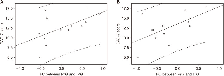
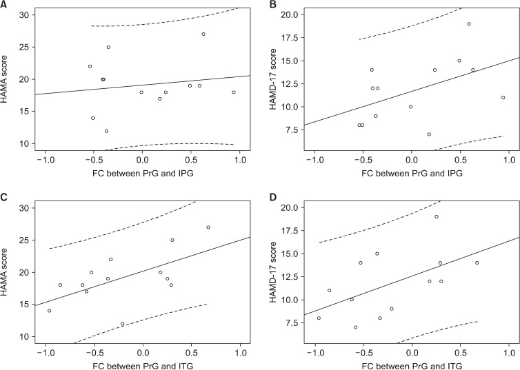
During the implicit memory encoding period, a positive correlation of strength of the FC of PrG-IPG with relevant GAD-7 scores was observed in patients with GAD (r2 = 0.324, p = 0.042) (Fig. 4A). During the explicit memory encoding period, we observed a positive correlations between the FC of PrG-ITG and scores of the GAD-7 (r2 = 0.378, p = 0.025) (Fig. 4B) in patients with GAD. However, there were no observed statistical significance with HAMA and HAMD-17 in implicit memory (HAMA: r2 = 0.028, p = 0.190; HAMD-17: r2 = 0.237, p = 0.333) (Fig. 5A, B) and explicit memory encoding (HAMA: r2 = 0.376, p = 0.312; HAMD-17: r2 = 0.317, p = 0.200) (Fig. 5C, D).
Fig. 4.
Correlation between the GAD-7 scores and the memory task-based functional connectivity (FC) of the precentral gyrus (PrG) to inferior parietal gyrus (IPG) in the implicit memory encoding process in patients with GAD (r2 = 0.324, p = 0.042) (A). Correlations between the FC of the PrG to inferior temporal gyrus (ITG) in the explicit memory encoding process with the score of GAD-7 (r2 = 0.378, p = 0.025) (B). The curved dotted line bands mean 95% confidence intervals.
Fig. 5.
There were no statistical significance in correlation between the clinical symptom severity scores (HAMA [r2 = 0.028, p = 0.190] [A], HAMD-17 [r2 = 0.237, p = 0.333] [B]) and the memory task-based functional connectivity (FC) of the precentral gyrus (PrG) to inferior parietal gyrus (IPG) in the implicit memory encoding process in patients with GAD. The correlations between the FC of the PrG to inferior temporal gyrus (ITG) in the explicit memory encoding process with the each scores of HAMA (r2 = 0.376, p = 0.312) (C) and HAMD-17 (r2 = 0.317, p = 0.200) (D) were not different too. The curved dotted line bands mean 95% confidence intervals.
DISCUSSION
Implicit Memory Task: Encoding vs. Retrieval
During the implicit memory encoding period, GAD patients showed differential brain activation patterns and the distinct FC with various seed ROIs. Compared with healthy control, GAD patients showed significantly higher activity in the PrG and this region was positively connected with IFG and IPG, respectively. Meanwhile, healthy controls showed a negative FC of PrG-SFG as well as a positive FC of PrG-MOs. These brain areas are the core center associated with verbal memory encoding [22,23]. A couple of studies [24,25] have demonstrated the neuro-anatomic and network abnormalities of the PrG in patients with GAD. Ma et al. [24] emphasized that GAD patients showed a negative resting-state FC of the PrG-IPG as well as a positive resting-state FC of the PrG-IFG. Also, other studies [26,27] have demonstrated the correlation of the increased brain activity in IFG under the negative emotional conditions such as aversion and social anxiety. In addition, activity of the IPG was closely related with anxiety severity [28,29]. Here, it is important to note that this current study showed the positive correlation between the FC of PrG-IPG with the GAD-7 scores during the implicit memory encoding period in GAD. Therefore, our findings support the evidence that anxiety symptoms in GAD are associated with both the abnormal FC of PrG-IPG and PrG-IFG, leading to memory dysfunction in the implicit memory encoding task under negatively controlled emotional condition.
During the implicit memory retrieval period, higher functional activities of the Th, MOG and Amg were observed in patients with GAD. Among them, the Amg showed differential FC between the two groups. In healthy controls, the Amg had positively connected with its contalateral Amg and Rt/Lt Cd and, respectively. Emotional arousal induces hyper-activity in the Amg, leading to mediating the modulatory effects of stress hormones and neurotransmitters on memory consolidation [30] and then, the activation of the Amg directly projected to both the Cd and hippocampus for the modulation of learning and memory [31-33]. Dissimilarly to the case of healthy controls, GAD patients showed a positive FC of Amg-GP as well as a negative FC of Amg-Cb. These brain areas are recognized as a key region of anxiety and emotion [34,35], where Cb has the morphological connection with fear and anxiety-related brain areas associated with modulation of emotional stress and memory formation [35,36]. An interesting study [34] reported that abnormal brain activation and functional connection were observed in these areas when unpleasant visual stimulation was given to the patients with social anxiety disorder. Therefore, it is suggested that abnormality patterns of the brain activation and FC are closely linked with malfunctioning of the memory formation and processing in GAD. As shown in our findings, distinct patterns of the higher activation of Amg and its FC of Amg-GP and Amg-Cb are presumably caused by abnormalities of the implicit memory retrieval processing under negatively controlled emotion.
Explicit Memory Task: Encoding vs. Retrieval
During the explicit memory encoding period, GAD patients showed lower brain activities in the MCG and PrG, where the PrG showed differential FC between the two groups. Healthy control showed a positive FC between PrG-SFG, which suggests a normal neuro-functional connection in the explicit memory encoding. However, patients with GAD showed a different pattern of FC of PrG-ITG. Previous studies [24,25,37] have demonstrated abnormalities of the PrG in the brain volume [37], functional activation [25] and neural network [24] in patients with GAD. The PrG is involved in the modulation of anticipatory threat [38] and anxious attachment styles [39]. Another neuroimaging study [23] demonstrated that the PrG and SFG play an important role in verbal encoding in memory process. Meanwhile, a recent nuroimaging study [24] found decreased volume of the PrG and negative FC of PrG-ITG, and these findings are in close relation with GAD symptoms with increased risk aversion and worry or anxiety. On the contrary, our current study showed a positive FC of PrG-ITG was positively correlated with the GAD-7 scores. Here, it should be noted that several other neuroimaging investigations [22,40,41] have demonstrated somewhat different neural networks linked with the prefrontal cortex and medial temporal regions in the similar verbal memory encoding studies. However, our findings in this explicit memory encoding task potentially support that the positive FC of PrG-ITG is an important neural connection of the emotional expression and regulation under negative emotion in the explicit memory encoding in GAD.
During the explicit memory retrieval period, patients with GAD showed higher brain activity in the ITG, MOs and ACG, and lower activity in the MOG. Among these brain areas, the ACG produced positive FC with OFG and STG, respectively, in healthy control. The ACG receives information on the emotional expression and memory formation followed by projection to the OFG and Amg [42]. Specifically, the ACG receives information on reward from the medial OFG as well as punishment and non-reward from the lateral OFG and the reward and punishment information elicit emotional responses [42]. Also, explicit memory retrieval process is associated with distinct FCs with different seed ROIs. The healthy controls showed the positive FC of ACG-STG and this network shares memory functioning [43]. Meanwhile, patients with GAD showed a positive FC of ACG-SFG and this connection is involved in abnormality of the emotional cognition in explicit memory retrieval [44].
As for the other FCs during the explicit memory retrieval process, healthy controls showed a positive FC of ITG-CcG and a negative FC of ITG-MOs. The CcG is known as the primary visual cortex (V1) involved in the projection of information on the object recognition and long-term memory to the ITG [45]. Particularly, the MOs is connected with the dorsolateral prefrontal cortex which shares cognitive function and working memory [46,47]. Abnormalities in the brain activity and volume alteration in the ITG were observed in patients with GAD [48,49]. Also, previous studies [50,51] have reported that abnormality of brain activation of the LiG and its volume alteration are associated with memory dysfunction in GAD. As shown in our findings, it is suggested that brain centers including the ITG, CcG and MOs work together for the explicit memory retrieval process. On the other hand, the FC of ITG-LiG is the general trend in GAD and this connection possibly leads to memory impairment. In addition, during the explicit retrieval period, healthy control showed a variety of the functional connections, for example, positive FC of MOs-its contralateral MOs and MOs-PoG. The PoG is located in the primary somatosensory cortex which is the main sensory receptive area of sense of touch. However, an animal study [52] suggested that somatosensory cells are involved in retaining information for nontactile-visual stimuli as well as tactile stimuli. Although activation of the PoG is not commonly observed in the human memory function study, it has been reported in the face-name encoding and working memory tasks [53,54]. Here, it should be noted that brain areas of the MOs and PoG collaborate for visual stimulation and memory retrieval. Dissimilarly to healthy controls, GAD patients showed a different pattern of the FC of MOG-SPG. White matter (WM) abnormalities in the MOG and SPG in GAD patients have been identified by a couple of diffusion tensor imaging or resting state fMRI based studies [55,56]. The WM abnormality is directly linked with the disruption of neural circuits [57,58], potentially leading to cognitive and memory impairment and malfunctioning of the behavior and cognitive control of emotion. As shown in our study, the distinct FC of MOG-SPG in GAD is assumed to be associated with abnormal cognitive control of emotion. These findings are also supported by a combined resting-state fMRI and DTI study [59] focusing on the evaluation of changes of the anatomical and functional connectivity in mental disorder.
There are several limits in our study. First, the population of subjects consisted of only thirteen GAD patients and thirteen normal controls. Therefore, future studies with larger population are needed to increase the statistic power. Second, this study has a likelihood of double-dipping problem [60] from small sample size and the use of one same data for selective analysis. Third, the possibility for the drug effects on the brain functional difference in patients with GAD was not considered. Thus, further study is needed to discriminate the effects on the memory functions before and after medication. Third, our study only included task-based FC. Therefore, the combined use of the task-based FC and the resting-state dynamic FC is required to expend our understanding of the neural networks on the implicit vs. explicit verbal memory functioning in GAD.
In summary, it is assumed that brain activation patterns and the task-based FC within the brain networks are potentially different in the implicit vs. explicit verbal memory performance underlying negative emotional feelings in GAD. In addition, the FC strength in patients with GAD had unique correlations with the relevant clinical symptom severities, especially in the memory encoding processing in explicit memory task.
This current study compared the differential patterns of brain activation and functional connectivity between the implicit and explicit verbal memory tasks underlying negative emotion in patients with GAD. These findings will be helpful for an understanding of the brain functional mechanisms associated with clinical symptom severities in GAD.
Footnotes
Funding
This work was supported by National Research Foundation (NRF) grants funded by Korea government (MSIT) (2018R1A2B2006260, 2019R1F1A1059029, 2020R1A6A3A01095786) and Chonnam National Research Fund for CNU Research Distinguished professor (2017-2022).
Conflicts of Interest
No potential conflict of interest relevant to this article was reported.
Author Contributions
Conceptualization: Jong-Chul Yang, Gwang-Woo Jeong. Data acquisition: Shin-Eui Park. Formal analysis: Shin-Eui Park. Funding: Jong-Chul Yang, Gwang-Woo Jeong. Supervision: Gwang-Woo Jeong. Writing—original draft: Shin-Eui Park. Writing—review & editing: Gwang-Woo Jeong, Jong-Chul Yang, Yun-Hyeon Kim.
References
- 1.Roemer L, Lee JK, Salters-Pedneault K, Erisman SM, Orsillo SM, Mennin DS. Mindfulness and emotion regulation difficulties in generalized anxiety disorder: preliminary evidence for independent and overlapping contributions. Behav Ther. 2009;40:142–154. doi: 10.1016/j.beth.2008.04.001. [DOI] [PMC free article] [PubMed] [Google Scholar]
- 2.Zainal NH, Newman MG. Executive function and other cognitive deficits are distal risk factors of generalized anxiety disorder 9 years later. Psychol Med. 2018;48:2045–2053. doi: 10.1017/S0033291717003579. [DOI] [PMC free article] [PubMed] [Google Scholar]
- 3.Caudle DD, Senior AC, Wetherell JL, Rhoades HM, Beck JG, Kunik ME, et al. Cognitive errors, symptom severity, and response to cognitive behavior therapy in older adults with generalized anxiety disorder. Am J Geriatr Psychiatry. 2007;15:680–689. doi: 10.1097/JGP.0b013e31803c550d. [DOI] [PubMed] [Google Scholar]
- 4.Mantella RC, Butters MA, Dew MA, Mulsant BH, Begley AE, Tracey B, et al. Cognitive impairment in late-life generalized anxiety disorder. Am J Geriatr Psychiatry. 2007;15:673–679. doi: 10.1097/JGP.0b013e31803111f2. [DOI] [PubMed] [Google Scholar]
- 5.Coles ME, Turk CL, Heimberg RG. Memory bias for threat in generalized anxiety disorder: the potential importance of stimulus relevance. Cogn Behav Ther. 2007;36:65–73. doi: 10.1080/16506070601070459. [DOI] [PubMed] [Google Scholar]
- 6.Yu F, Zhu C, Zhang L, Chen X, Li D, Zhang L, et al. The neural substrates of response inhibition to negative information across explicit and implicit tasks in GAD patients: electrophysiological evidence from an ERP study. Front Psychol. 2015;6:275. doi: 10.3389/fpsyg.2015.00275. [DOI] [PMC free article] [PubMed] [Google Scholar]
- 7.Moon CM, Jeong GW. Abnormalities in gray and white matter volumes associated with explicit memory dysfunction in patients with generalized anxiety disorder. Acta Radiol. 2017;58:353–361. doi: 10.1177/0284185116649796. [DOI] [PubMed] [Google Scholar]
- 8.Coles ME, Heimberg RG. Memory biases in the anxiety disorders: current status. Clin Psychol Rev. 2002;22:587–627. doi: 10.1016/S0272-7358(01)00113-1. [DOI] [PubMed] [Google Scholar]
- 9.MacLeod C, McLaughlin K. Implicit and explicit memory bias in anxiety: a conceptual replication. Behav Res Ther. 1995;33:1–14. doi: 10.1016/0005-7967(94)E0004-3. [DOI] [PubMed] [Google Scholar]
- 10.Mathews A, Mogg K, May J, Eysenck M. Implicit and explicit memory bias in anxiety. J Abnorm Psychol. 1989;98:236–240. doi: 10.1037/0021-843X.98.3.236. [DOI] [PubMed] [Google Scholar]
- 11.Price RB, Eldreth DA, Mohlman J. Deficient prefrontal attentional control in late-life generalized anxiety disorder: an fMRI investigation. Transl Psychiatry. 2011;1:e46. doi: 10.1038/tp.2011.46. [DOI] [PMC free article] [PubMed] [Google Scholar]
- 12.Blair K, Shaywitz J, Smith BW, Rhodes R, Geraci M, Jones M, et al. Response to emotional expressions in generalized social phobia and generalized anxiety disorder: evidence for separate disorders. Am J Psychiatry. 2008;165:1193–1202. doi: 10.1176/appi.ajp.2008.07071060. [DOI] [PMC free article] [PubMed] [Google Scholar]
- 13.Monk CS, Telzer EH, Mogg K, Bradley BP, Mai X, Louro HM, et al. Amygdala and ventrolateral prefrontal cortex activation to masked angry faces in children and adolescents with generalized anxiety disorder. Arch Gen Psychiatry. 2008;65:568–576. doi: 10.1001/archpsyc.65.5.568. [DOI] [PMC free article] [PubMed] [Google Scholar]
- 14.Moon CM, Yang JC, Jeong GW. Explicit verbal memory impairments associated with brain functional deficits and morphological alterations in patients with generalized anxiety disorder. J Affect Disord. 2015;186:328–336. doi: 10.1016/j.jad.2015.07.038. [DOI] [PubMed] [Google Scholar]
- 15.Nelson EE, McClure EB, Monk CS, Zarahn E, Leibenluft E, Pine DS, et al. Developmental differences in neuronal engagement during implicit encoding of emotional faces: an event-related fMRI study. J Child Psychol Psychiatry. 2003;44:1015–1024. doi: 10.1111/1469-7610.00186. [DOI] [PubMed] [Google Scholar]
- 16.Vinehout K, Schmit BD, Schindler-Ivens S. Lower limb task- based functional connectivity is altered in stroke. Brain Connect. 2019;9:365–377. doi: 10.1089/brain.2018.0640. [DOI] [PMC free article] [PubMed] [Google Scholar]
- 17.Whitfield-Gabrieli S, Nieto-Castanon A. Conn: a functional connectivity toolbox for correlated and anticorrelated brain networks. Brain Connect. 2012;2:125–141. doi: 10.1089/brain.2012.0073. [DOI] [PubMed] [Google Scholar]
- 18.Friston KJ. Functional and effective connectivity: a review. Brain Connect. 2011;1:13–36. doi: 10.1089/brain.2011.0008. [DOI] [PubMed] [Google Scholar]
- 19.Crocq MA. The history of generalized anxiety disorder as a diagnostic category. Dialogues Clin Neurosci. 2017;19:107–116. doi: 10.31887/DCNS.2017.19.2/macrocq. [DOI] [PMC free article] [PubMed] [Google Scholar]
- 20.Behzadi Y, Restom K, Liau J, Liu TT. A component based noise correction method (CompCor) for BOLD and perfusion based fMRI. Neuroimage. 2007;37:90–101. doi: 10.1016/j.neuroimage.2007.04.042. [DOI] [PMC free article] [PubMed] [Google Scholar]
- 21.Park SE, Kim BC, Yang JC, Jeong GW. MRI-based multimodal approach to the assessment of clinical symptom severity of obsessive-compulsive disorder. Psychiatry Investig. 2020;17:777–785. doi: 10.30773/pi.2020.0124. [DOI] [PMC free article] [PubMed] [Google Scholar]
- 22.Cabeza R, Nyberg L. Imaging cognition II: an empirical review of 275 PET and fMRI studies. J Cogn Neurosci. 2000;12:1–47. doi: 10.1162/08989290051137585. [DOI] [PubMed] [Google Scholar]
- 23.Baker JT, Sanders AL, Maccotta L, Buckner RL. Neural correlates of verbal memory encoding during semantic and structural processing tasks. Neuroreport. 2001;12:1251–1256. doi: 10.1097/00001756-200105080-00039. [DOI] [PubMed] [Google Scholar]
- 24.Ma Z, Wang C, Hines CS, Lu X, Wu Y, Xu H, et al. Frontoparietal network abnormalities of gray matter volume and functional connectivity in patients with generalized anxiety disorder. Psychiatry Res Neuroimaging. 2019;286:24–30. doi: 10.1016/j.pscychresns.2019.03.001. [DOI] [PubMed] [Google Scholar]
- 25.Strawn JR, Wehry AM, Chu WJ, Adler CM, Eliassen JC, Cerullo MA, et al. Neuroanatomic abnormalities in adolescents with generalized anxiety disorder: a voxel-based morphometry study. Depress Anxiety. 2013;30:842–848. doi: 10.1002/da.22089. [DOI] [PubMed] [Google Scholar]
- 26.Christopoulos GI, Tobler PN, Bossaerts P, Dolan RJ, Schultz W. Neural correlates of value, risk, and risk aversion contributing to decision making under risk. J Neurosci. 2009;29:12574–12583. doi: 10.1523/JNEUROSCI.2614-09.2009. [DOI] [PMC free article] [PubMed] [Google Scholar]
- 27.Kilts CD, Kelsey JE, Knight B, Ely TD, Bowman FD, Gross RE, et al. The neural correlates of social anxiety disorder and response to pharmacotherapy. Neuropsychopharmacology. 2006;31:2243–2253. doi: 10.1038/sj.npp.1301053. [DOI] [PubMed] [Google Scholar]
- 28.Grieder M, Homan P, Federspiel A, Kiefer C, Hasler G. Increased anxiety after stimulation of the right inferior parietal lobe and the left orbitofrontal cortex. Front Psychiatry. 2020;11:375. doi: 10.3389/fpsyt.2020.00375. [DOI] [PMC free article] [PubMed] [Google Scholar]
- 29.Irle E, Barke A, Lange C, Ruhleder M. Parietal abnormalities are related to avoidance in social anxiety disorder: a study using voxel-based morphometry and manual volumetry. Psychiatry Res. 2014;224:175–183. doi: 10.1016/j.pscychresns.2014.08.013. [DOI] [PubMed] [Google Scholar]
- 30.Buchanan TW, Lovallo WR. Enhanced memory for emotional material following stress-level cortisol treatment in humans. Psychoneuroendocrinology. 2001;26:307–317. doi: 10.1016/S0306-4530(00)00058-5. [DOI] [PubMed] [Google Scholar]
- 31.Cahill L, Haier RJ, Fallon J, Alkire MT, Tang C, Keator D, et al. Amygdala activity at encoding correlated with long-term, free recall of emotional information. Proc Natl Acad Sci U S A. 1996;93:8016–8021. doi: 10.1073/pnas.93.15.8016. [DOI] [PMC free article] [PubMed] [Google Scholar]
- 32.McGaugh JL. The amygdala modulates the consolidation of memories of emotionally arousing experiences. Annu Rev Neurosci. 2004;27:1–28. doi: 10.1146/annurev.neuro.27.070203.144157. [DOI] [PubMed] [Google Scholar]
- 33.Roozendaal B, McEwen BS, Chattarji S. Stress, memory and the amygdala. Nat Rev Neurosci. 2009;10:423–433. doi: 10.1038/nrn2651. [DOI] [PubMed] [Google Scholar]
- 34.Heitmann CY, Feldker K, Neumeister P, Zepp BM, Peterburs J, Zwitserlood P, et al. Abnormal brain activation and connectivity to standardized disorder-related visual scenes in social anxiety disorder. Hum Brain Mapp. 2016;37:1559–1572. doi: 10.1002/hbm.23120. [DOI] [PMC free article] [PubMed] [Google Scholar]
- 35.Farley SJ, Radley JJ, Freeman JH. Amygdala modulation of cerebellar learning. J Neurosci. 2016;36:2190–2201. doi: 10.1523/JNEUROSCI.3361-15.2016. [DOI] [PMC free article] [PubMed] [Google Scholar]
- 36.Moreno-Rius J. The cerebellum in fear and anxiety-related disorders. Prog Neuropsychopharmacol Biol Psychiatry. 2018;85:23–32. doi: 10.1016/j.pnpbp.2018.04.002. [DOI] [PubMed] [Google Scholar]
- 37.Wu JC, Buchsbaum MS, Hershey TG, Hazlett E, Sicotte N, Johnson JC. PET in generalized anxiety disorder. Biol Psychiatry. 1991;29:1181–1199. doi: 10.1016/0006-3223(91)90326-H. [DOI] [PubMed] [Google Scholar]
- 38.Drabant EM, Kuo JR, Ramel W, Blechert J, Edge MD, Cooper JR, et al. Experiential, autonomic, and neural responses during threat anticipation vary as a function of threat intensity and neuroticism. Neuroimage. 2011;55:401–410. doi: 10.1016/j.neuroimage.2010.11.040. [DOI] [PMC free article] [PubMed] [Google Scholar]
- 39.Suslow T, Kugel H, Rauch AV, Dannlowski U, Bauer J, Konrad C, et al. Attachment avoidance modulates neural response to masked facial emotion. Hum Brain Mapp. 2009;30:3553–3562. doi: 10.1002/hbm.20778. [DOI] [PMC free article] [PubMed] [Google Scholar]
- 40.Kapur S, Craik FI, Tulving E, Wilson AA, Houle S, Brown GM. Neuroanatomical correlates of encoding in episodic memory: levels of processing effect. Proc Natl Acad Sci U S A. 1994;91:2008–2011. doi: 10.1073/pnas.91.6.2008. [DOI] [PMC free article] [PubMed] [Google Scholar]
- 41.Buckner RL, Kelley WM, Petersen SE. Frontal cortex contributes to human memory formation. Nat Neurosci. 1999;2:311–314. doi: 10.1038/7221. [DOI] [PubMed] [Google Scholar]
- 42.Rolls ET. The cingulate cortex and limbic systems for emotion, action, and memory. Brain Struct Funct. 2019;224:3001–3018. doi: 10.1007/s00429-019-01945-2. [DOI] [PMC free article] [PubMed] [Google Scholar]
- 43.Rolls ET. The storage and recall of memories in the hippocampo-cortical system. Cell Tissue Res. 2018;373:577–604. doi: 10.1007/s00441-017-2744-3. [DOI] [PMC free article] [PubMed] [Google Scholar]
- 44.Palm ME, Elliott R, McKie S, Deakin JF, Anderson IM. Attenuated responses to emotional expressions in women with generalized anxiety disorder. Psychol Med. 2011;41:1009–1018. doi: 10.1017/S0033291710001455. [DOI] [PubMed] [Google Scholar]
- 45.Bitar AW, Mansour MM, Chehab A Computer Vision, Imaging and Computer Graphics Theory and Applications, editor. VISIGRAPP. Berlin, Germany: 2015. Algorithmic optimizations in the HMAX model targeted for efficient object recognition; pp. 374–395. Mar 11-14, 2015. [DOI] [Google Scholar]
- 46.Nachev P, Kennard C, Husain M. Functional role of the supplementary and pre-supplementary motor areas. Nat Rev Neurosci. 2008;9:856–869. doi: 10.1038/nrn2478. [DOI] [PubMed] [Google Scholar]
- 47.Cañas A, Juncadella M, Lau R, Gabarrós A, Hernández M. Working memory deficits after lesions involving the supplementary motor area. Front Psychol. 2018;9:765. doi: 10.3389/fpsyg.2018.00765. [DOI] [PMC free article] [PubMed] [Google Scholar]
- 48.Cui Q, Sheng W, Chen Y, Pang Y, Lu F, Tang Q, et al. Dynamic changes of amplitude of low-frequency fluctuations in patients with generalized anxiety disorder. Hum Brain Mapp. 2020;41:1667–1676. doi: 10.1002/hbm.24902. [DOI] [PMC free article] [PubMed] [Google Scholar]
- 49.Molent C, Maggioni E, Cecchetto F, Garzitto M, Piccin S, Bonivento C, et al. Reduced cortical thickness and increased gyrification in generalized anxiety disorder: a 3 T MRI study. Psychol Med. 2018;48:2001–2010. doi: 10.1017/S003329171700352X. [DOI] [PubMed] [Google Scholar]
- 50.Park JI, Kim GW, Jeong GW, Chung GH, Yang JC. Brain activation patterns associated with the effects of emotional distracters during working memory maintenance in patients with generalized anxiety disorder. Psychiatry Investig. 2016;13:152–156. doi: 10.4306/pi.2016.13.1.152. [DOI] [PMC free article] [PubMed] [Google Scholar]
- 51.Couvy-Duchesne B, Strike LT, de Zubicaray GI, McMahon KL, Thompson PM, Hickie IB, et al. Lingual gyrus surface area is associated with anxiety-depression severity in young adults: a genetic clustering approach. eNeuro. 2018;5 doi: 10.1523/ENEURO.0153-17.2017. ENEURO.0153-17.2017. [DOI] [PMC free article] [PubMed] [Google Scholar]
- 52.Zhou YD, Fuster JM. Visuo-tactile cross-modal associations in cortical somatosensory cells. Proc Natl Acad Sci U S A. 2000;97:9777–9782. doi: 10.1073/pnas.97.17.9777. [DOI] [PMC free article] [PubMed] [Google Scholar]
- 53.Herholz K, Ehlen P, Kessler J, Strotmann T, Kalbe E, Markowitsch HJ. Learning face-name associations and the effect of age and performance: a PET activation study. Neuropsychologia. 2001;39:643–650. doi: 10.1016/S0028-3932(00)00144-5. [DOI] [PubMed] [Google Scholar]
- 54.Grady CL, McIntosh AR, Bookstein F, Horwitz B, Rapoport SI, Haxby JV. Age-related changes in regional cerebral blood flow during working memory for faces. Neuroimage. 1998;8:409–425. doi: 10.1006/nimg.1998.0376. [DOI] [PubMed] [Google Scholar]
- 55.Liao M, Yang F, Zhang Y, He Z, Su L, Li L. White matter abnormalities in adolescents with generalized anxiety disorder: a diffusion tensor imaging study. BMC Psychiatry. 2014;14:41. doi: 10.1186/1471-244X-14-41. [DOI] [PMC free article] [PubMed] [Google Scholar]
- 56.Ayling E, Aghajani M, Fouche JP, van der Wee N. Diffusion tensor imaging in anxiety disorders. Curr Psychiatry Rep. 2012;14:197–202. doi: 10.1007/s11920-012-0273-z. [DOI] [PubMed] [Google Scholar]
- 57.Schmahmann JD, Smith EE, Eichler FS, Filley CM. Cerebral white matter: neuroanatomy, clinical neurology, and neurobehavioral correlates. Ann N Y Acad Sci. 2008;1142:266–309. doi: 10.1196/annals.1444.017. [DOI] [PMC free article] [PubMed] [Google Scholar]
- 58.Thomason ME, Thompson PM. Diffusion imaging, white matter, and psychopathology. Annu Rev Clin Psychol. 2011;7:63–85. doi: 10.1146/annurev-clinpsy-032210-104507. [DOI] [PubMed] [Google Scholar]
- 59.Liu H, Fan G, Xu K, Wang F. Changes in cerebellar functional connectivity and anatomical connectivity in schizophrenia: a combined resting-state functional MRI and diffusion tensor imaging study. J Magn Reson Imaging. 2011;34:1430–1438. doi: 10.1002/jmri.22784. [DOI] [PMC free article] [PubMed] [Google Scholar]
- 60.Kriegeskorte N, Simmons WK, Bellgowan PS, Baker CI. Circular analysis in systems neuroscience: the dangers of double dipping. Nat Neurosci. 2009;12:535–540. doi: 10.1038/nn.2303. [DOI] [PMC free article] [PubMed] [Google Scholar]


