Abstract
Co-crystallization of the antibacterial agents proflavine and methyl viologen with the inorganic salts CuCl, CuCl2 and AgNO3 results in enhanced antimicrobial activity with respect to the separate components.
Co-crystallization of the antibacterial agents proflavine and methyl viologen with the inorganic salts CuCl, CuCl2 and AgNO3 results in enhanced antimicrobial activity with respect to the separate components.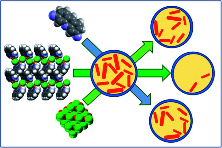
Introduction
The phenomenon of antimicrobial resistance (AMR) is one of the major medical challenges in most healthcare systems.1 The AMR has increased dramatically in recent years and represents a global public health threat.2 A number of diseases considered to be under control by the application of antibacterial remedies are acquiring resistance to these therapies. One of the main factors that prompted the development of resistance in microorganisms is the intensive overuse of antibiotics to control infection in human and animal diseases as well as in the agricultural sector.3 Actually, the reason why the AMR is a significant concern is the high mortality level attributed to resistance.4 Most of the common pathogenic strains already have antibiotic-resistant genes and, presumably, more antibiotic-resistant pathogens will emerge in the future.5 Besides the obvious need to optimize the use of antibiotics, one of the main routes to cope with the problem of antibacterial resistance is the quest for new antibacterial agents, which implies higher costs and long research times.6 For this reason there is a growing interest in alternatives to classical antibiotics. Metal-based antimicrobials have been gaining popularity as a new means of dealing with the AMR threat,7,8 although they have been used for millennia as antimicrobial agents. In this work we describe a crystal engineering9 approach to the obtainment of novel metal based antibacterial materials. Two known antibacterial agents, proflavine and methyl viologen dichloride10,11 (Scheme 1) have been co-crystallized with copper(i) and (ii) and silver(i), also known to exhibit antimicrobial activity,12 and the antimicrobial activity of the products has been tested. Indeed, co-crystallization as a way to synthesize new materials, or to enhance/improve the physico-chemical properties of active molecules,13 is a powerful application of crystal engineering principles, and in the last decade has been employed efficaciously in a number of diverse areas such as pharmaceutics,14 agrochemistry,15 high-energy materials,16 food,17etc. The basic idea is that formation of a stable, non-covalent association in the solid-state between an active ingredient and a solid co-former is a way to alter the physico-chemical properties of the active ingredient molecule crystal (solubility, dissolution rate, thermal stability, photoreactivity, hygroscopicity, etc.).18
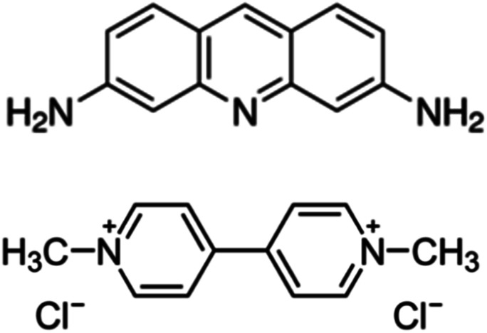
Scheme 1. Top: proflavine (PF); bottom: methyl viologen dichloride (MVCl2).
Furthermore, it has been shown that ionic co-crystals,19 whereby the active organic molecule (urea, thiourea) is co-crystallized with inorganic salts such as KCl, ZnCl2, can be successfully utilized to inhibit the activity of metalloenzymes involved in the degradation of urea.15,20
Neutral proflavine (acridine-3,6-diamine) and the dichloride salt of methyl viologen (1,1′-dimethyl-4,4′-bipyridinium dichloride) (see Scheme 1) have been chosen for this preliminary investigation, since promising results have been obtained by using quaternary-cation compounds (QCC) and metals mixed with extracted biosurfactants,21,22 where the mixtures had greater efficacy than each compound alone. Synthetic QCCs are widely used as antiseptics, cationic surfactants, disinfectants, herbicides and dyes in domestic, clinical and industrial setting.23
Results and discussion
Proflavine, obtained as a free base from the commercial sulphate salt, was co-crystallized with the inorganic salts AgNO3 and CuCl, both via mechanochemistry and via slurry in acetone (see ESI†). Slurry mediated reactions yielded higher purity target products, which were subsequently utilized for the investigation of the antimicrobial activity. It should be stressed here that the term co-crystallization, in this paper as in other related studies, is used to emphasize that the products are obtained via direct mixing of solid compounds that form stable crystalline phases at ambient conditions.
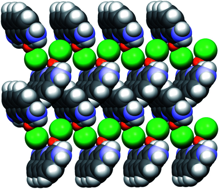
The structure of PF·CuCl (1a) was determined from X-ray powder diffraction (XRPD) data (see ESI†). The structure can be described as layers of PF molecules, arranged in a herring-bone fashion; the acridine nitrogens coordinate the copper(i) cations of the CuCl pairs, which in turn form a (Cu–Cl⋯Cu–Cl⋯)n 1D chain (Fig. 1).
Fig. 1. Relevant packing features in crystalline PF·CuCl (1a), showing the herring-bone arrangement of the proflavine molecules and the 1D (CuCl⋯CuCl⋯)n chains.
The reaction of PF and AgNO3, both by slurry and grinding, yielded the anhydrous, novel solid PF·AgNO3 (1b), with a consistent and reproducible XRPD pattern different from the starting material (see ESI†). Unfortunately, in spite of multiple crystallization attempts, the low quality of the diffraction patterns has thus far prevented a full structural characterization from powder data.
Co-crystallization of the MVCl2 salt with copper(i) and copper(ii) chlorides was also performed via slurry at ambient conditions (see ESI†). The reaction resulted in the formation of two solid compounds, 2a and 2b, whose XRPD patterns were found to match those of catena-(MV2+)tetrakis(μ2-chloro)-di-copper(i), (CSD refcode DMDPCU,24 obtained in different conditions in the original paper) and N,N′-dimethyl-4,4′-bipyridinium tetrachloro-copper(ii), (CSD refcode MBPYCU25 – no synthesis reported in the original paper), respectively (see ESI†).
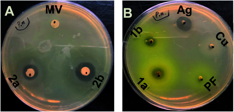
The organics PF and MV as well as salts of Cu and Ag have been shown to have antimicrobial activity. Therefore, we performed standard agar media plate antimicrobial assays to determine if the co-crystal of the compounds maintained or changed their antimicrobial efficacies. For the antimicrobial assays working stocks of 1a,b and 2a,b as well as of PF and MV and of the inorganic salts CuCl, CuCl2 and AgNO3 were made fresh at a concentration of 25 mg mL−1 in double distilled water producing solutions or slurries. Antimicrobial testing was performed using the pathogen indicator strains: Pseudomonas aeruginosa ATCC27853, Staphylococcus aureus ATCC25923, and Escherichia coli ATCC25922. Efficacy of antimicrobials used the established method of measuring a zone of growth inhibition which was performed either by adding the compounds to a well in the agar media or through soaking the compounds into an antimicrobial susceptibility filter disk. An example of zones of inhibition is shown in Fig. 2. In order to deal with subtle differences in biological growth between trials, zones in a given technical trial plate were normalized to allow more effective comparisons between trials and antimicrobial formulations.
Fig. 2. Examples of zones of clearing. (A): Representation of susceptibility filter disk application of MV and of 2a and 2b. (B): Representation of agar well application method of PF and of 1a and 1b. The silver and copper salts are also tested. One can see the yellow coloration in the plates of the diffusion of the proflavine from the application center well. Sample is for P. aeruginosa.
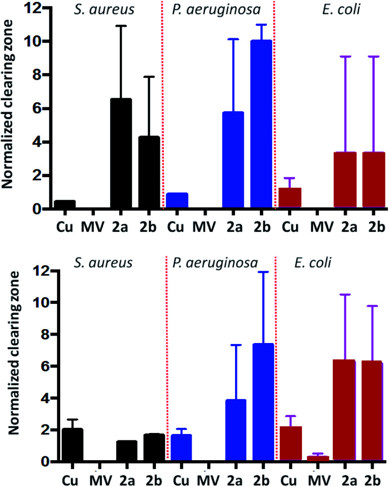
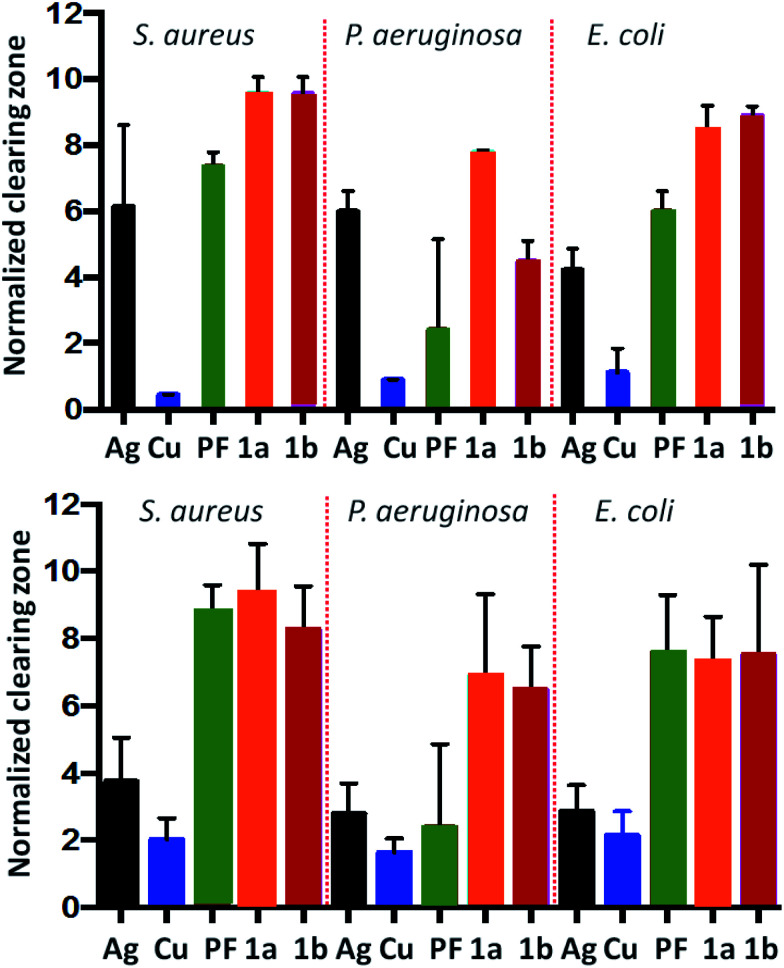
We evaluated first the MV compounds. MV salts with CuCl or CuCl2 showed maximum zones of inhibition only on the order of 10–12 mm. However, one can see in the normalized results (see Fig. 3) that the antimicrobial activity was greater than the metal salts or MV alone. This establishes that generating a crystal combining these two antimicrobial agents provides enhanced antimicrobial activity compared to Cu chlorides or MV alone towards all three bacteria.
Fig. 3. Efficacy of MV based compounds. Top: normalized zones of growth inhibition utilizing agar well method. Bottom: normalized zones of growth inhibition utilizing compound impregnated antimicrobial filter disks.
With the success of the MV based compounds we explored the antimicrobial efficacy of our new proflavine-based materials. These compounds showed more impressive antimicrobial activities. For the agar-well approach, zones of inhibition were as much as 150 mm in some experiments and with less variability between biological trials. Disk diffusion assays were more reproducible with zones of inhibition maxing out at 60 to 90 mm. The normalized data is compared in Fig. 4.
Fig. 4. Efficacy of PF based compounds. Top: normalized zones of growth inhibition with compounds deposited in agar well (method 1). Bottom: normalized zones of growth inhibition utilizing compound impregnated disks (method 2).
As expected, differences in susceptibility were observed between bacterial species, as seen with previous studies.26,27 Yet overall the co-crystals showed better efficacy than the metal salts on their own. Experiments using method 1 (see ESI†), where a physical mixture of PF and metal salt gave rise to a zone of inhibition on the same order as the most toxic compound of the two, suggest a degree of synergy of the components as a co-crystal. Although proflavine has poor efficacy on its own towards P. aeruginosa, it shows quite a good efficacy against S. aureus and E. coli on its own. But in the agar-well assay (Fig. 4 top), PF compounds 1a and 1b show a significant improved efficacy (p < 0.0001) over PF alone. For the complementary assay this improvement is lost. Regardless there is still improved efficacy against P. aeruginosa. These results imply delivery approach of the engineered crystals is an important variable when considering the target organism.
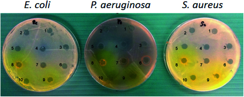
An alternative antimicrobial assay is to evaluate contact killing against an existing growth of bacteria. We tested to see if contact killing and cell lysis will occur, using a compound impregnated dried susceptibility disk applied to a robustly grown lawn of bacteria. We found that, under the media and growth conditions used here, all compounds tested demonstrated the ability to kill and lyse the existing lawn of bacteria, generating a zone of clearing beneath the disk (see Fig. 5). No zones of clearing beyond the disk were observed for E. coli lawns. However, for 1a, an additional 0.5–1.0 mm zone was observed in the S. aureus lawn. Several compounds showed additional zone of clearing beyond the disk against P. aeruginosa with the comparator compounds of AgNO3 and Ag2O nanoparticles showing the largest with a 3 mm zone. Compounds 1a, 1b, 2a and 2b all showed a 1 to 1.5 mm zone, however MV and PF also showed the same sized zone. This experiment demonstrated that our new formulations have contacting killing activity.
Fig. 5. Killing and lysing of established cultures (method 3, see ESI†). (1) MV; (2) 2a; (3) 2b; (4) CuSO4; (5) CuCl2; (6) AgNO3; (7) silver oxide; (8) PF; (9) 1a; (10) 1b. The clearing demonstrates the ability to kill and lyse a pre-grown culture of bacteria compared to the other methods that measure the ability of compounds to prevent growth.
Using such compounds led to experimental challenges between experiments. The issue was primarily producing reproducible slurries. Since it was noted that the silver based compounds were more photosensitive, all data shown is with fresh stocks prepared within 24 hours stored in the dark. Additionally, zones of inhibition were not always distinct, with two zones often observed, one of full clearing of growth and a second larger ring where growth was inhibited but not prevented.
Conclusions
In summary, our results show that co-crystallization of known QCC antibacterial compounds, e.g. proflavine and methyl viologen, with metal salts, such as CuCl, CuCl2 and AgNO3, obtained either mechanochemically or from solution, is a viable, eco-friendly, and inexpensive way to obtain new materials with enhanced antibacterial properties. In particular PF·CuCl and PF·AgNO3 appear to perform better than PF and the salts separately. Much less striking antimicrobial levels have been obtained when the MVCl2 salt is reacted with the inorganic salts, yet we still see an improvement of the overall association over the individual agents. Crystal engineering approaches can be used to devise materials for biological uses, in an area of increasing global importance, such as that of antibiotic resistance.
We envisage the use of our compounds as coating materials in the prevention of infectious disease transfer. The challenges that exist is producing a metal formulation that releases the metal at a level that sustains antimicrobial properties and that is stable to the physical manipulation of the material that has been coated. This opens avenues for development of unique metal chelates and organo-metal compounds.
Conflicts of interest
There are no conflicts to declare.
Supplementary Material
Acknowledgments
This material is based upon O. S. D. B., F. G., work supported by the University of Bologna (RFO scheme). NSERC supported the contribution of R. J. T. R. J. T. thanks Patrick Macrohon for his assistance. O. S. D. B., F. G. thank Aistė Miliūtė for her assistance.
Electronic supplementary information (ESI) available: Synthesis, DSC and TGA, XRPD. CCDC 1964562. For ESI and crystallographic data in CIF or other electronic format see DOI: 10.1039/c9ra10353h
Notes and references
- Theuretzbacher U. J Glob. Antimicrob. Resist. 2013;1:63–69. doi: 10.1016/j.jgar.2013.03.010. [DOI] [PubMed] [Google Scholar]
- Levy S. B. Marshall B. Nat. Med. 2004;10:S122–S129. doi: 10.1038/nm1145. [DOI] [PubMed] [Google Scholar]
- Brown E. D. Wright G. D. Nature. 2016;529:336–343. doi: 10.1038/nature17042. [DOI] [PubMed] [Google Scholar]
- de Kraker M. E. Davey P. G. Grundmann H. group B. s. PLoS Med. 2011;8:e1001104. doi: 10.1371/journal.pmed.1001104. [DOI] [PMC free article] [PubMed] [Google Scholar]
- Li X. Bai H. Yang Y. Yoon J. Wang S. Zhang X. Adv. Mater. 2019;31:e1805092. doi: 10.1002/adma.201805092. [DOI] [PubMed] [Google Scholar]
- Payne D. J. Gwynn M. N. Holmes D. J. Pompliano D. L. Nat. Rev. Drug Discovery. 2007;6:29–40. doi: 10.1038/nrd2201. [DOI] [PubMed] [Google Scholar]
- Turner R. J. Microb. Biotechnol. 2017;10:1062–1065. doi: 10.1111/1751-7915.12785. [DOI] [PMC free article] [PubMed] [Google Scholar]
- Mehra C. Gala R. Kakatkar A. S. Kumar V. Khurana R. Chatterjee S. Kumar N. N. Barooah N. Bhasikuttan A. C. Mohanty J. Chem. Commun. 2019;55:14275. doi: 10.1039/C9CC07378G. [DOI] [PubMed] [Google Scholar]
- Desiraju G. R. Parshall G. W. Mater. Sci. Monogr. 1989:54. [Google Scholar]
- Wainwright M. J. Antimicrob. Chemother. 2001;47:1–13. doi: 10.1093/jac/47.1.1. [DOI] [PubMed] [Google Scholar]
- Sulavik M. C. Houseweart C. Cramer C. Jiwani N. Murgolo N. Greene J. DiDomenico B. Shaw K. J. Miller G. H. Hare R. Shimer G. Antimicrob. Agents Chemother. 2001;45:1126–1136. doi: 10.1128/AAC.45.4.1126-1136.2001. [DOI] [PMC free article] [PubMed] [Google Scholar]
- Lemire J. A. Harrison J. J. Turner R. J. Nat. Rev. Microbiol. 2013;11:371–384. doi: 10.1038/nrmicro3028. [DOI] [PubMed] [Google Scholar]
- Desiraju G. R. CrystEngComm. 2003;5:466. doi: 10.1039/B313552G. [DOI] [Google Scholar]
- Vishweshwar P. McMahon J. A. Bis J. A. Zaworotko M. J. J. Pharm. Sci. 2006;95:499–516. doi: 10.1002/jps.20578. [DOI] [PubMed] [Google Scholar]
- Casali L. Mazzei L. Shemchuk O. Honer K. Grepioni F. Ciurli S. Braga D. Baltrusaitis J. Chem. Commun. 2018;54:7637–7640. doi: 10.1039/C8CC03777A. [DOI] [PubMed] [Google Scholar]
- Bolton O. Matzger A. J. Angew. Chem. 2011;50:8960–8963. doi: 10.1002/anie.201104164. [DOI] [PubMed] [Google Scholar]
- Oertling H. CrystEngComm. 2016;18:1676–1692. doi: 10.1039/C6CE00218H. [DOI] [Google Scholar]
- Duggirala N. K. Perry M. L. Almarsson O. Zaworotko M. J. Chem. Commun. 2016;52:640–655. doi: 10.1039/C5CC08216A. [DOI] [PubMed] [Google Scholar]
- Braga D. Grepioni F. Shemchuk O. CrystEngComm. 2018;20:2212–2220. doi: 10.1039/C8CE00304A. [DOI] [Google Scholar]
- Casali L. Mazzei L. Shemchuk O. Sharma L. Honer K. Grepioni F. Ciurli S. Braga D. Baltrusaitis J. ACS Sustainable Chem. Eng. 2018;7:2852–2859. doi: 10.1021/acssuschemeng.8b06293. [DOI] [PubMed] [Google Scholar]
- Harrison J. J. Turner R. J. Joo D. A. Stan M. A. Chan C. S. Allan N. D. Vrionis H. A. Olson M. E. Ceri H. Antimicrob. Agents Chemother. 2008;52:2870–2881. doi: 10.1128/AAC.00203-08. [DOI] [PMC free article] [PubMed] [Google Scholar]
- Rivardo F. Martinotti M. G. Turner R. J. Ceri H. Can. J. Microbiol. 2010;56:272–278. doi: 10.1139/W10-007. [DOI] [PubMed] [Google Scholar]
- McDonnell G. Russell A. D. Clin. Microbiol. Rev. 1999;12:147–179. doi: 10.1128/CMR.12.1.147. [DOI] [PMC free article] [PubMed] [Google Scholar]
- Prout C. K. Murray-Rust P. J. Chem. Soc. A. 1969:1520–1525. doi: 10.1039/J19690001520. [DOI] [Google Scholar]
- Russell J. H. Wallwork S. C. Acta Crystallogr., Sect. B: Struct. Crystallogr. Cryst. Chem. 1969;25:1691–1695. doi: 10.1107/S0567740869004572. [DOI] [Google Scholar]
- Gugala N. Vu D. Parkins M. D. Turner R. J. Antibiotics. 2019;8:51. doi: 10.3390/antibiotics8020051. [DOI] [PMC free article] [PubMed] [Google Scholar]
- Gugala N. Lemire J. A. Turner R. J. J. Antibiot. 2017;70:775–780. doi: 10.1038/ja.2017.10. [DOI] [PubMed] [Google Scholar]
Associated Data
This section collects any data citations, data availability statements, or supplementary materials included in this article.


