Many patients with hematological cancers are not completely protected after the initial dose or after both primary doses of the vaccines1,2 with most failing to seroconvert on completion of the two-dose vaccine schedule.2 These reports only included three patients with myelodysplastic syndrome (MDS). MDS represents a spectrum of clonal bone marrow neoplasms from lowrisk disease through to those transforming into acute myeloid leukemia. Patients with MDS, especially with lower-risk disease, many of whom are minimally treated and who might be expected to have a comparable immune response to healthy volunteers, and as such a better immune response to COVID-19 vaccines than other hematological cancers. Previous studies looking at the immune response to influenza vaccination in those with MDS had shown promising results with immune responses not differing from those of healthy family members.3 However, a recent study which included six MDS patients, reported poor seroconversion rates following a single dose of COVID-19 vaccine in a group of 60 myeloid cancer patients, including those who are not on cytoreductive treatments and those in complete hematological remission, suggesting a clear need for more detailed interrogation of COVID-19 vaccination in this group of patients.4 Here, we report the humoral and T-cell responses of 38 patients with MDS 2 weeks following completion of the second dose vaccine schedules of ChAdOx1 or BNT162b2 nCoV-19 vaccines.
Following approval by the Institutional Review Boards, patients with MDS (n=38) vaccinated with either BNT162b2 mRNA or ChAdOx1 nCoV-19 COVID-19 vaccine provided written informed consent. Eligibility criteria for the study included diagnosis of MDS as per the World Health Organization classification5 and age ≥18 years. The study also included healthy volunteers (HV) (mainly healthcare workers, n=30) serving as a reference group, included principally to provide an experimental control for study assays and facilitate their comparison with results of other studies of BNT162b2 in healthy populations. Plasma samples were tested for immunoglobulin G (IgG) binding the SARS-CoV-2 spike (S) protein and nucleoprotein (N) and neutralization assays against HIV-1 based virus particles pseudotyped with SARS-CoV-2 Wuhan strain (WT), variant of concern (VOC)B.1.1.7 (a) or VOC.B.1.617.2 (δ) spike as previously described.1,2,6 Cellular responses were assessed using interferon g (IFNg) ELISPOT and flow cytometry (CD25 and CD69 expression) after 24 hours of peptide stimulation. IFNg ELISpot analysis was performed ex vivo for assessment of T-cell response following stimulation with SARS-CoV-2 peptide pools, Cytomegalovirus (CMV), Epstein-Barr virus (EBV), and influenza virus positive control (CEF) peptides for 24 hours.
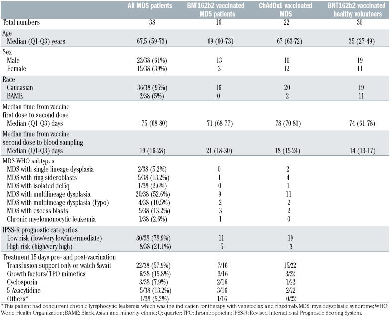
Thirty-eight MDS patients and 30 HV provided a blood sample 2 weeks following a second primary dose of their initial vaccine. Clinical characteristics along with median times to second dose are provided in Table 1. We observed significant differences between the ages of the HV and MDS cohorts (Student’s t-test, equal variance, P<0.001). 42% (n=16) of the MDS patients received BNT162b2 and 58% (n=22) received ChAdOx1 nCoV-19 vaccines. All HV received a delayed BNT162b2 second dose. As per UK government guidelines at the time of vaccination, individuals receiving BNT162b2 second doses received these between 8-12 weeks following the first dose, representing a delay compared to the licensed administration. Prior SARS-CoV-2 infection can influence the magnitude of the vaccine response,7 and as such we excluded two MDS and four HV based on being positive for nucleoprotein-specific IgG (IgG(N)) (representing response to prior infection) (Online Supplementary Figure S1A). We observed that the anti-S IgG titres at approximately 2 weeks following the second dose were within the upper quantile in these previously virus-exposed individuals (Online Supplementary Figure S1B, red dots). These were excluded from the overall immune efficacy analysis.
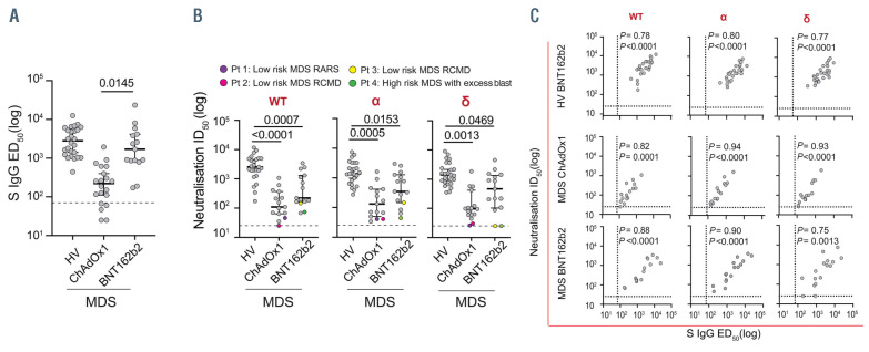
In the remaining (HV BNT162b2, n=26; MDS BNT162b2, n=15 and MDS ChAdOx1, n=21) cohort; we assessed the anti-S IgG titres following their second primary dose. Overall serological responses were: HV BNT162b2 100% (26/26); MDS BNT162b2 100% (15/15) and MDS ChAdOx1 76.2% (16/21) (Figure 1A); notably, the MDS ChAdOx1 cohort demonstrated significantly decreased serological titres to the MDS BNT162b2 cohort (Figure 1A). It is noteworthy that the median titre for the MDS BNT162b2-vaccinated patients is higher (>103) compared to the median reported in a heterogenous BNT162b2-vaccinated hematological cancer population (<103) observed in McKenzie et al.2 Of the five nonresponders within the MDS ChAdOx1, three patients were on disease-modifying treatments (5-azacytidine, venetoclax and danazol), with the patient on venetoclax/rituximab having a concurrent diagnosis of chronic lymphocytic leukemia (CLL). None of these patients were noted to be on steroid therapy around the time of vaccination; and no differences in the clinical white blood cells were observed between serological responders or non-responders (Online Supplementary Figure 1C). Similar to our previous reports1,2 there was no significant correlation between spike IgG titres and age or the time between the first and second doses of the vaccine in the two MDS cohorts (Online Supplementary Figure S1D).
Next, we assessed the functional implications of seroconversion by neutralization assays for SARS-CoV-2 WT and VOC a and δ (Figure 1B). All but four MDS patients (Figure 1B; colored dots) could neutralize all variant strains, but MDS cohorts showed significantly reduced median neutralizations for all three variant strains compared to HV (Figure 1B); importantly this was the case for both the MDS ChAdOx1 and MDS BNT162b2 cohorts. We acknowledge the younger age of the HV cohort may contribute to this reduction, although age was not a determinant of neutralization response in cancer patients in our previous reports.1,2 Review of the four MDS (2 BNT162b2 mRNA and two ChAdOx1 nCoV-19 COVID- 19-vaccinated) patients classified as non-responders by neutralization assay demonstrated that these patients were predominantly low risk MDS on no treatment, except one patient with excess of blasts on 5-azacytidine. These data clearly support the need for a third primary dose for this clinically vulnerable patient group irrespective of the seroconversion rates across cohorts. This is especially the case in those who have seroconverted but have a low anti-S IgG titre after the second dose. Third doses have demonstrated higher anti-S IgG titres in other hematological cohorts,8 and in keeping with our previous reports,1,2 anti-S IgG titres were highly correlated with neutralization among all cohorts (Figure 1C).
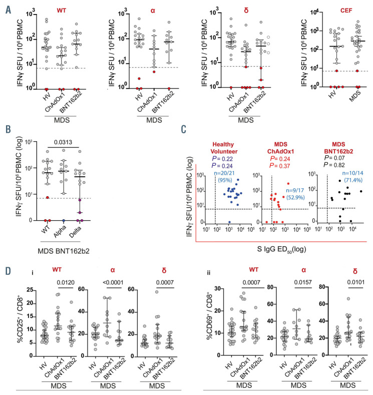
In order to measure functional SARS-CoV-2 T-cell responses to vaccination, peripheral blood mononuclear cells (PBMC) from our study participants were assessed by ELISpot assays as described. It is noteworthy that no differences in the percentages of T cells amongst the PBMC plated for ELISpot were observed across healthy and MDS cohorts (Online Supplementary Figure S1E). Using previously published thresholds for response,1,2 non-T cell responders were seen in all cohorts (Figure 2A; red dots). Specifically, SARS-CoV-2-specific IFNg T-cell responses against the δ variant were: HV BNT162b2 95% (20/21); MDS ChAdOx1 70.6% (12/17) and MDS BNT162b2 71.4% (10/14) (Figure 2A); in stark contrast to the comparable control CEF induced effector T-cell responses across healthy and MDS samples (Figure 2A). Interestingly, significantly reduced T-cell responses were seen in MDS BNT162b2-vaccinated patients when challenged with δ compared to wt variant strain (Figure 2B). Further, five MDS ChAdOx1 patients who did not have a serological response, were able to mount T-cell responses. Additionally, treatment with either azacytidine or calcineurin inhibitor cyclosporine did not impair appropriate T-cell responses. One high risk MDS BNT162b2 patient on 5-azacytidine, who showed no neutralizing activity, showed significantly reduced T-cell response to WT and a, but not to δ variant. During the study period, the δ variant was the predominant VOC in the UK. We observed non-significant but positive correlations between serological and IFNg T-cell responses against the δ variant within the MDS vaccinated cohorts (Figure 2C). Numbers of individuals who were both serological and Tcell responders were as follows: HV 95% (20/21), MDS BNT162b2 71.4% (10/14) and MDS ChAdOx1 52.9% (9/17) (Figure 2C). In order to further investigate the cellular readout of vaccine efficacy, we assessed the activation state of SARS-CoV-2 stimulated CD8 T cells, by measuring activation markers CD25 and CD69 cell surface expression by flow cytometry before and after in vitro stimulation. Despite the poorer humoral response observed in MDS-ChAdOx1 vaccinated individuals, we found significantly higher activated CD25+ and CD69CD8 T cells across all variants in this group of patients compared to those vaccinated with BNT162b2 vaccine (Figure 2Di&ii). These data are compelling and warrant further investigation with one hypothesis being the ChAdOx1 vector reveals an innate weakness in this patient group inducing a hyper-stimulated but poorly efficacious effector T-cell response.
Table 1.
Clinical characteristics of patients evaluable for analysis 2 weeks following two primary vaccine doses.
Figure 1.
Humoral responses to BNT162b2 COVID-19 and ChAdOx1 nCoV-19 in patients with myelodysplastic syndromes. (A) Serum concentrations of immunoglobulin G (IgG) antibodies reactive to the spike protein of SARS-CoV-2 (S IgG) with cases positive for nucleorprotein N IgG removed. Healthy volunteer (HV; n=26), myelodysplastic syndrome (MDS) patients vaccinated with ChAdOx1 (MDS ChAdOx1; n=20), MDS patients vaccinated with BNT162b2 (MDS BNT162b2; n=15). Mean (95% confidence interval [CI]): healthy volunteers (HV) 3,611 (2,455-4,768), MDS ChAdOx1 360.9 (149.9-572.2) and MDS BNT162b2 3781 (523.9-7,037). Dashed line represents seroconversion threshold. Tukey’s multiple comparison’s test. (B) Neutralization of variants (as indicated in red) by plasma antibodies. Dashed line represents neutralization threshold. Individual cases on the threshold line are colored as indicated, as are their matched responses to other variants. HV (n=26); MDS ChAdOx1 (n=15); MDS BNT162b2 (n=15). Tukey’s multiple comparison’s test. (C) Correlation matrices showing serum S IgG 50% effective dose (ED50) (log) against neutralization for each indicated variant in the MDS ChAdOx1 (n=20) and MDS BNT162b2 (n=15) cohorts. Correlation coefficients (rho;r) and P-values are given. Dashed lines represent threshold as previously described. Pearson’s correlation test. WT: Wuhan strain.
Figure 2.
Cellular responses to BNT162b2 COVID-19 and ChAdOx1 nCoV-19 in patients with myelodysplastic syndromes. (A) Interferon g (IFNg) spot-forming units (SFU) formed after stimulation of peripheral blood mononuclear cells (PBMC) from indicated cohorts in response to indicated variants. Samples were classed as responders if >7 cytokine secreting cells/106 PBMC after correcting for background; as indicated by dashed line. Non-responders are colored as indicated. Wuhan strain (WT); (healthy volunteers [HV] [n=26]; MDS ChAdOx1 [n=20]; MDS BNT162b2 [n=15]); B.1.1.7; (HV [n=11]; MDS ChAdOx1 [n=11]; MDS BNT162b2 [n=15]); B.1.617.2; (HV [n=21]; MDS ChAdOx1 [n=17]; myelodysplastic syndrome (MDS) BNT162b2 [n=14]). Tukey’s multiple comparison’s test. Influenza virus positive control (CEF)= Cytomegalovirus (CMV), Epstein-Barr virus (EBV) and influenza virus positive control peptides: (B) IFNγ SFU formed after stimulation of PBMC from MDS BNT162b2 cases to indicated variants. WT (n=15); B.1.1.7 (n=11); B.1.617.2 (n=14). Tukey’s multiple comparison’s test. (C) Correlation matrices showing IFNg SFU formed after PBMC were stimulated with the B.1.617.2 variant and paired S IgG 50% effective dose (ED50) values for indicated cohorts. Correlation coefficients (rho;r), P-values, n numbers and % double positivity are given. Dashed lines represent thresholds as previously described. Pearson’s correlation test. E (i&ii). CD8+CD25+ cells (i) and CD8+CD69+ cells (ii) within the live CD3+ population after stimulation of PBMC from indicated cohorts in response to indicated variants. HV (n=26); MDS ChAdOx1 (n=20); MDS BNT162b2 (n=15). Tukey’s multiple comparison’s test.
In totality, although ChAdOx1-treated MDS patients do mount both humoral and cellular immune responses, they are weak in comparison to BNT162b2. The overall serological responses in the MDS cohorts were 100% for those who had completed the two-dose BNT162b2 vaccine schedule compared to 76.2% of patients vaccinated with the ChAdOx1 vaccine. As such, it may be pertinent to advise the clinical community to administer MDS patients with an mRNA-based vaccine to promote enhanced immunity. Finally, we observed that neutralization in seroconverted patients was significantly weaker for both the ChAdOx-1 and BNT162b2 MDS cohorts compared to HV, highlighting the potential benefit of a third primary dose for this clinically vulnerable patient group, in addition to subsequent booster doses.
Supplementary Material
Acknowledgements
We thank patients and blood donors consenting in this. We thank members of the King’s College Hospital (KCH) trial teams who contributed to patient recruitment for the SOAP study at KCH hospitals.
Funding Statement
Funding: the SOAP study (IRAS 282337) is sponsored by King’s College London and GSTT Foundation NHS Trust. It is funded from grants from the KCL Charity funds to SI (PS10822), Cancer Research UK to S.I. (C56773/ A24869). This work was supported by Blood Cancer UK awarded to SI, AK and PP and the Leukaemia & Lymphoma Society (LLS) Award to SI and PP (6631-21).
References
- 1.Monin L, Laing AG, Munoz-Ruiz M, et al. Safety and immunogenicity of one versus two doses of the COVID-19 vaccine BNT162b2 for patients with cancer: interim analysis of a prospective observational study. Lancet Oncol. 2021;22(6):765-778. [DOI] [PMC free article] [PubMed] [Google Scholar]
- 2.McKenzie DR, Munoz-Ruiz M, Monin L, et al. Humoral and cellular immunity to delayed second dose of SARS-CoV-2 BNT162b2 mRNA vaccination in patients with cancer. Cancer Cell. 2021;39(11):1445-1447. [DOI] [PMC free article] [PubMed] [Google Scholar]
- 3.Vachhani P, Wiatrowski K, Srivastava P, et al. Quantification of humoral immune response to influenza vaccination in MDS. Blood. 2019;134(Suppl 1):S4756. [Google Scholar]
- 4.Chowdhury O, Bruguier H, Mallett G, et al. Impaired antibody response to COVID-19 vaccination in patients with chronic myeloid neoplasms. Br J Haematol. 2021;194(6):1010-1015. [DOI] [PMC free article] [PubMed] [Google Scholar]
- 5.Arber DA, Orazi A, Hasserjian R, et al. The 2016 revision to the World Health Organization classification of myeloid neoplasms and acute leukemia. Blood. 2016;127(20):2391-2405. [DOI] [PubMed] [Google Scholar]
- 6.Dupont L, Snell LB, Graham C, et al. Neutralizing antibody activity in convalescent sera from infection in humans with SARS-CoV-2 and variants of concern. Nat Microbiol. 2021;6(11):1433-1442. [DOI] [PMC free article] [PubMed] [Google Scholar]
- 7.Anichini G, Terrosi C, Gandolfo C, et al. SARS-CoV-2 antibody response in persons with past natural infection. N Engl J Med. 2021;385(1):90-92. [DOI] [PMC free article] [PubMed] [Google Scholar]
- 8.Greenberger LM, Saltzman LA, Senefeld JW, Johnson PW, DeGennaro LJ, Nichols GL. Anti-spike antibody response to SARSCoV- 2 booster vaccination in patients with B cell-derived hematologic malignancies. Cancer Cell. 2021;39(10):1297-1299. [DOI] [PMC free article] [PubMed] [Google Scholar]
Associated Data
This section collects any data citations, data availability statements, or supplementary materials included in this article.


