Abstract
Drug delivery technology has a wide spectrum, which is continuously being upgraded at a stupendous speed. Different fabricated nanoparticles and drugs possessing low solubility and poor pharmacokinetic profiles are the two major substances extensively delivered to target sites. Among the colloidal carriers, nanolipid dispersions (liposomes, deformable liposomes, virosomes, ethosomes, and solid lipid nanoparticles) are ideal delivery systems with the advantages of biodegradation and nontoxicity. Among them, nano-structured lipid carriers and solid lipid nanoparticles (SLNs) are dominant, which can be modified to exhibit various advantages, compared to liposomes and polymeric nanoparticles. Nano-structured lipid carriers and SLNs are non-biotoxic since they are biodegradable. Besides, they are highly stable. Their (nano-structured lipid carriers and SLNs) morphology, structural characteristics, ingredients used for preparation, techniques for their production, and characterization using various methods are discussed in this review. Also, although nano-structured lipid carriers and SLNs are based on lipids and surfactants, the effect of these two matrixes to build excipients is also discussed together with their pharmacological significance with novel theranostic approaches, stability and storage.
Drug delivery technology has a wide spectrum, which is continuously being upgraded at a stupendous speed.
Introduction
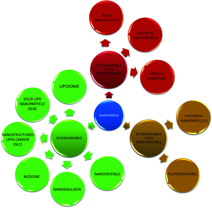
With the development of technology in the last two decades, the particle size of materials ranges from the micro- to nano-scale. The reduction in the particle size of materials at the nanometer scale increases their overall surface area by several orders of magnitude. Particles with a size in the range of 1 nm to 1000 nm are known as nanoparticles. The word “nano” can be easily defined, but it covers numerous areas of application. Fig. 1 represents several nano-based systems composed of different types of materials, which can be utilized as nanocarriers.
Fig. 1. Schematic diagram of the different types of nanoparticles.
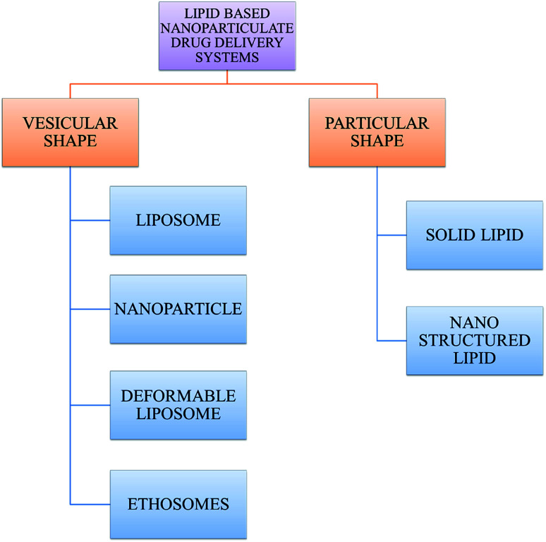
However, nanomaterials with excellent biodegradability and biocompatibility are considered to be the best vehicles for drug delivery systems in biomedical applications. Currently, scientists and researchers are focused on discovering new methods/routes to control the pharmacokinetics (ADME), pharmacodynamics, non-specific toxicity, immunogenicity, biorecognition, and drug efficacy of drugs. These new strategies are often called novel drug delivery systems (NDDS) and are based on interdisciplinary approaches that combine polymer science, pharmaceutics, bioconjugate chemistry, and molecular biology. Some of the different approaches for novel drug delivery include transdermal patches, sustained and controlled release by polymeric and magnetic control, liposomes, hydrogels, implants, microspheres, erythrocytes, and nanoparticles. Nanoparticular drug delivery systems are a successful approach in the treatment of chronic human diseases, which have excellent function in satisfying the biopharmaceutical and pharmacological considerations. The emergence of nanotechnology and the growing capabilities of functional proteomics, genomics, and bioinformatics combined with combinatorial chemistry have driven scientists to become more enthusiastic to express their technical expertise to discover, invent and explore novel approaches for drug delivery systems through new techniques. Novel drug delivery systems remain the foundation to deliver drugs having complications that cannot be minimized by conventional drug delivery systems, where the therapeutic effectiveness of drugs depends on their pharmacokinetics and site of administration. Pharmacokinetics are also based on physico-chemical properties such as solubility, crystallinity, toxicity, and HLB value. After understanding the biopharmaceutics and pharmacokinetics, the administration route, absorptive surface area, and transportation of drugs in the body are the key points for their absorption and distribution. Furthermore, metabolism and elimination depend on the aforementioned properties.1 The formulation design has a major impact on the effective delivery of the active pharmaceutical ingredient (API), and thus all the above parameters are crucial challenges. Drugs based on the HLB scale are categorized into two classes, hydrophilic and lipophilic molecules. Lipophilic molecules exhibit very poor solubility, and depending on this, they produce a great challenge to design safe, efficacious, and cost-effective drug delivery systems and have been a source of frustration for pharmaceutical scientists.2 Lipophilic molecules allow the design of formulations for hydrophobic drug molecules, and despite all the problems confronted by pharmaceutical scientists, the current solid lipid nanoparticles are the result of their great effort. Traditionally, lipid-based novel drug delivery systems have focused on the delivery of lipophilic molecules, but recently, lipoid drug delivery systems have received attention due to their inherent properties such as biocompatibility, self-assembly capabilities, ability to cross the blood brain barrier, particle size variability and finally cost effectiveness, making lipid-based delivery systems much more attractive.3 Lipid-based nanoparticles can also be subcategorized as follows in Chart 1.
Chart 1. Classification of lipid-based nanoparticle drug delivery systems.
Over the past few years, nanomaterials have emerged as drug carriers. Liposomes are important biological molecules, which have been used for many years, but currently, there are various alternative molecules. Niosomes are one of the promising economical alternatives to liposomes. Niosomes are highly stable and slightly more leaky than liposomes. The size of niosomes decreases substantially upon freezing in liquid nitrogen and subsequent thawing, as evident by cryo-EM and dynamic light scattering. The successful delivery of drugs through nanoparticles depends on their ability to penetrate barriers, continuously release drugs and their stability. However, the scarcity of regulatory approved polymers, i.e. the Food and Drug Administration (FDA), and their expensive costs have limited their clinical application.4 Thus, to overcome these limitations, scientists and researchers have proposed lipids as alternative carriers. These lipid-based nanoparticles are known as solid lipid nanoparticles (SLNs), which have attracted worldwide interest due to their advantages (Table 1).5
Advantages of SLNs over liposomes and polymeric nanoparticles.
| Issue | Advantages of SLNs over liposomes | Advantages of SLNs over polymeric nanoparticles |
|---|---|---|
| Avoidance of organic solvents | Avoidance of organic solvents when desired | Avoidance of organic solvents when desired |
| Preparation and reproducibility | Excellent reproducibility and feasible large-scale production | Excellent reproducibility and feasible large-scale production with cost-effective high-pressure homogenization method as the preparation method6 |
| Stability | Increased stability of the active ingredient because of the rigid core lipid matrix8 | Increased product stability of about 3 years7 |
| Biodegradability | Both liposomes and SLNs are biodegradable | Lipids of SLNs are physiological and biodegradable, and hence have better biocompatibility and sterilization. On the other hand, polymeric nanoparticles may accumulate undesirably in the liver, spleen etc.9 |
| Binding, entrapment and release | SLNs impose greater entrapment efficiency for hydrophobic drugs (since they do not contain an aqueous core with lipid bilayer like liposomes) | Drug delivery is extremely site specific for SLNs, whereas polymeric nanoparticles may produce non-specific drug delivery or show unpredictable release towards siRNAs10 |
| Ability to allow controlled release (similar to polymeric nanoparticles) and drug targeting by coating/attaching ligands to SLNs12 | 11 |
Solid lipid nanoparticle overview
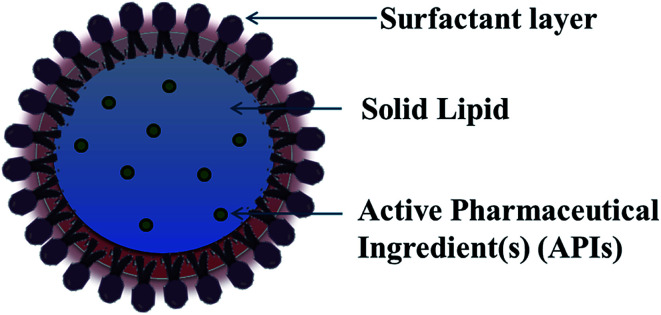
For lipid and lipid-based drug delivery systems, phospholipids are an important constituent because of their various properties, such as amphiphilic nature, biocompatibility and multifunctionality. However, liposomes, lipospheres, and microsimulation carrier systems have many drawbacks such as their complicated production method, low percentage entrapment efficiency (% EE), difficult large-scale manufacture, and thus the SLN delivery system has emerged.13,14 SLNs are commonly spherical in shape with a diameter in the range of 50 to 1000 nm. The key ingredients of SLN formulations include lipids, which are in the solid state at room temperature, emulsifiers and sometimes a mixture of both, active pharmaceutical ingredients (APIs) and an adequate solvent system (Fig. 2). Nanocarrier-based drug delivery systems can be subcategorized in many aspects depending on the route of administration, degree of degradability, etc. The route of administration includes nanoparticles for parenteral administration, oral administration, ocular administration, and topical administration, and nanoparticles for protein peptide delivery. Nanocarrier systems can also be subcategorized based on the degree of their degradability as follows.
Fig. 2. Schematic presentation of the complete structure of solid lipid nanoparticles.
An ideal nanoparticulate drug delivery system must contain the following characteristics:
(1) Maximum drug bioavailability.
(2) Tissue targeting.
(3) Controlled release kinetics.
(4) Minimal immune response.
(5) Ability to deliver traditionally difficult drugs such as lipophiles, amphiphiles and biomolecules.
(6) Sufficient drug loading capacity.
(7) Good patient compliance.
Solid lipid nanoparticles have changed the dimension of drug delivery by combining all the advantageous characteristics of polymeric nanoparticles, liposomes and microemulsion.15 All the properties of lipid nanoparticles are upgraded with surface modification, better pharmacokinetic acceptability, formation of inclusion complexes, improved stability pattern and incorporation of chemotherapeutic agents. SLNs are appropriate for intravenous applications because of their effortless dispersion in solution, which are aqueous or aqueous-surfactant. Nanoparticles undergo phagocytic uptake,16 and thus by surface modification, their phagocytic uptake can be minimized.17 A pharmacokinetic study also showed a good increase in the of concentration doxorubicin in with solid lipid nanoparticles compared with conventional commercial drug solutions, and it was found that the drug concentrations were higher in the lungs, spleen and brain of rats.18 In drug delivery technology, cyclodextrin is used as a complex agent, which can be used to increase aqueous solubility, bioavailability and improve the physicochemical properties of drugs by forming inclusion complexes. The incorporation of these inclusion complexes into solid lipid nanoparticles increases their release profile compared to solid lipid nanoparticles without cyclodextrin.19 Furthermore, the stability pattern of solid lipid nanoparticles (SLNs) is more attractive than that of other nanoparticulate formulations. Aqueous SLNs can be stored for up to 3 years or longer, and their gelling tendency due to long term storage and light exposure can be stabilized by inhibiting the transitions by lipid modification.20 The major aim of solid lipid nanoparticles (SLN) in terms of drug delivery is to enhance the bioavailability and efficacy of drugs, and control the non-specific toxicity, immunogenicity, pharmacokinetics and pharmacodynamics of drugs. This review focuses on the potential of SLNs in various types of chemotherapy such as cancer, where conventional chemotherapy is hindered by different obstacles such as drug resistance, low specificity and poor stability of chemotherapeutic compounds.9 These issues may be partly overcome by encapsulating drugs as SLNs. The new generations of SLN such as nanostructured lipid carriers (NLC), lipid drug conjugates (LDC), polymeric lipid hybrid nanoparticles (PLN), and long-circulating SLNs, improve the role of SLNs as versatile drug carriers for various types of chemotherapy, and treatment of parasitic infections and tuberculosis.14,17 Cell line studies have shown that SLNs can be easily internalized and may be designed as surrogate colloidal drug carriers for the administration of chemotherapeutic agents, especially for the treatment of malignant melanoma and colorectal cancer.21 Besides their antitumor activities, SLNs are also capable of hindering the adhesive interactions between cancerous cells (resulting from human breast, prostate cancers, melanoma, etc.) with the cells present on human umbilical vein endothelium.22 Furthermore, since SLNs are based on nontoxic and non-irritating materials, they are ideal for use in topical formulations.23 Accordingly, there has been extensive research on the topical applications of SLNs (containing lipids such as glyceryl palmitostearate and glyceryl behenate) to treat several skin diseases since SLNs adhere strongly because of their greater surface area as a result of their smaller sizes.24,25 The coenzyme Q10 penetrated the stratum corneum more effectively as SLNs in comparison with liquid paraffin and isopropanol.26 The extent of drug release was higher and more rapid for SLNs of Compritol®(Retinol-loaded) compared to conventional carriers.27,28 Also, SLNs were found to be significant vehicles for numerous sunscreen agents.29,30
The delivery of genetic material via nanotechnology is now gaining significant attention. Cationically modified SLNs can effectively deliver DNA to binding sites, where the transfection efficiency and cytotoxicity are also very low.31 Furthermore, solid lipid nanoparticles (SLNs) and nanostructured lipid carriers (NLCs) have been considered as effective and safe alternatives to potentially treat both genetic and non-genetic diseases. Lipid nanoparticles (LNs) easily overcome the main biological barriers for cell transfection, including degradation by nucleases, cell internalization, intracellular trafficking, and selective targeting to a specific cell type. SLNs and NLCs can effectively be used for gene therapy, and the treatment of ocular diseases, infectious diseases, and lysosomal storage disorders. SLNs and NLCs have been established to be very effective in the topical delivery of antifungals such as clotrimazole and ketoconazol. Various studies have shown that because of several factors such as stability, complete release, and low toxicity, SLNs can also be considered as new potential vehicles for the pulmonary delivery of antitubercular drugs.32
Claus-Michael Lehr and co-workers showed that a two-tail cationic lipid had a greater transfection efficiency than a one-tail cationic lipid, and concluded that higher tolerability and transfection efficiency can be achieved with SLNs.33 Ocular drug delivery is one of the most critical drug delivery technologies, which is still lacking regarding sensitivity. Accordingly, since SLNs contain no inflammatory lipid material, they may be suitable for ocular drug delivery. Tobramycin was incorporated in SLNs and compared to a reference eye drop, showing a 1.5-fold and 8-fold increase in Cmax and tmax value with respect to the reference solution. SLNs show occlusive properties and UV blocking potential, which are ideal for cosmetic preparation, resulting in excellent skin hydration.34,35 Thus, SLNs are interesting for drug delivery, where they mostly cover all the sites for drug delivery and have numerous applications with respect to the route of administration. Furthermore, stability-related issues are not a major problem, and drugs, proteins and peptides can also be deliverable to the target site. Thus, SLNs are potential carriers for bioactive materials.
Principle of lipid nanoparticle formulation36
General ingredients
SLNs are comprised of a phospholipid-coated solid hydrophobic core matrix (containing the hydrophobic tails of the phospholipid section) (Fig. 2). Also, SLNs consist mainly of solid lipid(s), emulsifiers together with APIs such as drugs, genes, DNA, plasmid, and proteins. The lipids utilized in the formation of SLNs are surfactant stabilized, and thus solid at both physiological and room temperature. Depending on their structure, lipids are mainly divided into fatty acids, fatty esters, fatty alcohols, triglycerides, and partial glycerides. Ionic and nonionic polymers (Pluronic® such as F-68 and F127), surfactants, and organic salts are used as emulsifiers. However, their physicochemical characteristics also affect the behavior of the corresponding SLNs in both in vivo and in vitro release. The formation of colloidal nanoparticles depends on the interfacial tension and surface tension between two liquids. Thus, the main principle for the formation of solid lipid nanoparticles is the adhesive forces between two liquids. Normally, the interfacial tension between two liquids is less than their surface tension because of the weaker adhesive forces compared to that with gas. Molecules at the interface constitute surface free energy of interfacial tension, while they undergo agitation and form a spherical system to minimize the surface free energy.
To increase the surface of the dispersed particles, the amount of work needed to be done is as follows:W = γ × ΔAwhere W = work in ergs, γ = surface tension in dynes/cm2, and ΔA = increase in surface area in cm2.
Surfactant selection also based on the HLB scale, as described by Griffin, where a high value denotes hydrophilic molecule and low value indicates a hydrophobic molecule.
In the case of non-ionic surfactants whose hydrophilic portion is only polyoxyethylene where E is the % by weight of ethylene oxide.
In the case of polyhydric alcohol fatty acid esters
SLNs are very similar to emulsions, where solid lipids are used as a substitute for the oil phase and melted and mixed with the aqueous phase. Agitation at high speed is applied to this mixture, which results in the formation of fine droplets of dispersed phase in the dispersion medium. By adding a surfactant as a third substance, the interfacial tension between the two liquids is reduced, thereby is also reducing the surface energy, and stable SLNs are formed.
Surfactant literally means ‘surface-active agent’. Surfactants lower the surface tension between the contact surface of two or more substances existing in the same or different physical states. Surfactants enhance the drug loading capacity and stability of SLNs. For example, CPC, Poloxamer 407 and Tween-80 are widely used surfactants to increase the efficacy of SLNs during drug delivery.
Techniques for the fabrication of SLNs
Preparation method
1. High pressure homogenization or HPH (hot/cold)69
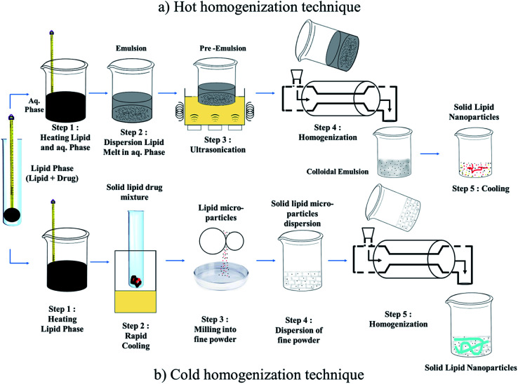
HPH is a technique in which high pressure (100 to 2000 bar) is used to push a liquid or dispersion through a gap of few micrometers to produce submicron size particles. A high shear stress and cavitational forces break down the particles, resulting in a decrease in particle size. HPH can be performed either at high temperature or below room temperature, called hot-HPH and cold-HPH, respectively (Fig. 3).70 In the first step of both processes, the lipid(s) and drug(s) are heated to about 5–10 °C higher than the melting point of the lipid so that the drug is dissolved or dispersed in the melted lipid.71 Generally, the concentration range of lipid is between 5% to 20% w/v. In the second step of the HPH technique, the aqueous phase containing the amphiphile molecules is added to the lipid phase (at the same temperature as the lipid melting) and the hot pre-emulsion is obtained using a high-speed stirring device. The lipid (more added for homogenization) is forced at high pressure (100–1000 bar) through a narrow space (few μm) for 3–5 times, which depends on the formulation and required product. Before homogenization the drug is dispersed or dissolved in the lipid melt. However, there are certain drawbacks to this method as follows: (1) it cannot be used for heat-sensitive drugs because of their degradation and (2) an increase in the number of rotations or pressure of homogeneity often results in an increase in particle size.72 However, these limitation can be overcome using cold-HPH to prepare SLNs. As discussed earlier, the first step involves the formation of a suspension of melting lipids and drugs, followed by rapid cooling in dry ice and liquid nitrogen. In the third step, the powder is converted into micro-particles by milling. Then, the micro-particles are dissolved cold aqueous surfactant solution. In the last step, to create SLNs, homogenization is usually performed for 5 cycles at 500 bars.73
Fig. 3. Homogenization technique: (a) Hot homogenization technique and (b) Cold homogenization technique.
2. Oil/water (o/w) microemulsion breaking technique
This method was invented by Gasco, as shown in Chart 2. Firstly, the microemulsion is prepared by mixing the lipid melt with the drug, surfactant and co-surfactant mixture preheated to a temperature equal to the melting point of the lipid, and then the obtained microemulsion is dispersed in water at a temperature between 2–10 °C.
Chart 2. Preparation of solid lipid nanoparticles by oil/water (o/w) microemulsion method.
3. Solvent-emulsification diffusion technique74
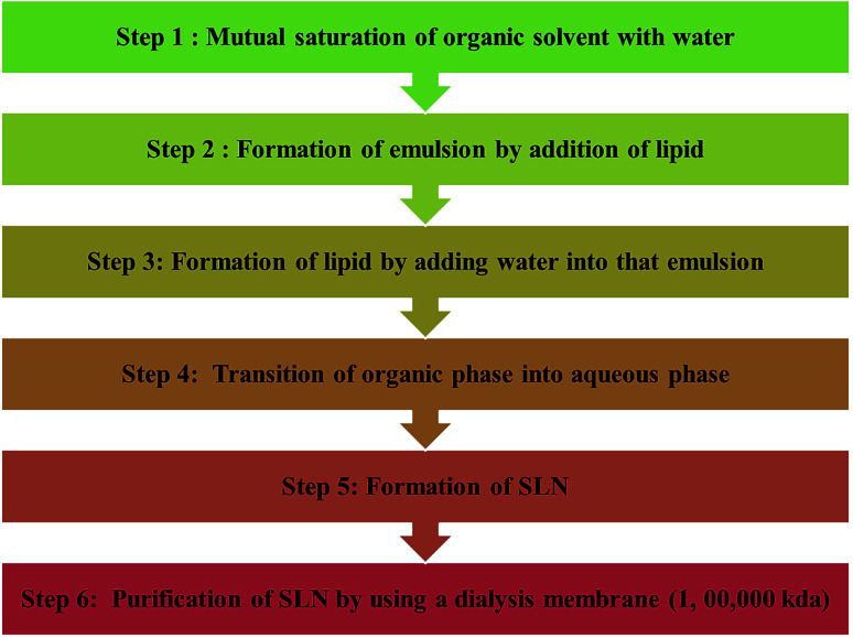
Chart 3 shows the solvent-emulsification diffusion technique for the synthesis of solid lipid nanoparticles. In this method, the lipid is dissolved in an organic solvent saturated with water, and the obtained solution is further emulsified with water and saturated with organic solvent with constant stirring. Lipid nanoparticles are obtained by adding water to the prepared emulsion, which later results in the diffusion of the organic phase into the continuous phase. The SLN dispersion can be purified by ultra-filtration using a dialysis membrane with a cut-off of approximately 100 000 kDa (Chart 4).
Chart 3. Solvent-emulsification diffusion technique for the synthesis of solid lipid nanoparticles.
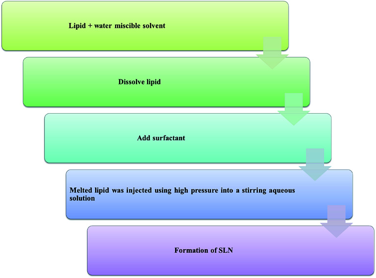
Chart 4. Solvent injection method for the synthesis of solid lipid nanoparticles.
4. Solvent injection method75
In this method the lipids are dissolved in a water-miscible solvent and the dissolved lipids are injected through an injection needle into a stirring aqueous solution with or without surfactant. The parameters of the process for the synthesis of nanoparticles in this method include the nature of the injected solvent, lipid concentration, injected amount of lipid solution, viscosity and the diffusion of the lipid solvent phase into the aqueous phase.
5. Water/oil/water (w/o/w) double emulsion method76
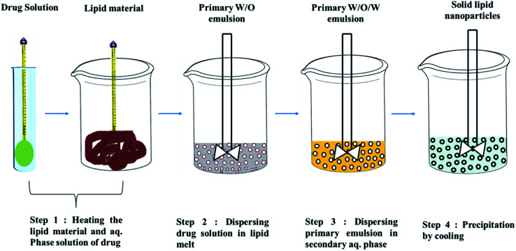
Fig. 4 shows the double emulsion technique to prepare SLNs. This method is mainly used for the preparation SLNs loaded with hydrophilic drugs and some biological molecules such as peptides and insulin.77 SLNs are produced from w/o/w multiple emulsions via the solvent in water emulsion diffusion technique, insulin is dissolved in the inner acidic phase of the w/o/w multiple emulsion and lipids dissolved in the water-miscible organic phase, and then SLNs are produced by diluting the w/o/w emulsion in water. This results in the diffusion of the organic solvent into the aqueous phase and precipitation of the SLNs. The nature of the solvent and interaction of the hydrophilic drug with the solvent and excipients affect the preparation process using this method.
Fig. 4. w/o/w double emulsion technique for the preparation of solid lipid nanoparticles.
6. Ultrasonication76
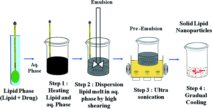
This method is based on the principle of particle size reduction by applying sound waves. In this method, homogenization with high pressure and ultrasonication are simultaneously used to prepare SLNs with a size in the range of 80–800 nm (Fig. 5).
Fig. 5. Ultrasonication technique for the preparation of solid lipid nanoparticles.
Some other advanced techniques have also been introduced to formulate SLNs.
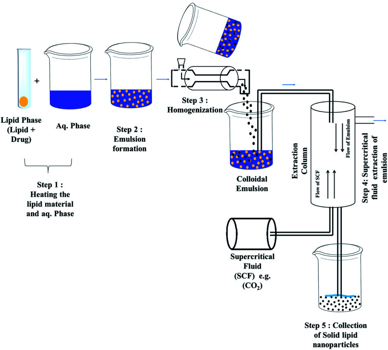
7. Super critical fluid technique78
Super critical carbon dioxide tends to dissolve lipophilic drugs, and combined with the ultrasonication technique, can be used to prepare SLNs. Xionggui-loaded SLNs have been prepared using super critical carbon dioxide fluid extraction and ultrasonication (Fig. 6).
Fig. 6. Super critical fluid technique for the preparation of solid lipid nanoparticles.
8. Membrane contractor technique79
In this method, a membrane contactor is used to prepare SLNs, where a lipid is pressed at a temperature above its melting point through the membrane pores, and water circulated beyond the pores flow with the produced droplets of melted lipid, which is further cooled at room temperature.
9. Electrospray technique80
It is the recent novel technique for the preparation of SLNs, electrodynamic atomization is used to produce narrowly dispersed spherical SLNs less than 1 μm size. In this method, SLNs are directly obtained in powder form.
10. Preparation of semisolid solid lipid nanoparticles
A more effective and faster single-step process was developed for the production of SLNs, especially semisolid formulations. The process is performed by melting a lipid and then dispersing it in hot surfactant solution whose temperature is ca. 10 °C above its melting point and rotated at 9500 rpm for 1 min. Three cycles of dispersion are then performed at 85 °C and 500 bar pressure. After the completion of the first cycle, the dispersion becomes viscous and is further used for the remaining two cycles. Finally, the hot viscous nanoemulsion is cooled at room temperature. The lipid droplets recrystallize and form a gel network, and therefore the SLNs become semi-solid compatible. A 30–50% w/v lipid concentration is required for this process.81
The conversion of liquid lipid nanoparticles into a solid plays a pivotal role in enhancing the stability and safe storage of drug delivery systems. Besides spray drying, lyophilization is also suitable for converting nanolipid dispersions into dry, solid particles. Among these techniques, spray drying is cost-effective and can be used beneficially for large-scale purposes. Spray drying of lipid nanoparticles is a very sensitive process since low melting temperature lipids are used in the formulation. Some studies36,41 demonstrated the use of an organic solvent to reduce the processing temperature and facilitate the drying of heat-sensitive materials. The removal of organic solvents from the lipid nanoparticle matrix again requires exposure to high temperatures, which is not always advisable.
Effect of lipids and surfactants
The process for the production of SLNs is not responsible for any chemical instability. Obviously, the concentration of lipid used may be the special consideration that can alter the stability. It is reported that the maximum lipid degradation is around 10% over 2 years of storage, and SLNs prepared with triglycerides are more stable than SLNs prepared with mono and diglycerides.82 The melting point of the lipid is also a point of discussion regarding the particle size distribution.83 Thus, for the preparation of SLNs, lipids that do not undergo hydrolyzation with the aqueous phase should be chosen, and for SLNs prepared from natural lipids, the addition of preservatives can stabilize the microbial contamination.84 Amphiphilic molecules such as surfactants and block copolymers are used as stabilizing agents, emulsifier, and co-emulsifiers in the preparation of SLNs. Some examples including phospholipids (tricaprin),85 ethylene oxide or propylene oxide copolymers (poloxamer 188 or Pluronic® 68),86 sorbitan ethylene oxide or propylene oxide copolymers (Tween 80 and Tween 20),87,88 bile salts (sodium taurocholate)89 and others are listed in Table 2.
SLN formulations reported by different researchers.
| Drug | Lipid | Surfactant/emulsifier | Co-Surfactant | Method for preparation of SLNs | Techniques for characterization of SLNs | Size (nm) | Ref. |
|---|---|---|---|---|---|---|---|
| Amphotericin B | Compritol® ATO 888, Precirol ATO 5 and stearic acid, | Pluronic® F-68, Pluronic® F-127, | Solvent diffusion method | DLS, DSC, zeta potential | 111–415.8 | 37 | |
| Compritol® ATO 888 (glycerylbehenate), glycerylpalmitostearate (Precirol® ATO 5), medium chain triglyceride | Tween 20, Pluronic® F-127, Cremophor RH40, polyoxyethylene (40) stearate (Myrj 52) | HPH | DLS, zeta potential, HPLC, TEM, FTIR, DSC, PXRD, 1H NMR | 90–260 | 38 | ||
| Baclofen | Stearic acid | Epikuron 200 (92% phosphatidylcholine) | Propionic acid, butyric acid, and sodium taurocholate | Multiple (w/o/w) warm, microemulsion | DLS | 161.4 | 39 |
| BuspironeHCl | Cetyl alcohol, Spermaceti | Pluronic® F-68, Tween 80 | Emulsification-evaporation followed by ultrasonication | DLS | 86–123 | 40 | |
| Camptothecin | Soybean lecithin, stearic acid | Pluronic® F-68, Tween 80 | Glycerol, PEG 400, PPG | Hot HPH | TEM | 196.8 | 41 |
| Carvedilol | Stearic acid | Pluronic® F-68 | Sodium taurocholate and ethanol | Microemulsion | TEM, DLS | 120–200 and 600–800 | 42 |
| Clozapine | Trimyristin, tripalmitin, tristearin, soy phosphatidylcholine | Pluronic® F-68 | Ultrasonication method | DLS, zeta potential | 96.7 ± 3.8 to 163.3 ± 0.7 | 43 | |
| Crypto-Tanshinone | Glycerylmonostearate, Compritol 888 ATO | Soy lecithin, Tween 80, sodium dehydrocholate | Ultrasonic and high-pressure homogenization method | TEM, DLS, DSC | 121.4 ± 6.3 and 137.5 ± 7.1 | 44 | |
| Curcumin | Compritol 888 ATO | Soy lecithin, Tween 80 | Microemulsion | DLS, TEM | 134.6, 40–120 | 45 | |
| Tristearin | Polyoxyethylene (10) stearyl ether (Brij®S10), polyoxyethylene (100) stearyl ether (Brij® S100) | Oil-in-water emulsion technique | PCS, zeta potential | 111–350 | 45 | ||
| Cyclosporine A | Imwitor® 900 | Tagat®S, sodium cholate | HPH, hot HPH | DLS | 157, 143 | 46 and 47 | |
| Diazepam | Compritol 888 ATO, Imwitor® 900 | Pluronic® F-68, Tween 80 | Ultrasound techniques modified high-shear homogenization and | TEM | <500 | 48 | |
| Doxorubicin hydrochloride | Glycerylcaprate | Polyethylene glycol 660 hydrox-ystearate (Solutol®HS15) | Ultrasonic homogenization | DLS, zeta potential, DSC | 199 | 49 | |
| Fenofibrate | Vitamin E TPGS, Vitamin E 6–100 | Hot HPH | DLS | 58 | 50 | ||
| Hydrocortisone | Precirol® ATO 5, Compritol® 888 ATO, Rylo TM MG 14 Pharma, Dynasan® 114 Dynasan® 118, Tegin® 4100 | Tween 80 | Hot high pressure homogenization | DLS, DSC | 150–220 | 51 | |
| Ibuprofen | Trilaurin, tripalmitin, stearic acid | Pluronic®F127, sodium taurocholate | Solvent-free high-pressure homogenization (HPH) | DLS, X-ray powder diffraction, DSC, AFM | 111–121 (empty SLN) 175–189 (loaded sample) | 52 | |
| Idarubicin | Stearic acid | Epikuron 200 (soy phosphatidylcholine 95%) | Taurocholate sodium salt | Microemulsion | PCS, 90 PLUS | 80 ± 10((loaded sample)) | 53 |
| Emulsifying wax | Polyoxyl 20-stearyl ether (Brij 78), D-alpha-tocopheryl polyethylene glycol succinate (vitamin E TPGS),DSPE-PEG3000 | Sodium taurodeoxycholate (STDC), sodium tetradecylsulfate (STS) | PCS, Zetasizer nano Z | 94.4 (blank), 80–104 (loaded sample) | 54 | ||
| Ketoprofen | Beeswax and carnauba wax | Tween 80, egg lecithin | Microemulsion technique | PCS, DSC | 65–250 (loaded sample) | 55 | |
| Lopinavir | Compritol 888 ATO (glycerylbehenate) | Pluronic®F127 | Hot homogenization, ultrasonication | DLS, zeta potential, HPLC, DSC, WAXS, AFM | 230 | 56 | |
| Lovastatin | Triglyceride, and phosphatidylcholine 95% | Pluronic®F68 | Hot homogenization ultrasonication | DLS, HPLC, DSC, PXRD, LC-MS/MS | 60–119 | 57 | |
| Methotrexate | Stearic acid, monostearin, tristearin, and Compritol 888 ATO | l-α-Soya lecithin, and Sephadex G-50 | Solvent diffusion method | DLS, zeta potential, TEM | 120–167 | 58 | |
| Nevirapine | Steric acid, Compritol 888 ATO | Dimethyldioctadecyl ammonium bromide (DODAB), Tween 80, Lecithin | 1-Butanol | Microemulsion | DLS, zeta potential, field emission scanning electron microscopy (FE-SEM), DSC | 153.1 | 59 |
| Nitrendipine | triglyceride and phosphatidylcholine | Pluronic®F68 | Hot homogenization ultrasonication method | DLS, zeta potential, scanning electron microscopy (SEM) | 110–140 | 60 | |
| Octadecylamine-fluorescein isothiocyanate | Stearic acid | Otcadecylamine, polyethylene glycol monostearate (PEG2000-SA) | Solvent diffusion | DLS, zeta potential | 203 | 61 | |
| Pentoxifylline | Stearic acid, cetyl alcohol, soy lecithin, | Tween 20, Pluronic F®68 | Homogenization followed by the ultrasonication | DLS, zeta-potential | 255–4000 | 62 | |
| Praziquantel | Hydrogenated castor oil | Poly vinyl alcohol (PVA) | Hot homogenization and ultrasonication | DLS, zeta-potential, SEM | 344.0 | 63 | |
| Puerarin | Monostearin, and soy lecithin | Pluronic F®68 | Solvent injection method | DLS and zeta-potential | 160 | 64 | |
| Quercetin | Glycerylmonostearat, soy lecithin | Tween-80 and PEG 400 | Emulsification-solidification | DLS, zeta-potential, TEM | 65 | ||
| Rifampicin | Stearic acid | PVA | Emulsion-solvent diffusion | 66 | |||
| Tobramycin | Stearic acid | Epikuron 200 | Sodium taurocholate | Microemulsion | DLS, TEM | 70–100 | 67 |
| Vinpocetine | Glycerylmonostearat, soy lecithin, polyoxyethylene hydrogenated castor oil | Tween 80 | Ultrasonic-solvent emulsification | DLS, TEM | 70–170 | 68 |
The general mechanism of all these surfactants is to reduce the interfacial tension between the lipid and aqueous phase by applying their amphiphilic nature. Previous studies have shown that the use of surfactant together with a co-surfactant is likely to result in a smaller particle size.90 Recrystallization of the lipid phase results in the rapid growth of particle size, and thereby the long-term stability of the aqueous SLN dispersion is reduced.91 The surfactant structure and interaction between lipid molecules are also responsible for the crystallization process,92 and thus the impact of the lipid and surfactant with or without a co-surfactant and their concentration are significant.
SLN characterization
Physical and chemical characterization are also required after the preparation of SLNs. Due to the particle size, complexity and dynamic nature of the delivery system, the characterization of SLNs is a serious challenge. The parameters needed to evaluate SLNs include particle size, zeta potential, degree of crystallinity, drug release, entrapment efficiency (% EE) and surface morphology. Particle size, polydispersity index and charge analysis can be measured by photon correlation spectroscopy (PCS), dynamic light scattering (DLS) and quasi-elastic light scattering (QELS).93 The main advantage of these techniques is that they are not time-consuming, with speedy analysis and high sensitivity.94 The crystallinity of lipid or polymorphic modifications can be analyzed via differential scanning calorimetric analysis (DSC).95 The crystallinity within nanoparticles is measured by the function of the glass and melting point temperature associated with the enthalpies. Nuclear magnetic resonance (NMR) can also be used to determine the size and qualitative nature of nanoparticles. Changes in their chemical shift are related to the molecular dynamics, which provide information about the physicochemical state of the constituents inside the nanoparticles. Electron microscopy is an advanced technique that can offer a direct way of observing nanoparticles. The size, surface topography, stability and structural changes of SLNs with time can be better investigated using scanning electron microscopy (SEM) and transmission electron microscopy (TEM). However, cryo-microscopic analysis involves rapid freezing, and thus the specimen is preserved in its hydrated state. Cryoelectron microscopy (cryo-EM) such as cryo-TEM and cryo-FESEM provides 3D images of stable frozen-hydrated particles.96 Atomic force microscopy (AFM) is more advanced than TEM and SEM. This method allows atomic-level resolution to be accessed together with size, colloidal attraction and resistance to deformation, making AFM an important tool. The surface distribution of surfactant molecules, bio-conjugation confirmation in case of cationic SLNs, and functionalization of nanoparticles can be estimated by X-ray photoelectron spectroscopy (XPS).97,98 SLN entrapment can be measured by either centrifugation or micro-centrifugation techniques. The samples are centrifuged at high rpm, and the amount of free compound is determined by UV-Visible spectroscopy or high-performance liquid chromatography in a clear supernatant.99–101 The drug loading and release profile or release kinetics of SLNs depend on the crystalline state and melting behavior of the lipid.102
Pharmacological performance of SLNs
Nanoparticles for drug delivery or nanotechnology-induced drug delivery systems are going to be the most innovative and crucial cornerstones in the pharmaceutical research area with a great economic impact.103 Gradually, novel SLNs will be widely accepted pharmaceutical carriers for drug delivery to a specific site with increasing interest and improved pharmacokinetic profiles compared to traditional drug delivery.104 Targeted dug delivery, oral administration, topical administration, cosmetics, intravenous administration, protein peptide delivery and ocular delivery are the areas covered by SLNs.84,105–113 Targeting the brain for the successful delivery of pharmaceutical actives is a challenging part of NDDS since 98% of drugs cannot cross the blood brain barrier (BBB).114 Accordingly, SLNs demonstrate a potential approach due to their lipid behavior and effective nanometer size range for targeted drug delivery.
Stability issue and storage conditions of SLNs
It has already been reported that SLNs are stable for more than three years. The stability of SLNs is mainly associated with their lipid material, surfactant concentration, and temperature optimization during their preparation. Thus, all these parameters should be considered for their stability and storage. Triglycerides undergo α (alpha), β (beta) and β′ (beta prime) crystal modification during their preparation and storage.115 The kinetics of their polymorphic transitions largely depend on their chain length, where the crystallization process is slower for longer chain than shorter chain triglycerides.116 Sometimes SLNs undergo gel formation, and their gelling tendency strongly depends upon β′ modification due to exposure to light, temperature and shear force.7 Also, the size of the particles can vary because of exposure to light.117
In a study, SLNs were exposed to various destabilizing factors, and it was found that gelation occurred and their zeta potential decreased.118 However, SLNs have several stability issues and the drug may be hydrolyzed in aqueous dispersion. Thus, drying is a necessary option for the prolonged storage of SLNs. Freeze drying, spray drying, and lyophilization are techniques for drying. Recently, the electrospray method was employed to prepare SLNs, where a dry SLN powder was obtained directly.119 The formulation of SLNs in a powder form, which may be loaded into pellets, capsules, or tablets, makes these materials highly advantageous for drug delivery. On the other hand, the applications of SLN formulations may be restricted due to their uncontrolled particle growth through coagulation or agglomeration, generating very swift “burst release” of the drug.112 SLNs possess perfect crystal lipid matrices, which carry the loaded drug in its molecular form between fatty acid chains.76,83 The formation and uncontrolled, unwanted enhancement of the crystal structure during both the production and storage of SLNs often result in the release of the loaded drug solution, which is a huge drawback of SLNs.98
Applications in drug delivery
SLNs have been widely applied for various medical applications due to their flexible surface topology and versatile properties (Table 2).
Novel theranostic approach
Recently, the emerging trends of nanoparticulate drug delivery systems include nanotherapeutics with diagnostic imaging on the same platform based on image-guided drug delivery to track the pharmacokinetics and biodistribution of the therapeutic agent in real time. Thus, the in vivo theranostics approach can be a great dimensional change in drug delivery systems and diagnostic imaging.120 Image-guided drug delivery systems include the combination of disease diagnosis and therapy, bio-distribution tracking, drug distribution at the target site, drug response prediction, drug efficacy, monitoring and quantification.121 Nanotheranostics or drug delivery together with diagnostic imaging using nanoparticles has a great impact on localizing the target site, and the disease-specific targeting of active pharmaceutical ingredients can also be monitored.122 Since nanoparticles possess dimensions similar to that of various biomolecules such as DNA, RNA, and proteins, they can play a crucial role in surgery together with drug delivery and imaging.123 Nowadays, polymeric nanoparticles, liposomal drug delivery systems, dendrimers and silica nanoparticles are used in the theranostics approach.124–128 Furthermore, pH-triggered nanoparticles, and magnetic and photo-responsive theranosomes are also included in image-guided drug delivery systems for cancer therapy.129,130 Also, SLNs inserted with prostacyclin (PGI2) can be used for the image-guided treatment of atherosclerosis by inhibiting platelet aggregation.131
Lipid vesicles can be used as a theranostics platform for non-invasive drug delivery and imaging, and since SLNs are lipid nanovesicles, they have potential for application in the theranostic approach.
Conclusion
Solid lipid nanoparticles are colloidal dispersions with modified properties of other nanoparticles such as microemulsions, suspensions, liposomes and polymeric nanoparticles. The major problems encountered with nanoparticles can be successively avoided using SLNs, and finally a chemically stable and physiologically suitable drug delivery system can be achieved with less limitations. Only their gelation tendency seems to be the main problem, but nanostructured lipid carriers are a possible way to overcome this problem. In addition, the active component, i.e. the drug, may be degraded during their production based on the hot homogenization method because of the generated heat and stress. Thus, choosing an appropriate production method is crucial. Several other difficulties such as particle size, coexistence of various colloidal forms, different shapes and drug ejection from the lipid matrix also need to be addressed.132 The various well-established methods for the bulk production of the SLN matrix and its characterization were discussed. Drugs with physicochemical incompatibility, lower pharmacokinetic profile, and thermolabile drugs can be delivered to the target site via SLNs. Protein and peptide delivery with a higher degree of efficiency and lower toxicity can also be achieved with SLNs. Thus, the addition of the theranostics approach with SLNs can take therapeutics and diagnostics in a new direction.
Conflicts of interest
The authors declare no competing interests.
Supplementary Material
Acknowledgments
This work was supported by National Natural Science Foundation of China (Project No. 81903623), the China Postdoctoral Science Foundation (No. 2019M652586), and the Postdoctoral Research Grant in Henan Province (Project No. 19030008).
References
- Mycek M. J., Harvey R. A. and Champe P. C., Pharmacology, Lippincott Williams & Wilkins, Philadelphia, P.A., 2000 [Google Scholar]
- Muller R. H. Mader K. Gohla S. Eur. J. Pharm. Biopharm. 2000;50:161–177. doi: 10.1016/S0939-6411(00)00087-4. [DOI] [PubMed] [Google Scholar]
- Salunkhe S. S. Bhatia N. M. Bhatia M. S. Drug Deliv. 2016;23:1306–1316. doi: 10.3109/10717544.2014.943337. [DOI] [PubMed] [Google Scholar]
- Scheffel U. Rhodes B. A. Natajaran T. K. Wagner H. N. J. Nucl. Med. 1970;13:498–503. [PubMed] [Google Scholar]
- Jumaa M. Muller B. W. Eur. J. Pharm. Sci. 2000;9:285–290. doi: 10.1016/S0928-0987(99)00071-8. [DOI] [PubMed] [Google Scholar]
- Gohla S. H. Dingler A. Pharmazie. 2001;56:61–63. [PubMed] [Google Scholar]
- Freitas C. Müller R. H. Eur. J. Pharm. Biopharm. 1999;47:125–132. doi: 10.1016/S0939-6411(98)00074-5. [DOI] [PubMed] [Google Scholar]
- Lockman P. R. Oyewumi M. O. Koziara J. M. Roder K. E. Mumper R. J. Allen D. D. J. Controlled Release. 2003;93:271–282. doi: 10.1016/j.jconrel.2003.08.006. [DOI] [PubMed] [Google Scholar]
- Schwarz C. J. Microencapsul. 1999;16:205–213. doi: 10.1080/026520499289185. [DOI] [PubMed] [Google Scholar]
- Kammari R., Das N. G. and Das S. K., Emerging Nanotechnologies for Diagnostics, Drug Delivery and Medical Devices Micro and Nano Technologies, 2017, pp. 105–144 [Google Scholar]
- Mehnert W. Mader K. Adv. Drug Delivery Rev. 2001;47:165–196. doi: 10.1016/S0169-409X(01)00105-3. [DOI] [PubMed] [Google Scholar]
- Mehnert W. Mader K. Adv. Drug Deliv. Rev. 2001;47:165–196. doi: 10.1016/S0169-409X(01)00105-3. [DOI] [PubMed] [Google Scholar]
- Lim S. B. Banerjee A. Önyüksel H. J. Controlled Release. 2012;163:34–45. doi: 10.1016/j.jconrel.2012.06.002. [DOI] [PubMed] [Google Scholar]
- Dhakad R. S. Tekade R. K. Jain N. K. Curr. Drug Delivery. 2013;10:477–491. doi: 10.2174/1567201811310040012. [DOI] [PubMed] [Google Scholar]
- Yang S. C. Lu L. F. Cai Y. Zhu J. B. Liang B. W. Yang C. Z. J. Controlled Release. 1999;59(3):299–307. doi: 10.1016/S0168-3659(99)00007-3. [DOI] [PubMed] [Google Scholar]
- Gustafson H. H. Holt-Casper D. Grainger D. W. Ghandehari H. Nano Today. 2015;10:487–510. doi: 10.1016/j.nantod.2015.06.006. [DOI] [PMC free article] [PubMed] [Google Scholar]
- Bocca C. Caputo O. Cavalli R. Gabriel L. Miglietta A. Gasco M. R. Int. J. Pharm. 1998;175(2):185–193. doi: 10.1016/S0378-5173(98)00282-8. [DOI] [Google Scholar]
- Zara G. P. Cavalli R. Fundaro A. Bargoni A. Caputo O. Gasco M. R. Pharmacol. Res. 1999;40:281–286. doi: 10.1006/phrs.1999.0509. [DOI] [PubMed] [Google Scholar]
- Cavalli R. Peira E. Caputo O. Gasco M. R. Int. J. Pharm. 1999;182:59–69. doi: 10.1016/S0378-5173(99)00066-6. [DOI] [PubMed] [Google Scholar]
- Freitas C. Müller R. H. Eur. J. Pharm. Biopharm. 1999;47:125–132. doi: 10.1016/S0939-6411(98)00074-5. [DOI] [PubMed] [Google Scholar]
- Miglietta A. Cavalli R. Bocca C. Gabriel L. Gasco M. R. Int. J. Pharm. 2000;210:61–67. doi: 10.1016/S0378-5173(00)00562-7. [DOI] [PubMed] [Google Scholar]
- Dianzani C. Zara G. P. Maina G. Pettazzoni P. Pizzimenti S. Rossi F. Gigliotti C. L. Ciamporcero E. S. Daga M. Barrera G. Drug delivery nanoparticles in skin cancers. BioMed Res. Int. 2014:895–986. doi: 10.1155/2014/895986. [DOI] [PMC free article] [PubMed] [Google Scholar]
- Jenning V. Gysler A. Schäfer-Korting M. Gohla S. H. Eur. J. Pharm. Biopharm. 2000;49:211–218. doi: 10.1016/S0939-6411(99)00075-2. [DOI] [PubMed] [Google Scholar]
- Muller-Goymann C. C. Eur. J. Pharm. Biopharm. 2004;58:343–356. doi: 10.1016/j.ejpb.2004.03.028. [DOI] [PubMed] [Google Scholar]
- Schafer-Korting M. Mehnert W. Korting H.-C. Adv. Drug Delivery Rev. 2007;59:427–443. doi: 10.1016/j.addr.2007.04.006. [DOI] [PubMed] [Google Scholar]
- Muller R. H. Radtke M. Wissing S. A. Adv. Drug Delivery Rev. 2002;54:S131–S155. doi: 10.1016/S0169-409X(02)00118-7. [DOI] [PubMed] [Google Scholar]
- Jenning V. Gysler A. Schafer-Korting M. Gohla S. H. Eur. J. Pharm. Biopharm. 2000;49:211–218. doi: 10.1016/S0939-6411(99)00075-2. [DOI] [PubMed] [Google Scholar]
- Jenning V. Schafer-Korting M. Gohla S. J. Controlled Release. 2000;66:115–126. doi: 10.1016/S0168-3659(99)00223-0. [DOI] [PubMed] [Google Scholar]
- Wissing S. A. Muller R. H. Adv. Drug Delivery Rev. 2001;23:233–243. [Google Scholar]
- Wissing S. A. Lippacher A. Muller R. H. J. Cosmet. Sci. 2001;52:313–323. [PubMed] [Google Scholar]
- Olbrich C. Bakowsky U. Lehr C. M. Müller R. H. Kneuer C. J. Controlled Release. 2001;77:345–355. doi: 10.1016/S0168-3659(01)00506-5. [DOI] [PubMed] [Google Scholar]
- Gaspar D. P. Vasco F. Goncalves L. M. D. Taboad P. Remunan-Lopez C. Almeida A. Int. J. Pharm. 2016;497:199–209. doi: 10.1016/j.ijpharm.2015.11.050. [DOI] [PubMed] [Google Scholar]
- Tabatt K. Sameti M. Olbrich C. Müller R. H. Lehr C. M. Eur. J. Pharm. Biopharm. 2004;57:155–162. doi: 10.1016/j.ejpb.2003.10.015. [DOI] [PubMed] [Google Scholar]
- Wissing S. A. Muller R. H. Int. J. Pharm. 2003;254:65–68. doi: 10.1016/S0378-5173(02)00684-1. [DOI] [PubMed] [Google Scholar]
- Wissing S. A. Muller R. H. J. Pharmacokinet. Biopharm. 2003;56:67–72. doi: 10.1016/S0939-6411(03)00040-7. [DOI] [PubMed] [Google Scholar]
- Kumar S. Randhawa J. K. Mater. Sci. Eng., C. 2013;33:1842–1852. doi: 10.1016/j.msec.2013.01.037. [DOI] [PubMed] [Google Scholar]
- Butani D. Yewale C. Misra A. Colloids Surf., B. 2016;139:17–24. doi: 10.1016/j.colsurfb.2015.07.032. [DOI] [PubMed] [Google Scholar]
- Jansook P. Fulop Z. Ritthidej G. C. Drug Dev. Ind. Pharm. 2019;45:560–567. doi: 10.1080/03639045.2019.1569023. [DOI] [PubMed] [Google Scholar]
- Priano L. Zara G. P. El-Assawy N. Cattaldo S. Muntoni E. Milano E. Serpe L. Musicanti C. Pérot C. Gasco M. R. Eur. J. Pharm. Biopharm. 2011;79:135–141. doi: 10.1016/j.ejpb.2011.02.009. [DOI] [PubMed] [Google Scholar]
- Varshosaz J. Tabbakhian M. Mohammadi M. Y. J. Liposome Res. 2010;20:286–296. doi: 10.3109/08982100903443065. [DOI] [PubMed] [Google Scholar]
- Yang S. Zhu J. Lu Y. Liang B. Yang C. Pharm. Res. 1999;16:751–757. doi: 10.1023/A:1018888927852. [DOI] [PubMed] [Google Scholar]
- Sanjula B. Shah F. M. Javed A. Alka A. J. Drug Target. 2009;17:249–256. doi: 10.1080/10611860902718672. [DOI] [PubMed] [Google Scholar]
- Manjunath K. Venkateswarlu V. J. Controlled Release. 2005;107:215–228. doi: 10.1016/j.jconrel.2005.06.006. [DOI] [PubMed] [Google Scholar]
- Hu L. Xing Q. Meng J. Shang C. AAPS PharmSciTech. 2010;11:582–587. doi: 10.1208/s12249-010-9410-3. [DOI] [PMC free article] [PubMed] [Google Scholar]
- Kakkar V. Singh S. Singla D. Kaur I. P. Mol. Nutr. Food Res. 2011;55:495–503. doi: 10.1002/mnfr.201000310. [DOI] [PubMed] [Google Scholar]
- Muller R. Runge S. Ravelli V. Mehnert W. Thunemann A. Souto E. Int. J. Pharm. 2006;317:82–89. doi: 10.1016/j.ijpharm.2006.02.045. [DOI] [PubMed] [Google Scholar]
- Muller R. Runge S. Ravelli V. Thunemann A. Mehnert W. Souto E. Eur. J. Pharm. Biopharm. 2008;68:535–544. doi: 10.1016/j.ejpb.2007.07.006. [DOI] [PubMed] [Google Scholar]
- Abdelbary G. Fahmy R. H. AAPS PharmSciTech. 2009;10:211–219. doi: 10.1208/s12249-009-9197-2. [DOI] [PMC free article] [PubMed] [Google Scholar]
- Subedi R. K. Kang K. W. Choi H.-K. Eur. J. Pharm. Sci. 2009;37:508–513. doi: 10.1016/j.ejps.2009.04.008. [DOI] [PubMed] [Google Scholar]
- Hanafy A. Spahn-Langguth H. Vergnault G. Grenier P. Grozdanis M. T. Lenhardt T. Langguth P. Adv. Drug Delivery Rev. 2007;59:419–426. doi: 10.1016/j.addr.2007.04.005. [DOI] [PubMed] [Google Scholar]
- Jensen L. B. Magnussson E. Gunnarsson L. Vermehren C. Nielsen H. M. Petersson K. Int. J. Pharm. 2010;390:53–60. doi: 10.1016/j.ijpharm.2009.10.022. [DOI] [PubMed] [Google Scholar]
- Potta S. G. Minemi S. Nukala R. K. Peinado C. Lamprou D. A. Urquhart A. Douroumis D. J. Microencapsul. 2011;28:74–81. doi: 10.3109/02652048.2010.529948. [DOI] [PubMed] [Google Scholar]
- Zara G. P. Bargoni A. Cavalli R. Fundarò A. Vighetto D. Gasco M. R. J. Pharm. Sci. 2002;91:1324–1333. doi: 10.1002/jps.10129. [DOI] [PubMed] [Google Scholar]
- Ma P. Dong X. Swadley C. L. Gupte A. Leggas M. Ledebur H. C. Mumper R. J. J. Biomed. Nanotechnol. 2009;5:151–161. doi: 10.1166/jbn.2009.1021. [DOI] [PMC free article] [PubMed] [Google Scholar]
- Kheradmandnia S. Vasheghani-Farahani E. Nosrati M. Atyabi F. Nanomed. Nanotechnol. Biol. Med. 2010;6:753–759. doi: 10.1016/j.nano.2010.06.003. [DOI] [PubMed] [Google Scholar]
- Alex M. A. Chacko A. Jose S. Souto E. Eur. J. Pharm. Sci. 2011;42:11–18. doi: 10.1016/j.ejps.2010.10.002. [DOI] [PubMed] [Google Scholar]
- Suresh G. Manjunath K. Venkateswarlu V. Satyanarayana V. AAPS PharmSciTech. 2007;8:E162–E170. doi: 10.1208/pt0801024. [DOI] [PMC free article] [PubMed] [Google Scholar]
- Paliwal R. Rai S. Vaidya B. Khatri K. Goyal A. K. Mishra N. Mehta A. Vyas S. P. Nanomed. Nanotechnol. Biol. Med. 2009;5:184–191. doi: 10.1016/j.nano.2008.08.003. [DOI] [PubMed] [Google Scholar]
- Kuo Y.-C. Chung J.-F. Colloids Surf., B. 2011;83:299–306. doi: 10.1016/j.colsurfb.2010.11.037. [DOI] [PubMed] [Google Scholar]
- Manjunath K. Venkateswarlu V. J. Drug Targeting. 2006;14:632–645. doi: 10.1080/10611860600888850. [DOI] [PubMed] [Google Scholar]
- Yuan H. Chen J. Du Y.-Z. Hu F.-Q. Zeng S. Zhao H.-L. Colloids Surf., B. 2007;58:157–164. doi: 10.1016/j.colsurfb.2007.03.002. [DOI] [PubMed] [Google Scholar]
- Varshosaz J. Minayian M. Moazen E. J. Liposome Res. 2010;20:115–123. doi: 10.3109/08982100903161456. [DOI] [PubMed] [Google Scholar]
- Xie S. Pan B. Wang M. Zhu L. Wang F. Dong Z. Wang X. Zhou W. Nanomedicine. 2010;5:693–701. doi: 10.2217/nnm.10.42. [DOI] [PubMed] [Google Scholar]
- Luo C.-F. Yuan M. Chen M.-S. Liu S.-M. Zhu L. Huang B.-Y. Liu X.-W. Xiong W. Int. J. Pharm. 2011;410:138–144. doi: 10.1016/j.ijpharm.2011.02.064. [DOI] [PubMed] [Google Scholar]
- Li H. Zhao X. Ma Y. Zhai G. Li L. Lou H. J. Controlled Release. 2009;133:238–244. doi: 10.1016/j.jconrel.2008.10.002. [DOI] [PubMed] [Google Scholar]
- Pandey R. Sharma S. Khuller G. Tuberculosis. 2005;85:415–420. doi: 10.1016/j.tube.2005.08.009. [DOI] [PubMed] [Google Scholar]
- Cavalli R. Bargoni A. Podio V. Muntoni E. Zara G. P. Gasco M. R. J. Pharm. Sci. 2003;92:1085–1094. doi: 10.1002/jps.10368. [DOI] [PubMed] [Google Scholar]
- Luo Y. Chen D. Ren L. Zhao X. Qin J. J. Controlled Release. 2006;114:53–59. doi: 10.1016/j.jconrel.2006.05.010. [DOI] [PubMed] [Google Scholar]
- Gardouh A. R. Gad S. Ghonaim H. M. Ghorab M. M. Br. J. Pharm. Res. 2013;3:326–346. doi: 10.9734/BJPR/2013/2770. [DOI] [Google Scholar]
- Schwarz C. Mehnert W. Lucks J. Müller R. J. Controlled Release. 1994;30:83–96. doi: 10.1016/0168-3659(94)90047-7. [DOI] [Google Scholar]
- Emeje M. O., Akpabio E. I., Obidike I. C. and Ofoefule S. I., Nanotechnology in Drug Delivery. INTECH Open Access Publisher, 2012 [Google Scholar]
- Waghmare A. Grampurohit N. Gadhave M. Gaikwad D. Jadhav S. Int. Res. J. Pharm. 2012;3:100–107. [Google Scholar]
- Shah C. Shah V. Upadhyay U. Curr. Pharma Res. 2011;1:351–368. doi: 10.33786/JCPR.2011.v01i04.009. [DOI] [Google Scholar]
- Trotta M. Debernardi F. Caputo O. Int. J. Pharm. 2003;257:153–160. doi: 10.1016/S0378-5173(03)00135-2. [DOI] [PubMed] [Google Scholar]
- Schubert M. A. Müller-Goymann C. C. Eur. J. Pharm. Biopharm. 2003;55:125–131. doi: 10.1016/S0939-6411(02)00130-3. [DOI] [PubMed] [Google Scholar]
- Naguib Y. W. Rodriguez B. L. Li X. Hursting S. D. Williams III R. O. Cui Z. Mol. Pharmaceutics. 2014;11:1239–1249. doi: 10.1021/mp4006968. [DOI] [PMC free article] [PubMed] [Google Scholar]
- Svilenov H. Tzachev C. Solid lipid nanoparticles–apromising drug delivery system. Nanomedicine. 2014:187–237. [Google Scholar]
- Chen Y. J. Jin R. X. Zhou Y. Q. Zeng J. Zhang H. Feng Q. R. China J. Chin. Mater. Med. 2006;31(5):376–379. [PubMed] [Google Scholar]
- Charcosset C. El-Harati A. Fessi V. J. Controlled Release. 2005;108:112–120. doi: 10.1016/j.jconrel.2005.07.023. [DOI] [PubMed] [Google Scholar]
- Sridhar R. Ramakrishna S. Biomatter. 2013;3:e24281. doi: 10.4161/biom.24281. [DOI] [PMC free article] [PubMed] [Google Scholar]
- Abdel-Salam F. S. Elkheshen S. A. Mahmoud A. A. Ammar H. O. Bull. Fac. Pharm. Cairo Univ. 2016;54:1–7. doi: 10.1016/j.bfopcu.2015.11.002. [DOI] [Google Scholar]
- Radomska-Soukharev A. Adv. Drug Delivery Rev. 2007;59:411–418. doi: 10.1016/j.addr.2007.04.004. [DOI] [PubMed] [Google Scholar]
- Siekmann B. Westesen K. Pharm. Pharmacol. Lett. 1992;1:123–126. [Google Scholar]
- Skotland T. Sagini K. Sandvig K. Llorente A. Adv. Drug Delivery Rev. 2020 doi: 10.1016/j.addr.2020.03.002. [DOI] [PubMed] [Google Scholar]
- Castelli F. Puglia C. Sarpietro M. G. Rizza L. Bonina F. Int. J. Pharm. 2005;304:231–238. doi: 10.1016/j.ijpharm.2005.08.011. [DOI] [PubMed] [Google Scholar]
- Schwarz C. Mehnert W. Int. J. Pharm. 1997;157:171–179. doi: 10.1016/S0378-5173(97)00222-6. [DOI] [PubMed] [Google Scholar]
- Liu J. Hu W. Chen H. Ni Q. Xu H. Yang X. Int. J. Pharm. 2007;328:191–195. doi: 10.1016/j.ijpharm.2006.08.007. [DOI] [PubMed] [Google Scholar]
- Awad T. S. Helgason T. Weiss J. Decker E. A. McClements D. J. Cryst. Growth Des. 2009;9:3405–3411. doi: 10.1021/cg8011684. [DOI] [Google Scholar]
- Cavalli R. Gasco M. R. Chetoni P. Burgalassi S. Saettone M. F. Int. J. Pharm. 2002;238:241–245. doi: 10.1016/S0378-5173(02)00080-7. [DOI] [PubMed] [Google Scholar]
- Cavalli R. Pharmazie. 1998;53:392–396. [Google Scholar]
- Muhlen Z., Feste Lipid Nanopartikel mit prolongierter Wirkstoffliberation. Herstellung, Langzeitstabilitat, Charakterisierung, Freiset zungsverhalten und -mechanismen. PhD thesis, Freie Universitat Berlin, 1996. – from 1992-2 ref. no 2 [Google Scholar]
- Kovacevic A. Savic S. Vuleta G. Müller R. H. Keck C. M. Int. J. Pharm. 2011;406:163–172. doi: 10.1016/j.ijpharm.2010.12.036. [DOI] [PubMed] [Google Scholar]
- Alexander M. Dalgleish D. G. Food Biophys. 2006;1:2–13. doi: 10.1007/s11483-005-9000-1. [DOI] [Google Scholar]
- Mukherjee S. Ray S. Thakur R. S. Indian J. Pharm. Sci. 2009;71:349. doi: 10.4103/0250-474X.57282. [DOI] [PMC free article] [PubMed] [Google Scholar]
- Dale P. J. Kijlstra J. Vincent B. Langmuir. 2005;21:12250–12256. doi: 10.1021/la052141q. [DOI] [PubMed] [Google Scholar]
- Saupe A. Gordon K. C. Rades T. Int. J. Pharm. 2006;314:56–62. doi: 10.1016/j.ijpharm.2006.01.022. [DOI] [PubMed] [Google Scholar]
- Venishetty V. K. Samala R. Komuravelli R. Kuncha M. Sistla R. Diwan P. V. Nanomed. Nanotechnol. Biol. Med. 2013;9:388–397. doi: 10.1016/j.nano.2012.08.004. [DOI] [PubMed] [Google Scholar]
- Venishetty V. K. Komuravelli R. Kuncha M. Sistla R. Diwan P. V. Nanomed. Nanotechnol. Biol. Med. 2013;9:111–121. doi: 10.1016/j.nano.2012.03.003. [DOI] [PubMed] [Google Scholar]
- Jain N. K. Tekade R. K. Drug Deliv. 2013:373–409. doi: 10.2174/1567201811310040012. [DOI] [PubMed] [Google Scholar]
- Kayat J. Gajbhiye V. Tekade R. K. Jain N. K. Nanomedicine. 2011;7:40–49. doi: 10.1016/j.nano.2010.06.008. [DOI] [PubMed] [Google Scholar]
- Tekade R. K. Dutta T. Gajbhiye V. Jain N. K. J. Microencapsul. 2009;26:287–296. doi: 10.1080/02652040802312572. [DOI] [PubMed] [Google Scholar]
- Zur Muhlen A. Schwarz C. Mehnert W. Eur. J. Pharm. Biopharm. 1998;45:149–155. doi: 10.1016/S0939-6411(97)00150-1. [DOI] [PubMed] [Google Scholar]
- Sahoo S. K. Labhasetwar V. Drug discovery Today. 2003;8:1112–1120. doi: 10.1016/S1359-6446(03)02903-9. [DOI] [PubMed] [Google Scholar]
- Fundaro A. Cavalli R. Bargoni A. Vighetto D. Zara G. P. Paolo G. Gasco M. R. Pharmacol. Res. 2000;42:337–343. doi: 10.1006/phrs.2000.0695. [DOI] [PubMed] [Google Scholar]
- Martins S. Tho I. Reimold I. Fricker G. Souto E. Ferreira D. Brandl M. Int. J. Pharm. 2012;439:49–62. doi: 10.1016/j.ijpharm.2012.09.054. [DOI] [PubMed] [Google Scholar]
- Zhang C. Gu C. Peng F. Liu W. Wan J. Xu H. Yang X. Molecules. 2013;18:13340–13356. doi: 10.3390/molecules181113340. [DOI] [PMC free article] [PubMed] [Google Scholar]
- Liu J. Hu W. Chen H. Ni Q. Xu H. Yang X. Int. J. Pharm. 2007;328:191–195. doi: 10.1016/j.ijpharm.2006.08.007. [DOI] [PubMed] [Google Scholar]
- Wissing S. A. Muller R. H. Int. J. Pharm. 2003;254:65–68. doi: 10.1016/S0378-5173(02)00684-1. [DOI] [PubMed] [Google Scholar]
- Carrillo C. Sanchez-Hernandez N. Garcia-Montoya E. Perez-Lozano P. Sune-Negre J. M. Tico J. R. Sune C. Minarro M. Eur. J. Pharm. Sci. 2013;49:157–165. doi: 10.1016/j.ejps.2013.02.011. [DOI] [PubMed] [Google Scholar]
- Del Pozo-Rodríguez A. Delgado D. Solinís M. Á. Pedraz J. L. Echevarría E. Rodríguez J. M. Gascon A. R. Int. J. Pharmacother. 2010;385:157–162. doi: 10.1016/j.ijpharm.2009.10.020. [DOI] [PubMed] [Google Scholar]
- Joshi M. D. Müller R. H. Eur. J. Pharm. Biopharm. 2009;71:161–172. doi: 10.1016/j.ejpb.2008.09.003. [DOI] [PubMed] [Google Scholar]
- Cortesi R. Campioni M. Ravani L. Drechsler M. Pinotti M. Esposito E. N. Biotech. 2014;31:44–54. doi: 10.1016/j.nbt.2013.10.001. [DOI] [PubMed] [Google Scholar]
- Fangueiroa J. F. Andreania T. Egead M. A. Garciad M. L. Souto S. B. Silva A. M. Soutoa E. B. Int. J. Pharmacother. 2014;461:64–73. doi: 10.1016/j.ijpharm.2013.11.025. [DOI] [PubMed] [Google Scholar]
- Pardridge W. M. Alzheimer's Dement. 2009;5:427–432. doi: 10.1016/j.jalz.2009.06.003. [DOI] [PMC free article] [PubMed] [Google Scholar]
- Luzzati V. Tardieu A. Gulik-Krzywicki T. Nature. 1968;217:1028–1030. doi: 10.1038/2171028a0. [DOI] [PubMed] [Google Scholar]
- Bunjes H. Westesen K. Koch M. H. Int. J. Pharm. 1996;129:159–173. doi: 10.1016/0378-5173(95)04286-5. [DOI] [Google Scholar]
- Muller R. H. et al. . Die Pharmazeutische Industrie. 1997;59(7):614–619. [Google Scholar]
- Freitas C. Müller R. H. Int. J. Pharm. 1998;168:221–229. doi: 10.1016/S0378-5173(98)00092-1. [DOI] [Google Scholar]
- Wu Y. Yu B. Jackson A. Zha W. Lee L. J. Wyslouzil B. E. Mol. Pharmaceutics. 2009;6:1371–1379. doi: 10.1021/mp9000348. [DOI] [PubMed] [Google Scholar]
- Yi-Ping H. Leong K. W. Nanoscale. 2010;2:60–68. doi: 10.1039/B9NR00178F. [DOI] [PMC free article] [PubMed] [Google Scholar]
- Lammers T. Kiessling F. Hennink W. E. Storm G. Mol. Pharmaceutics. 2010;7:1899–1912. doi: 10.1021/mp100228v. [DOI] [PubMed] [Google Scholar]
- Lammers T. Aime S. Hennink W. E. Storm G. Kiessling F. Acc. Chem. Res. 2011;44:1029–1038. doi: 10.1021/ar200019c. [DOI] [PubMed] [Google Scholar]
- Gendelman H. E. et al. . Nanomed. Nanotechnol. Biol. Med. 2015;11:751–767. doi: 10.1016/j.nano.2014.12.014. [DOI] [PMC free article] [PubMed] [Google Scholar]
- Luk B. T. Fang R. H. Zhang L. Theranostics. 2011;2:1117–1126. doi: 10.7150/thno.4381. [DOI] [PMC free article] [PubMed] [Google Scholar]
- Li S. Goins B. Zhang L. Bao A. Bioconjugate Chem. 2012;23:1322–1332. doi: 10.1021/bc300175d. [DOI] [PMC free article] [PubMed] [Google Scholar]
- Chen N. T. Cheng S. H. Souris J. S. Chen C. T. Mou C. Y. Lo L. W. J. Mater. Chem. B. 2013:3128–3135. doi: 10.1039/C3TB20249F. [DOI] [PubMed] [Google Scholar]
- Krasia-Christoforou T. Georgiou T. K. J. Mater. Chem. B. 2013;24:3002–3025. doi: 10.1039/C3TB20191K. [DOI] [PubMed] [Google Scholar]
- Lo S. T. Kumar A. Hsieh J. T. Sun X. Mol. Pharmaceutics. 2013;10:793–812. doi: 10.1021/mp3005325. [DOI] [PMC free article] [PubMed] [Google Scholar]
- Silva A. K. Kolosnjaj-Tabi J. Bonneau S. Marangon I. Boggetto N. Aubertin K. Wilhelm C. ACS Nano. 2013;7:4954–4966. doi: 10.1021/nn400269x. [DOI] [PubMed] [Google Scholar]
- Chen H. Moore T. Qi B. Colvin D. C. Jelen E. K. Hitchcock D. A. Anker J. N. ACS Nano. 2013;7:1178–1187. doi: 10.1021/nn304369m. [DOI] [PMC free article] [PubMed] [Google Scholar]
- Oumzil K. Ramin M. A. Lorenzato C. Hémadou A. Laroche J. Jacobin-Valat M. J. Mornet S. Roy C. E. Kauss T. Gaudin K. Clofent-Sanchez G. Barthelemy P. Bioconjugate Chem. 2016;27:569–575. doi: 10.1021/acs.bioconjchem.5b00590. [DOI] [PubMed] [Google Scholar]
- Mehnert W. Meader K. Adv. Drug Deliv. Rev. 2001;47:165–196. doi: 10.1016/S0169-409X(01)00105-3. [DOI] [PubMed] [Google Scholar]


