Abstract
Objectives
In this study, we aimed to assess the effectiveness of negative pressure wound therapy (NPWT) in a five-year patient cohort and to discuss the results in the light of literature data.
Patients and methods
Between January 2012 and December 2016, a total of 74 patients (35 males, 39 females; median age: 60 years; range, 20 to 95 years) who received NPWT were retrospectively analyzed. The patients included 49 orthopedic and traumatology, 12 vascular surgery, and 13 general surgery patients. The efficacy of wound healing, bacterial load, and the impact of comorbidities on wound healing were examined.
Results
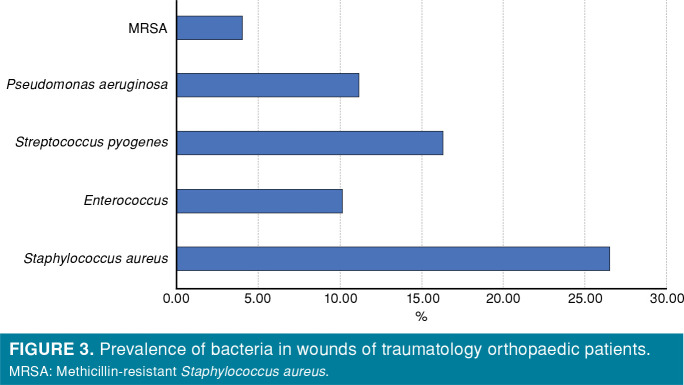
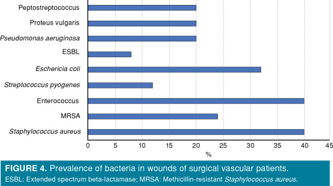
The distribution of wound types varied very widely. Certain comorbidities affected wound healing. In orthopedictraumatology patients, we observed mainly skin flora infection (57.14%), while in surgical and vascular patients, mixed flora (80%) and in many cases poly-resistant pathogens were present (methicillin-resistant Staphylococcus aureus 24%) A total of 43.3% of wounds were completely closed, while 44.6% of patients had a wound healing. Successful skin grafting was performed in 75% of wounds.
Conclusion
This technique may be used as widely and as early as possible. However, further large-scale, multi-center, randomized clinical trials are needed worldwide to find a place for this technique in wound care and even in primary care.
Keywords: Efficacy, negative pressure wound therapy, wound management.
Introduction
In the last decades, negative pressure wound therapy (NPWT) treatment has opened great possibilities in wound management. First used in the Russo-Afghan War in 1985 by Nail Bagaoutdinov,[1] modern vacuum treatment was, then, introduced by Louis Aregenta[1] and Michael Morykwas[1] in 1990 using a combination of polyurethane foam and a mechanical vacuum machine.[1]
Use of the technique in everyday care began in 1993. In 2017, the textbook Negative Pressure Therapy: Theory and Practice was published in Hungarian, and in 2019 in English, with clinical studies, but largely based on individual experiences.[2] Medical sites dealing with the technique include Web of Science 1,251, PubMed 4,590, Google Scholar 14,900 hits currently.
It has the advantages of increasing local blood flow, reducing edema, promoting granulation tissue formation, facilitating cell proliferation, removing soluble wound healing inhibitors from the wound, reducing bacterial load, and bringing wound edges closer together.[2] It is recommended for acute and chronic traumatic wounds, secondary wounds, wound healing disorders, dehiscence, even with stoma, pressure ulcers, open abdomen, abdominal compartment syndrome, diabetes-foot syndrome, skin grafts and tissue grafts, burns, sternotomy coverage as a preventive measure.[2]
In the present study, we aimed to evaluate the effectiveness of treatments in our own patient database five years after the introduction of NPWT in hospitals. We also aimed to compare how the use of NPWT changed compared to the initial ad hoc applications. Furthermore, open access studies published in English over the last five years were reviewed to draw attention to the evidence emerging in some specialties for the treatment options and the effectiveness of the therapy today.
Patients and Methods
This single-center, retrospective, clinical study was conducted at St. George’s Hospital, Department of Traumatology-Orthopaedic, Surgery and Vascular Surgery between January 1st, 2012 and December 31st, 2016. After the introduction of NPWT, five years of patient records were processed. A total of 74 patients (35 males, 39 females; median age: 60 years; range, 20 to 95 years) received NPWT. Of these, 49 were orthopedic and traumatology, 12 vascular surgery, and 13 general surgery patients. We examined the percentage of acute and chronic wounds, transplant ability, rate of healing and outcome. We outlined the proportion of any underlying conditions or medications that contributed to or hindered wound healing. The bacterial flora that dominated the infected wounds was determined by wound fluid cultures. Exclusion criteria included incomplete documentation. All treatments were performed at a single institution. All physicians received a detailed information and education on the course of NPWT therapy. A written informed consent was obtained from each patient. The study protocol was approved by the St George’s Hospital Institutional Review Board (IRB No: 01.28.2022). The study was conducted in accordance with the principles of the Declaration of Helsinki.
Before starting NPWT and at each dressing change, wound fluid was cultured. Before starting treatment, trauma-orthopedic patients usually received prophylactic antibiotic treatment (cefazolin). Surgical patients also received antibiotic therapy before treatment. In all cases, they were switched to targeted antibiotic therapy based on the culture results. Changes in patients' laboratory parameters were monitored continuously, with particular attention to changes in blood count, inflammatory parameters, liver and kidney functions. The NPWT was initially set to -125 mmHg in continuous mode according to the standard and then changed to intermittent mode depending on wound healing. The treatment was monitored 24 h, such as the wound area or the amount of wound exudate. Dressings were applied and changed in sterile operating theatre conditions every three to five days depending on the condition of the wound. After the wound was cleaned and dressed, the wound was covered with skin. Partial or total skin grafting was performed during surgery. After skin grafting, repeated NPWT was applied to the grafted area for 48 h at -125 mmHg pressure in continuous mode. After the treatment, the wound status was recorded at hospital discharge and a smart dressing was applied at home, if necessary.
Statistical analysis
Statistical analysis was performed using the Microsoft Excel for Microsoft 365 MSO (2112 build version 16.0.14729.20254). Descriptive data were expressed in median (min-max) or number and frequency, where applicable. Normality tests were used to assess the normality of the variables. A one-dimensional contingency table was used to present our discrete variables.
Results
Of 74 patients, 66.2% received traumatologyorthopedic care. Of these, 73.46% were under 60 years old and healthy and 22.5% had a comorbidity or several comorbidities. The most common were hypertension, diabetes and cardiovascular disease. Although there was no significant difference in sex in the proportion of traumatic wounds, the literature suggests that limb soft tissue injuries are more common in men due to traffic and domestic accidents.[2]
Surgical (17.6%) and vascular (12.6%) patients accounted for the other half of the 74 patients; i.e., 33.8%. 72% were between 40 and 80 years old, while the percentage of patients over 80 years old was 20%. In terms of sex, 76.9% of surgical patients were female, while 75% of vascular surgery patients were male.
Eligibility can be affected by previous internal medical conditions such as cardiac arrhythmia, severe heart disease, cardiac decompensation, renal failure, diabetes, steroid treatment and immunosuppressed states. The patient's medication may also affect the success of the treatment, taking antiplatelet agents, anticoagulants may increase the risk of bleeding and should be switched to low-molecular-weight heparin. Elderly patients with multiple comorbidities are more likely to develop prolonged wound healing or wounds that are difficult to heal. In their case, the use of negative pressure therapy can speed up wound healing and healing.[2] In our study, among comorbidities, the prevalence of hypertension (50.75%), cardiovascular disease (28.8%) or diabetes (34.82%) was 77.5% in total. In some cases, chronic renal insufficiency, dysplasia or chronic anemia also colored the picture.
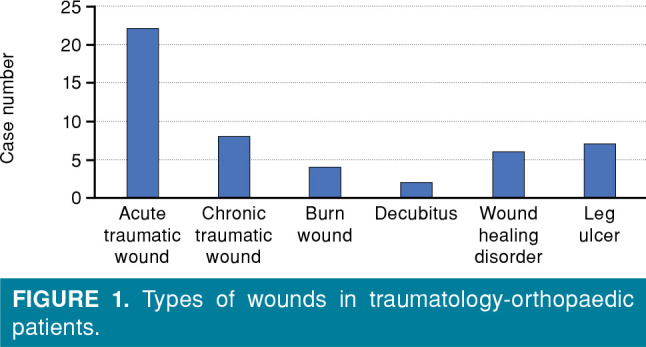
The distribution of wound types in the trauma group varied (Figure 1). The wounds included soil contaminated leg wounds, lacerations, gunshot wounds, skin and subcutaneous defects caused by necrotizing fasciitis, and burns. Chronic wounds included prolonged empyema thoracis, pressure ulcers, and leg ulcers.
Figure 1. Types of wounds in traumatology-orthopaedic patients.
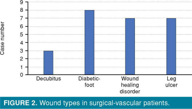
In surgical patients, the most common symptoms were wound healing disorder, wound dehiscence, leg ulcer, and diabetic foot syndrome. Vascular surgical patients presented with endothelial necrosis due to vascular stenosis, malum perforans and, in many cases, vascular reconstruction was part of the treatment for optimal wound healing (Figure 2).
Figure 2. Wound types in surgical-vascular patients.
Superficial and deep inflammation of the skin and subcutaneous tissues were the most common types of wound infections, accounting for 57.14% of all cases. In traumatic, acute patients, the infection was mainly from the skin flora. In chronic traumatic wounds, the bacterial flora was already Gram-negative. Diabetes mellitus, as an underlying disease when present, had a distinctly mixed bacterial flora (Figure 3).
Figure 3. Prevalence of bacteria in wounds of traumatology orthopaedic patients. MRSA: Methicillin-resistant Staphylococcus aureus.
Bacterial contamination in the surgical - vascular surgery group was quite different from the previous one, as a mixed flora was usually found and a higher degree of polyresistance was observed (Figure 4).
Figure 4. Prevalence of bacteria in wounds of surgical vascular patients. ESBL: Extended spectrum beta-lactamase; MRSA: Methicillin-resistant Staphylococcus aureus.
Negative pressure therapy cannot eliminate the bacterial flora from the wound; however, it can significantly reduce its volume through continuous drainage of phlegm. This made the wound suitable for graft adhesion after preparation, although the wound bed was not completely bacteria-free.
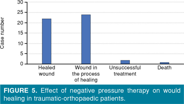
Regarding the effect of negative pressure therapy on wounds, 44.9% of the wounds in the traumatology group healed, and 48.9% of the wounds healed, but did not close. Skin grafts were successfully performed in 81.6%. The treatment of chronic osteomyelitis was the longest and most complex task. It also took longer to treat infections affecting joints compared to only soft tissue inflammation. Treatment was clearly beneficial in 93.8% of patients treated. We lost one elderly patient in a fallen state during the treatment and two patients (4.08%) were not successfully treated, the patient was not able to bear the dressing due to his disturbed state (Figure 5).
Figure 5. Effect of negative pressure therapy on would healing in traumatic-orthopaedic patients.
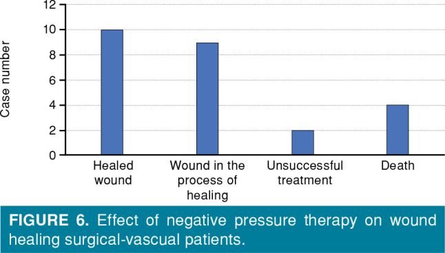
In the surgical-vascular group, 40% of the wounds closed and 36% of them healed. Unfortunately, two treatments were unsuccessful (8%), and these vascular surgery patients were embarrassed by several times removing the dressing from themselves or the circulation in the limb was insufficient for treatment, eventually requiring amputation. Four (16%) patients were lost during treatment, in which case the treatment was effective, but their disease was so severe or their vascular disease so advanced that they died despite treatment (Figure 6). Negative pressure therapy had no effect on their deaths. In terms of outcome, treatment was successful in 76% of patients. In 60% of patients, the wound could be closed.
Figure 6. Effect of negative pressure therapy on wound healing surgical-vascual patients.
Discussion
The primary finding of the current study was the high rate of wound healing after NPWT.[3]
While evaluating the recent literature regarding the NPWT, it is perhaps most widely used in traumatology at present, but for which wounds and when it is effective is not yet a settled fact. In primary, acute care, polytrauma patients often present with open bone injuries, which are always at a high risk of infection. The study by Liu et al.[4] confirmed that NPWT resulted in significantly lower infection rates, shorter wound healing times and hospital stays, and fewer amputations in accidental wounds. Our studies showed similar results in orthopedic-traumatology patients, and 93.8% of the patients treated had a clear benefit and 81.6% had a successful skin graft.
According to the Godina study, NPWT has a clear benefit for wounds left open for more than 72 h.[5] We mainly used it for large defects where primary coverage was not an option. The NPWT not only helped to extend the time for subsequent skin coverage, but also greatly helped in keeping the area clean, reducing the possibility of overinfection and treating other lesions.
One of the major problems in the surgical field is the multiple surgical repairs of abdominal hernias. In many cases, despite the utmost care, wound healing, necrosis, dehiscence and mesh rejection can be observed after surgery, particularly in the presence of multiple abdominal operations, high body mass index or malnutrition, and much comorbidity. Gök et al.[6] showed that NPWT significantly reduced swelling and wound tension after hernia surgery. The incidence of dehiscence was 3% compared to 25% for conventional wound closure and 20% for drained wounds.
A total of 15% of patients with diabetes would develop a wound or ulcer on the foot during their lifetime. A 2020 meta-analysis found a 51% complete wound healing rate and a reduction in major amputations.[7] However, in a 2018 Cochrane review, there is low-level evidence that NPWT can increase the rate of healed wounds and reduce the time to healing in postoperative foot wounds and foot ulcers in patients with diabetes mellitus compared to wound dressings.[8] In our study, the treatment of diabetic legs had good results, although in our cases, the primary treatment was to reduce the size of the wound and avoid amputation. Skin grafting was not successful in many cases due to their underlying disease and bacterial flora.
Treatment of large-pouch wounds at the base of decubitus ulcers with dressings is often impossible. A meta-analysis published in 2021 investigated the efficacy of NPWT and conventional dressings in the treatment of Stage II/IV decubitus wounds. Patients treated with NPWT had significantly shorter wound healing time and significantly lower cost of care.[9] Both groups of patients were treated with calcaneal and sacral decubitus. Our results are in line with those of large studies.
Wound infections continue to be a problem in all professions today. A study by Wang et al.[10] showed that NPWT was significantly more effective than conventional wound dressings in cases of deep[10] SSI, superficial SSI, and wound dehiscence.[10] In our study, superficial and deep inflammation of the skin and subcutaneous tissues was the most common type of wound infection, accounting for 57.14% of all cases in orthopedic-traumatology patients. Nevertheless, the treatment achieved a skin coverage of over 80% and a beneficial effect of over 90%.
A Cochrane study published in 2020 found that NPWT after primary wound closure had a greater reduction in the incidence of SSI compared to standard dressings.[11] In our study, the rate of wound infections was much higher for surgical-vascular patients, with pathogens in all wounds. After NPWT, 76% of the wounds responded well to treatment and 60% were closed or covered.
The risk of wound infection and wound healing disturbance is quite high in the case of endovascular reconstruction. A multi-center clinical trial published in 2015 found that NPWT significantly contributed to the salvage of grafts and helped to repair deep soft tissue infections. It accelerated granulation formation, even over the grafts, significantly contributing to secondary wound closure.[12] Kwon et al.[13] investigated the efficacy of NPWT in 119 patients undergoing lower limb vascular surgery with femoral exploration in terms of SSI and cost-effectiveness. It significantly reduced major wound complications and costs in the high-risk group. We found similarly good results in our vascular surgery patients, although adding that this is perhaps the group with the highest rate of recovery.
In skin grafting, whether used to pre-graft the area or to graft the mesh, partial-thickness skin for better adhesion, NPWT is excellent in both cases. In a 2017 article, NPWT was used to pre-treat chronic wounds, a mesh graft was, then, placed over the defect and vacuum therapy was also used to promote adhesion.[14] The infection was resolved in almost all patients. The study found that the use of NPWT over mesh skin grafting is significantly effective, particularly for wounds resistant to conventional therapies, thereby improving skin graft adherence rates. In our study, we favored mesh skin grafting over open wound treatment in both groups of patients. The NPWT could significantly increase the adherence rate. While the grafting rate in the orthopedic-traumatology group was 81.6%, and it was around 60% in the surgical-vascular group.
New articles have recently appeared questioning the effectiveness of NPWT. The study by Jensen et al.[15] did not recommend using of NPWT directly for internal osteosynthesis. The study by Älgå et al.[16] found no economic benefit of NPWT for open lower extremity fractures in terms of wound management. However, the study raises aspects such as wound healing and improvement in quality of life or reduction in pain that cannot be measured financially. A new technique such as NPWT with continuous wound irrigation is also looking for a place in the treatment palette according to the article by Wu et al.[17]
One of the limitations of the study is that, due to impracticality, it was often difficult to place and vacuum the grafts onto the tendons. In addition, the small number of cases did not allow us to draw conclusions about wound healing due to wound fluid cultures. Due to the lack of experience at the beginning, the start of NPWT was often delayed or only applied on the second or even third round. Over the years, with increasing experience at home and abroad, we now use it daily.
In our study, all patient groups benefited from the treatment. Traumatology patients, in particular, benefited from faster wound healing, especially if wound-free skin could be covered. In surgical patients, it is mainly beneficial in the case of swallowing of abdominal meshes, as the treatment can avoid the need to remove the mesh. In vascular surgery patients, the preservation of the implanted vascular prosthesis or the avoidance of graft replacement is an advantage.
In conclusion, the primary finding of the current study was the high rate of wound healing after NPWT and, given the findings of the current study, we recommend that this technique may be used as widely and as early as possible.
Footnotes
Conflict of Interest: The authors declared no conflicts of interest with respect to the authorship and/or publication of this article.
Financial Disclosure: The authors received no financial support for the research and/or authorship of this article.
References
- 1.Szabóné E. Révész: History of the development of wound care. KH. 2021;11:506–517. [Google Scholar]
- 2.Rached A, Bábel Z, Balog K, Balogh G, Bánky B, Bánvölgyi A, et al. Biatorbágy: The Negative Pressure Therapy for Wound Healing Association. 2019. Negative pressure therapy: theoretical knowledge and practical application. [Google Scholar]
- 3.Atik OŞ. What are the expectations of an editor from a scientific article. Jt Dis Relat Surg. 2020;31:597–598. doi: 10.5606/ehc.2020.57896. [DOI] [PMC free article] [PubMed] [Google Scholar]
- 4.Liu X, Zhang H, Cen S, Huang F. Negative pressure wound therapy versus conventional wound dressings in treatment of open fractures: A systematic review and meta-analysis. Int J Surg. 2018;53:72–79. doi: 10.1016/j.ijsu.2018.02.064. [DOI] [PubMed] [Google Scholar]
- 5.Qiu E, Kurlander DE, Ghaznavi AM. Godina revisited: A systematic review of traumatic lower extremity wound reconstruction timing. J Plast Surg Hand Surg. 2018;52:259–264. doi: 10.1080/2000656X.2018.1470979. [DOI] [PubMed] [Google Scholar]
- 6.Gök MA, Kafadar MT, Yeğen SF. Comparison of negativepressure incision management system in wound dehiscence: A prospective, randomized, observational study. J Med Life. 2019;12:276–283. doi: 10.25122/jml-2019-0033. [DOI] [PMC free article] [PubMed] [Google Scholar]
- 7.Rys P, Borys S, Hohendorff J, Zapala A, Witek P, Monica M, et al. NPWT in diabetic foot wounds-a systematic review and meta-analysis of observational studies. Endocrine. 2020;68:44–55. doi: 10.1007/s12020-019-02164-9. [DOI] [PubMed] [Google Scholar]
- 8.Liu Z, Dumville JC, Hinchliffe RJ, Cullum N, Game F, Stubbs N, et al. Negative pressure wound therapy for treating foot wounds in people with diabetes mellitus. CD010318Cochrane Database Syst Rev. 2018;10 doi: 10.1002/14651858.CD010318.pub3. [DOI] [PMC free article] [PubMed] [Google Scholar]
- 9.Song YP, Wang L, Yuan BF, Shen HW, Du L, Cai JY, et al. Negative-pressure wound therapy for III/IV pressure injuries: A meta-analysis. Wound Repair Regen. 2021;29:20–33. doi: 10.1111/wrr.12863. [DOI] [PubMed] [Google Scholar]
- 10.Wang C, Zhang Y, Qu H. Negative pressure wound therapy for closed incisions in orthopedic trauma surgery: A metaanalysis. J Orthop Surg Res. 2019;14:427–427. doi: 10.1186/s13018-019-1488-z. [DOI] [PMC free article] [PubMed] [Google Scholar]
- 11.Norman G, Goh EL, Dumville JC, Shi C, Liu Z, Chiverton L, et al. Negative pressure wound therapy for surgical wounds healing by primary closure. CD009261Cochrane Database Syst Rev. 2020;6 doi: 10.1002/14651858.CD009261.pub6. [DOI] [PMC free article] [PubMed] [Google Scholar]
- 12.Verma H, Ktenidis K, George RK, Tripathi R. Vacuumassisted closure therapy for vascular graft infection (Szilagyi grade III) in the groin-a 10-year multi-center experience. Int Wound J. 2015;12:317–321. doi: 10.1111/iwj.12110. [DOI] [PMC free article] [PubMed] [Google Scholar]
- 13.Kwon J, Staley C, McCullough M, Goss S, Arosemena M, Abai B, et al. A randomized clinical trial evaluating negative pressure therapy to decrease vascular groin incision complications. J Vasc Surg. 2018;68:1744–1752. doi: 10.1016/j.jvs.2018.05.224. [DOI] [PubMed] [Google Scholar]
- 14.Maruccia M, Onesti MG, Sorvillo V, Albano A, Dessy LA, Carlesimo B, et al. An alternative treatment strategy for complicated chronic wounds: Negative pressure therapy over mesh skin graft. Biomed Res Int. 2017;2017:8395219–8395219. doi: 10.1155/2017/8395219. [DOI] [PMC free article] [PubMed] [Google Scholar]
- 15.Jensen NM, Steenstrup S, Ravn C, Schmal H, Viberg B. The use of negative pressure wound therapy for fracturerelated infections following internal osteosynthesis of the extremity: A systematic review. J Clin Orthop Trauma. 2021;24:101710–101710. doi: 10.1016/j.jcot.2021.101710. [DOI] [PMC free article] [PubMed] [Google Scholar]
- 16.Älgå A, Löfgren J, Haweizy R, Bashaireh K, Wong S, Forsberg BC, et al. Cost analysis of negative-pressure wound therapy versus standard treatment of acute conflictrelated extremity wounds within a randomized controlled trial. World J Emerg Surg. 2022;17:9–9. doi: 10.1186/s13017-022-00415-1. [DOI] [PMC free article] [PubMed] [Google Scholar]
- 17.Wu L, Wen B, Xu Z, Lin K. Research progress on negative pressure wound therapy with instillation in the treatment of orthopaedic wounds. Int Wound J. 2022 doi: 10.1111/iwj.13741. [DOI] [PMC free article] [PubMed] [Google Scholar]


