
Figure 17:
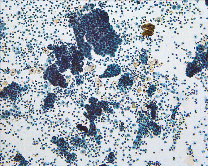
Groups of atypical cells interpreted as ‘low-grade serous tumor’ are present in this washing from a woman with intraperitoneal micropapillary serous tumor. While the cytologic pattern is that of a neoplasm, the precise categorization is difficult by cytology alone. Indeed, it is unclear whether this lesion is a well-differentiated carcinoma or a tumor of low malignant potential (borderline). (Modified PAP stain, 20X.)