Abstract
For antibiotic resistance to arise, new resistant mutants must establish in a bacterial population before they can spread via natural selection. Comprehending the stochastic factors that influence mutant establishment is crucial for a quantitative understanding of antibiotic resistance emergence. Here, we quantify the single-cell establishment probability of four Escherichia coli strains expressing β-lactamase alleles with different activity against the antibiotic cefotaxime, as a function of antibiotic concentration in both unstructured (liquid) and structured (agar) environments. We show that concentrations well below the minimum inhibitory concentration (MIC) can substantially hamper establishment, particularly for highly resistant mutants. While the pattern of establishment suppression is comparable in both tested environments, we find greater variability in establishment probability on agar. Using a simple branching model, we investigate possible sources of this stochasticity, including environment-dependent lineage variability, but cannot reject other possible causes. Lastly, we use the single-cell establishment probability to predict each strain's MIC in the absence of social interactions. We observe substantially higher measured than predicted MIC values, particularly for highly resistant strains, which indicates cooperative effects among resistant cells at large cell numbers, such as in standard MIC assays.
Keywords: establishment probability, antibiotic resistance evolution, minimum inhibitory concentration, social interactions
1. Introduction
A classical question in population genetics addresses the fate of a new adaptive allele in a population. Any new allele is initially present at a low frequency in the population and thus prone to extinction by genetic drift, even if the allele is beneficial [1,2]. In 1927, Haldane stated that a novel beneficial allele has to overcome drift loss and establish in the population before it can be picked up by selection; the probability of which he estimated to be roughly two times its selective benefit [3]. Since then, Haldane's approximation has been generalized and extended by a great number of models, but none of them has been tested empirically until about a decade ago [1]. With advances in genomic techniques, the issue of fixation probability has more recently been studied in experimental populations of microorganisms (e.g. [3,4]). These studies looked at the fate of beneficial alleles once they reach a relatively high frequency in the population, finding that their fixation strongly depends on the competition with further beneficial alleles that appear via mutation over time. Only a small number of empirical studies has investigated the process of establishment of an initially rare beneficial allele arisen by de novo mutation in fungi [6], bacteria [7,8] and nematodes [9]. Overall, these studies found that the establishment process is stochastic and that the probability for a beneficial allele to establish in a population depends on the alleles' initial frequency, its selective benefit (e.g. in response to the concentration of the selective agent), and the size of the population.
Whether a beneficial allele establishes successfully in a population is of particular relevance in cases of unwanted evolution, such as the evolution of antibiotic resistance. The worldwide growing concern about failing treatments of infections due to the emergence of resistant bacterial pathogens has led to the awareness that this problem must be tackled from different angles, including a better understanding of the emergence and evolution of antibiotic resistance [2,10–12]. Resistance alleles can be acquired via horizontal gene transfer or de novo mutation, resulting in the resistant allele initially being present in one or a few individuals within a susceptible population. But how likely is it that this single genotype can establish in the population and what factors influence this process? Using time-lapse microscopy, Coates et al. [13] followed the growth of single Escherichia coli cells on agar, supplemented with different antibiotics. They found that the establishment of a single cell, determined by its growth into a visible macrocolony, was a highly stochastic process driven by fluctuations in cell death and birth, leading to the extinction of microcolonies at various time points. Similar results were found for two Pseudomonas aeruginosa strains, resistant to streptomycin or meropenem [14]. By seeding one to a few cells of a resistant strain in liquid medium with increasing antibiotic concentrations, it was found that at concentrations as low as one-eighth of the minimum inhibitory concentration (MIC) of this strain, its establishment probability was only 5% and that establishment of one cell was independent of the presence of other cells. The latter finding, however, may differ in systems where the antibiotic is broken down in the environment, leading to positive social interactions [15].
A common resistance mechanism potentially allowing for social effects involves β-lactamase enzymes. β-lactamases hydrolyse the lactam ring of β-lactam antibiotics, which target penicillin-binding proteins (PBPs) that are involved in cell wall synthesis of gram-negative bacteria, thereby leading to loss of cell wall integrity and eventual cell lysis [16,17]. A well-studied and clinically relevant example is TEM-1 β-lactamase, which has high activity towards first-generation penicillins, but low activity towards third-generation β-lactams, including the cephalosporin cefotaxime (CTX). However, the TEM-1 allele is the ancestor of a large family of extended-spectrum β-lactamases that have acquired mutations causing enhanced activity against more recently introduced penicillins and cephalosporins, including CTX [18–20]. As β-lactamases are expressed in the periplasmic space of gram-negative pathogens like E. coli, this system has been recognized as potentially cooperative, since the enzymatic breakdown in the periplasm reduces the antibiotic concentration in the environment [21], thereby cross-protecting susceptible bacteria nearby [15]. Such a cooperative behaviour can be expected to have more pronounced consequences on agar due to the local breakdown of the antibiotic, creating microenvironments that may allow for the coexistence of strains with different levels of antibiotic resistance [22–25].
Here, we examined the single-cell establishment probability of antibiotic-degrading bacteria under a range of antibiotic concentrations in both a structured (agar) and an unstructured (liquid) environment. We used four E. coli strains expressing β-lactamase alleles with different activities towards CTX, and hence different levels of resistance. Each strain was tested separately at low cell densities in both liquid and on agar medium with increasing CTX concentrations. To determine the single-cell establishment probability from the data, we developed a simple branching model, followed by a more detailed investigation of the cause of differences in stochasticity between the two environments. We show that environmental structure and cooperative behaviour play at most a minor role in the establishment of resistant cells at low initial densities. Based on our results, we further introduce a new measure for resistance level, MICq(N), that takes inoculum size and stochasticity into account, thereby allowing for the detection of collective resistance. Application of the new measure to our data indicates substantial cooperative effects at higher cell densities. We therefore propose the new measure to be used for determining resistance levels as it overcomes the limitations of standard MIC assays.
2. Results and discussion
In this study, we aimed at quantifying how the establishment of single bacterial cells with different levels of antibiotic resistance is affected by: (i) the antibiotic concentration, (ii) the environmental structure and (iii) positive social interactions via the breakdown of the antibiotic in the environment. To determine single cell establishment probabilities, we performed one experiment in liquid (unstructured environment) and one on agar medium (structured environment). In each experiment, four E. coli strains were tested separately under exposure to a range of CTX concentrations: the TEM-1 Ancestor with very low β-lactamase activity, leaving this strain rather susceptible towards CTX, and three TEM mutant strains (referred to as Single mutant, Double mutant, Triple mutant; cf. table 1) with increasing levels of CTX resistance conferred by different antibiotic-hydrolysing β-lactamase enzymes. The tested concentrations differed between strains and were chosen to display relative establishment probabilities between 0 and 1 (i.e. relative to the absence of antibiotic; see Material and methods; electronic supplementary material, table S1). We use the notation pe(x) to denote the probability that a single cell establishes a macroscopic population (i.e. turbidity in liquid or colony formation on agar, visible to the naked eye) at CTX concentration x. When the antibiotic concentration is zero, a population should establish with maximum probability. Likewise, when the concentration is above the strains’ minimum inhibitory concentration (MIC), establishment is expected to be prevented. Strongly stochastic outcomes therefore arise only at intermediate concentrations.
Table 1.
Overview of the used strains with their respective measured minimum inhibitory concentration (MIC) and null-model likelihood of the corresponding predicted MICs (see main text).
| wild-type straina (without TEM) | TEM amino acid substitution | strain name (as used here) | MIC for Cefotaximeb (µg ml−1) | null-model likelihood Lc (%) |
|---|---|---|---|---|
| MG1655 galK::SYFP2-CAT | none (TEM-1) | ancestor | 0.08 | >99.99 |
| G238S | single mutant | 0.64 | 1.1 | |
| E104 K/G238S | double mutant | 10.24 | <0.01 | |
| E104 K/M182 T/G238S | triple mutant | 81.92 | <0.01 |
aMIC of the wild-type strain for CTX = 0.04 µg ml−1.
bAs determined prior to the study by standard MIC assays (see Material and methods). Note that the true MIC lies between this value and half of this concentration (due to the 2-fold dilution during the setup).
cAssuming that the inoculum establishes at the highest concentration at which growth is observed in the MIC assay (see the last section of Results and Discussion); values that show significant differences between the measured and predicted MICs are depicted in bold.
(a) . Establishment probability declines well below the MIC in liquid
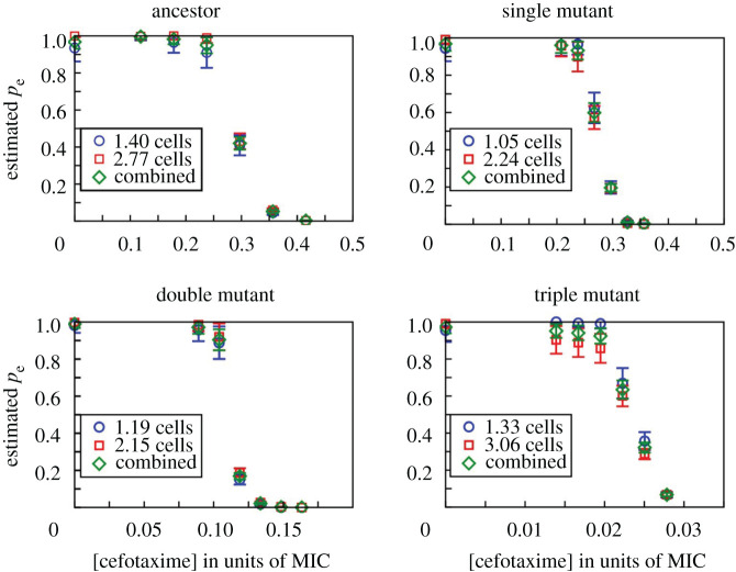
In the liquid medium experiment, we grew bacteria in multi-well plates and observed their establishment as a function of CTX concentration. Per strain, inoculum size (about 1 and 3 cells on average) and antibiotic concentration, a total of 285 replicate wells were tested, spread across three plates, including one medium control well per plate. Estimates of the viable cell numbers present in the inocula for evaluating the probabilities were obtained by applying cell culture aliquots on agar, taken from the culture dilutions that were used for inoculating the well plates. Using a bootstrapping algorithm and assuming a Poisson distribution of independently acting cells in each inoculum (details in electronic supplementary material, S2), we estimated the single-cell establishment probability pe(x) for all strains and conditions. The results in figure 1 show that the establishment probability nears zero well below each strain's MIC and that this effect is more pronounced for strains with higher resistance levels.
Figure 1.
Single-cell establishment probabilities, pe, in liquid medium, estimated for the four tested strains as a function of antibiotic concentration, shown as a fraction of each strain's measured MIC (cf. table 1). For each strain, two inocula were used, one of approximately 1 cell (blue circles) and another of 2–3 cells (red squares); the average inoculum size estimated from separate CFU counts is shown. The inferred single-cell establishment probabilities are nearly identical for the two cases, implying that the establishment probability of a cell is not influenced by the presence of other cells at these low initial cell densities. The green diamonds are weighted averages over the two inoculum sizes (see electronic supplementary material, supplement S2 for details on the method used to estimate pe). (Online version in colour.)
The observation that the establishment probability of a single cell nears zero at antibiotic concentrations well below the strain's MIC has been shown before in E. coli for various bactericidal antibiotics [13] and in P. aeruginosa for streptomycin and meropenem [14]. These studies showed that the underlying causes for this are rooted in stochastic effects in population dynamics due to among-cell variability in death rate [13,14] and lag-phase [14], which become more pronounced with increasing antibiotic concentration. The establishment probability is higher with increasing initial cell numbers, simply because the probability is higher for at least one cell to survive and outgrow into a population [13,14]. This explains the decreased establishment probability of single cells at lower antibiotic concentrations compared to each strain's MIC, as the latter is measured for inocula containing hundreds of thousands of cells per ml. While we confirm the results of this general pattern in our study, we extend the findings by using four E. coli strains with the same genetic background but different sensitivities towards the β-lactam antibiotic CTX. As these differences in sensitivity are based on differences in the level of β-lactamase activity (i.e. enzymatic capacity in breaking down CTX), our system provides the opportunity to study the effect of positive social interactions during establishment. Indeed, the increasingly pronounced discrepancy between the single-cell establishment probability and standard MIC estimates that is observed with increasing resistance level is, at least partially, attributed to an increased collective breakdown at high cell densities (see section ‘MIC and the inoculum effect’ for details).
(b) . Cells do not interact in liquid medium during establishment
One complication in estimating the single-cell establishment probability in liquid medium is that multiple cells may be present initially in some wells, and these cells may in principle interact, for example through competition for resources or the breakdown of antibiotic molecules. In that case, the establishment probabilities of individual cells would no longer be independent, biasing our estimates of pe(x). To test whether cells interact within initially small inocula, we conducted the liquid experiment with two different mean inoculum sizes (with approximately 1 and 3 cells, respectively; see section 4(b)) and estimated pe(x) for both cases under the assumption that there is no interaction (details in electronic supplementary material, S2). If interactions occurred, the inferred values should disagree. However, figure 1 shows that they agree closely, indicating that interactions between cells are insignificant at these low cell densities in unstructured environments.
The absence of social interactions in liquid medium at low cell density was also found in the study with P. aeruginosa, exposed to streptomycin or meropenem under similar conditions regarding culture volume and inoculum size [14]. In that system, positive social interactions are less likely to occur, because streptomycin resistance does not involve antibiotic-degrading enzymes. Our E. coli β-lactamase system on the contrary involves the potential for cooperative effects due to an active breakdown of the antibiotic in the environment. The absence of an effect in our data confirms that cell-to-cell interactions (both positive and negative) play an insignificant role during the establishment of single cells at such low densities in liquid medium and further shows that this observation is independent of the resistance mechanism.
(c) . Establishment probability shows moderately higher stochasticity on agar
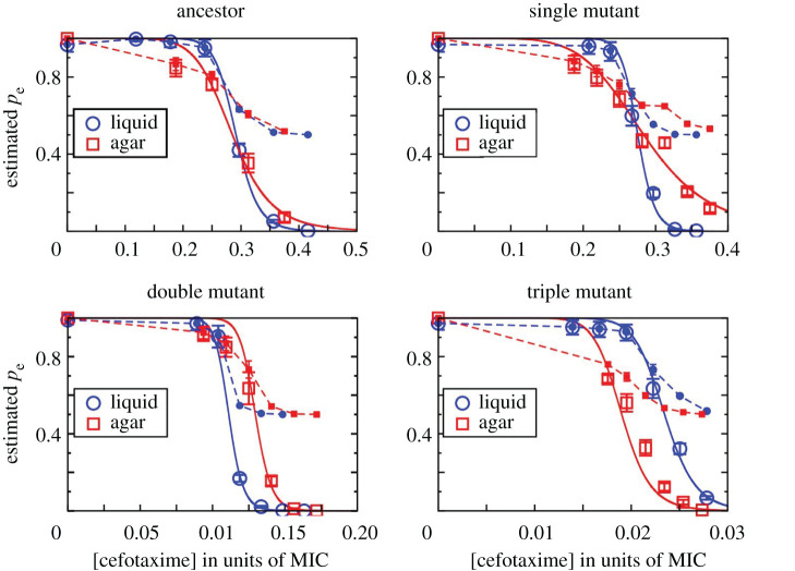
In the previous section, we found that the establishment probability of one cell was not influenced by the presence of other cells in liquid medium at very low densities. But how is this on agar? Previous studies have shown that evolutionary outcomes can largely differ depending on the environmental structure in the absence of migration [26–28]. For instance, while conditions in unstructured (liquid) environments commonly select for one strain to dominate the population, structured (agar) environments can maintain various strains due to the creation of microenvironments [29–31]. One condition that can be affected locally on agar is the concentration of the antibiotic due to enzymatic breakdown. While it has been shown that this can result in cooperative effects, where a resistant strain protects more sensitive ones [15,32] and such cooperation is more prominent in structured environments [25], it is, to the best of our knowledge, unknown whether such social interactions can influence the establishment of single antibiotic-resistant cells. To determine the effect of environmental structure on the establishment of β-lactamase-expressing cells, we plated about 200 cells of each E. coli strain on CTX-containing agar (eight plates per condition), followed by counts of colony-forming units (CFUs) after incubation. As for the experiment in liquid medium, the tested CTX concentration range depended on the strain to display establishment probabilities between 0 and 1 (see Material and methods; electronic supplementary material, table S1). The establishment probability was estimated as the CFU count relative to that at no CTX. The patterns of pe as a function of CTX concentration are broadly similar to those in liquid medium (figure 2), indicating a minor role of environmental structure being involved in mutant establishment at low cell densities.
Figure 2.
The single-cell establishment probability, pe, is shown as open symbols for the four tested strains in liquid (blue circles) and agar environments (red squares) for different CTX concentrations as the fraction of each strain's measured MIC. For liquid medium, the combined results from both tested inoculum sizes (cf. figure 1) are shown. The solid lines are fits performed with the Hill function pe(x) = (1 + (x/x0)n)−1 with x0 and n as parameters, where x0 is the concentration at which pe drops to 1/2, and n indicates the steepness of the curve. The values of the inferred parameters are given in electronic supplementary material, table S2. The filled symbols connected by dashed lines show the cell division probability pd inferred from a simple branching model for the data points where pe > 0 (see main text). (Online version in colour.)
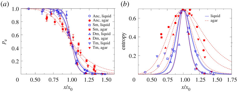
Although we found a general agreement of the establishment probability pattern between the liquid and agar experimental data, a closer comparison of the estimates of both environments shows that the decline of pe with CTX concentration is less steep on agar than in liquid (figure 3a). This implies a higher degree of stochasticity on agar, since pe remains between 0 and 1 over a larger range of concentrations. We quantified this effect by calculating the Shannon entropy for the establishment probabilities in both environments and all four strains. Shannon entropy is an information-theoretic measure that indicates the degree of stochasticity associated with a set of probabilities. In our context, the measure has a value of 0 (no stochasticity) in the extreme cases of pe = 0 or pe = 1, and is maximized at the intermediate value of pe = 0.5. Figure 3b shows our finding that the entropy, and therefore the stochasticity associated with the outcome of the establishment process, is higher on agar.
Figure 3.
(a) Establishment probabilities for all the strains in both liquid and agar environments as a function of CTX concentration (Anc: Ancestor, Sm: Single mutant, Dm: Double mutant, Tm: Triple mutant), scaled by the corresponding estimate x0 (i.e. the concentration at which pe drops to 0.5). The curves for agar medium (dashed red lines) are less steep than those for liquid medium (blue solid lines). (b) The Shannon entropy is calculated and plotted for the pe values, using the standard expression S = −pelnpe − (1 − pe)ln(1 − pe). It is seen to be higher for agar (dashed red lines and filled symbols) compared to liquid (solid blue lines and open symbols), as expected from the shallower slopes of the pe(x) curves for agar in panel (a). (Online version in colour.)
Next, using a modelling approach we explored possible underlying mechanisms for stochasticity in the system. The simplest model is a branching process, where every cell divides with a fixed probability pd, or dies otherwise; the population either continues to grow indefinitely (with probability pe) or eventually goes extinct (with probability 1 − pe; see electronic supplementary material, S3 for details). This model allows us to estimate the division probability pd using the measured pe values. Figure 2 shows that under the simple branching model, the division probability appears to approach the value ½ asymptotically as opposed to the natural expectation of approaching zero asymptotically (see electronic supplementary material, figure S1 for an illustration of the possible shapes of the pe and pd curves in the simple branching model). This is a clear indication that the simple model does not fully capture the establishment process. One way to modify the model is to introduce variability in pd among cell lineages, and this fits the data reasonably well (see electronic supplementary material, S4). Within this framework, the higher establishment stochasticity on agar could be due to small differences in pd of different cells on the agar surface (see electronic supplementary material, S4 and figure S3 for details). However, other possibilities remain; for instance, the cell division probability may change with time due to accumulating damage in cell walls under antibiotic action, changing rates of antibiotic and enzyme flux or changing CTX concentration over time at the location of a growing colony. Distinguishing conclusively between these alternatives is not possible based on our data, and we do not explore this issue any further here.
(d) . MIC and the inoculum effect
Results from our experiments in liquid medium have shown a discrepancy between the CTX concentration where the single-cell establishment probability, pe, nears zero and the standard MIC value. As mentioned above, the reasons for this lie in the fact that standard MIC values are measured from large inocula of 5 × 10⁵ cells ml−1, while establishment is a highly stochastic process at the cellular level. The stochastic nature of MIC estimates can be seen from the limited reproducibility of MIC assays, despite their coarse scale [13].
A conceptually cleaner approach to determine the MIC of a strain would therefore be to focus on a quantity that considers the role of stochasticity and initial inoculum size in the establishment of a population. For this purpose, we define the measure MICq(N) as the minimum concentration at which an inoculum of initially N cells has a probability q of not being established (see electronic supplementary material, S6). We suggest a null-model for this quantity, which assumes that the fates of the lineages arising from the N initial cells are independent, and therefore the extinction probability of the entire population equals (1 − pe)N. Predictions based on this null-model can be used to test for the presence of interactions in the establishment process. For example, in a traditional MIC assay with an inoculum of 105 cells/200 µl, the null model predicts that has the values of 0.053 (±3.5%), 0.31 (±4.7%), 2.0 (±3.6%) and 4.5 (±6.6%) µg ml−1 for the Ancestor, Single mutant, Double mutant and Triple mutant, respectively (see Supplement S6 for further details). The differences between these predicted values and the experimentally determined MICs (cf. table 1) become increasingly pronounced with the level of resistance of the mutants. A choice of q other than 0.5 produces only modest changes in the predicted values (unless q is very close to the limits 0 or 1; see electronic supplementary material, S6), and given the limited resolution of the MIC assay with two-fold increases in concentration, the precise choice of q does not affect the conclusion. The pattern of differences between the null-model MIC1/2 prediction and the measured MICs prompted us to calculate the likelihood, L, based on the null-model, that the inoculum establishes at the highest concentration at which growth is observed in the MIC assay (i.e. at half the reported MIC, due to the two-fold dilution steps). The results are shown in table 1. While L is high for the Ancestor, it is low for all the mutants (L < 2%), indicating that the measured MIC for the mutants is significantly above what we predict in the absence of social interactions (table 1). This suggests that positive interactions among cells present at high density substantially enhance the collective establishment of mutant cell populations.
The effect of inoculum size on the MIC through social interactions has been recognized before [33,34] and causes have been suggested to include the binding of the antibiotic to cellular components [35] or the expression of proteins that inactivate the antibiotic [22,36]. The four E. coli strains used here are genetically identical apart from the point mutations in the TEM allele, meaning that the number of penicillin-binding-proteins (PBPs), which are the main binding target of CTX [37,38], are comparable between them. Thus, binding of CTX molecules, although possibly contributing to the discrepancy between measured and predicted MICs, cannot explain the increasingly pronounced differences between the two resistance measures with increasing resistance levels of the mutant strains. As the level of resistance is conferred by increased enzyme activity against CTX with each additional point mutation [39], whereas the β-lactamase activity of the Ancestor is very low, the collective breakdown of the antibiotic in the classical MIC assay is a likely explanation for the observed effect. Overall, our results on the discrepancy between the standard MIC value and the single-cell establishment probability are in line with the findings of other studies that criticized the MIC assay for dosage determination for the treatment of antibiotic-resistant infections [14,40,41].
3. Conclusion
This study advances our understanding of the stochastic nature of the establishment of initially rare adaptive mutants (e.g. due to de novo mutation), a topic that has been under much theoretical scrutiny, but has rarely been investigated empirically. We find that the establishment of E. coli single mutant cells is negatively affected by CTX concentrations well below each of the four tested strain's MIC, consistent with other recent findings [14].
One aim of our study was to test if cooperative behaviour via collective antibiotic breakdown among cells of the same strain, would affect the establishment probability and whether this would differ between unstructured and structured environments. Despite higher stochasticity on agar, our data show a reasonable agreement between the establishment in liquid and agar environments, indicating that environmental structure plays only a modest role, and that cooperative behaviour has a negligible effect on the establishment probability of single resistant mutant cells in isolation. However, the process of establishment of antibiotic resistant mutants is particularly relevant for de novo mutants arising in populations of relatively susceptible bacteria, and in recent work, we demonstrated positive net effects on mutant establishment from susceptible cells in the same experimental system [42].
Lastly, we point out limitations of the traditional MIC measurements that make MIC values highly sensitive to the assay conditions. We suggest a more robust measure of resistance, MICq(N), which includes both inoculum size and probabilistic effects, for which we constructed a null model based on the single-cell establishment probability, pe, by assuming homogeneous cell populations and no social interactions across lineages. The null-model prediction disagrees with the measured MIC in systematic ways, indicating strong cooperative effects at high cell densities [42]. We propose that this measure may be used as a reference for detecting conditions of collective resistance for other bacterial strains and antibiotics.
4. Material and methods
(a) . Strains and culture conditions
Four strains were used for the experiments, derived from Escherichia coli strain MG1655 galK::SYFP2-CAT (a kind gift from the laboratory of Dan Andersson via Peter Lind). This strain (DA28100) had previously been modified with a YFP (yellow fluorescent protein) marker cassette, containing a resistance gene for chloramphenicol [43]. For the current study, the chloramphenicol resistance was removed, and each one of four TEM variants was inserted into the galK locus, together with the pTac promoter from plasmid pACTEM [44]: the ancestral TEM-1 allele (referred to as ‘Ancestor’), and three mutant alleles with either 1, 2 or 3 point mutations in the TEM allele (referred to as ‘Single mutant’, ‘Double mutant’ and ‘Triple mutant’, respectively). While the ancestral TEM-1 allele confers only low activity towards the β-lactam antibiotic cefotaxime (CTX), the three mutants show increasing CTX resistance with each additional substitution (table 1). All TEM loci are under the control of the LacI repressor and are expressed by adding 50 µM Isopropyl β-D-1-thiogalactopyranoside (IPTG) to the growth medium. The minimum inhibitory concentration (MIC) of each strain for CTX had previously been determined in duplicates (table 1), using 2-fold increases in CTX concentration in microtiter plates filled with 200 µl M9 minimal medium (containing 0.4% glucose, 0.2% casaminoacids, 2 µg ml−1 uracil and 1 µg ml−1 thiamine) and 50 µM IPTG, inoculated with 105 cells and incubated for 24 h at 37°C.
For culturing, all strains were first recovered from glycerol stocks by streaking them out on LB (Luria-Bertani) agar plates and incubated overnight at 37°C. From those plates, one colony was picked and introduced into 1 ml M9 medium (as above) and incubated overnight at 37°C, 250 rpm. The cultures were then serially diluted with phosphate-buffered saline (PBS) to the density needed for the particular experiment (see below), assuming an initial density of 2.5 × 10⁹ cells ml−1.
(b) . Liquid experiment
Each of the four strains was tested separately in the absence of CTX and six CTX concentrations (electronic supplementary material, table S1), where the particular tested concentrations depended on the strains' resistance level. The range of concentrations was chosen to display establishment probabilities between 0 and 1, based on pilot experiments to find the right conditions (data not shown).
Per strain and CTX concentration, on average either about 1 or 2–3 cells were seeded into 285 wells across three 96-well microtiter plates. For this, 190 µl M9 medium with the respective cefotaxime concentration and 50 µM IPTG were pipetted into all wells. Overnight cultures of the strains were diluted to 200 and 100 cells ml−1 with PBS. 10 µl of the dilutions were pipetted into the wells. One well per plate served as medium control, mock-inoculated with 10 µl PBS. The plates were incubated for about 40 h at 37°C (static). After incubation, OD at 600 nm of all wells was measured in a plate reader (Victor3, PerkinElmer) without the plate lid. All medium controls across the experiment showed no sign of growth; the OD ranged from 0.033 to 0.038. Thus, a threshold of greater than 0.05 was applied to determine whether a population has been established successfully.
To determine the mean number of seeded cells in culture dilutions, aliquots of the dilutions were dropped on LB agar plates (120 × 120 mm). Per dilution and strain, a total of 96 10 µl drops were applied across 3 plates (32 drops each), let dry for about 10 to 20 min, incubated at 37°C for about 2–3 h until leaving, and then moved to 30°C overnight to prevent overgrowth, followed by counting the number of colonies per droplet.
(c) . Agar experiment
As in the liquid experiment, all strains were tested separately and the CTX concentration range depended on the strain. Five to eight concentrations (including a 0 CTX treatment to estimate the relative CFU count) were tested per strain (electronic supplementary material, Supplement S1, table S1). The overnight cultures were diluted to 4000 cells ml−1. 50 µl (approx. 200 cells) of the dilutions were spread onto 92 mm agar plates containing M9 medium (as above) with 1.5% agar, 50 µM IPTG and the respective CTX concentration, using a bacterial spreader. Per strain and cefotaxime treatment, eight replicate plates were used. The plates were incubated at 37°C until colonies were big enough to count them unambiguously (20 to 48 h). Each colony is regarded as a successfully established single cell.
Acknowledgements
We greatly thank Andrew Farr for providing the fluorescently labelled MG1655 strain (which were a generous gift from the Dan Andersson laboratory) with the removed chloramphenicol resistance as well as the protocol for inserting the TEM alleles; Helen Alexander for fruitful discussions of her work, advice on the liquid experiment set-up and valuable input on an earlier version of the manuscript; and Tobias Bollenbach for advice on the pe model.
Contributor Information
Manja Saebelfeld, Email: msaebelfeld@gmail.com.
Suman G. Das, Email: sdas3@uni-koeln.de.
Data accessibility
Supplementary information is provided in electronic supplementary material [45].
Authors' contributions
M.S.: conceptualization, data curation, investigation, methodology, supervision, writing—original draft; S.G.D.: conceptualization, formal analysis, investigation, validation, visualization, writing—original draft; A.H.: conceptualization, investigation, methodology, writing—review and editing; J.K.: conceptualization, data curation, funding acquisition, resources, supervision, validation, writing—review and editing; J.A.G.M.d.V.: conceptualization, data curation, funding acquisition, resources, supervision, validation, writing—review and editing.
All authors gave final approval for publication and agreed to be held accountable for the work performed therein.
Conflict of interest declaration
We declare we have no competing interests.
Funding
This work was supported by the German Research Foundation (DFG) [SFB grant CRC 1310 Predictability in Evolution, 2018], granted to J.K. and J.A.G.M.d.V.
References
- 1.Patwa Z, Wahl LM. 2008. The fixation probability of beneficial mutations. J. R Soc. Interface 5, 1279-1289. ( 10.1098/rsif.2008.0248) [DOI] [PMC free article] [PubMed] [Google Scholar]
- 2.Baquero F, Martínez JL, Lanza VF, Rodríguez-Beltrán J, Galán JC, San Millán A, Cantón R, Coque TM. 2021. Evolutionary pathways and trajectories in antibiotic resistance. Clin. Microbiol. Rev. 34, e00050-19. ( 10.1128/CMR.00050-19) [DOI] [PMC free article] [PubMed] [Google Scholar]
- 3.Haldane JBS. 1927. A mathematical theory of natural and artificial selection, part V: selection and mutation. Math. Proc. Cambridge Philos. Soc. 23, 838-844. ( 10.1017/S0305004100015644) [DOI] [Google Scholar]
- 4.Lang GI, Botstein D, Desai MM. 2011. Genetic variation and the fate of beneficial mutations in asexual populations. Genetics 188, 647-661. ( 10.1534/genetics.111.128942) [DOI] [PMC free article] [PubMed] [Google Scholar]
- 5.Frenkel EM, Good BH, Desai MM. 2014. The fates of mutant lineages and the distribution of fitness effects of beneficial mutations in laboratory budding yeast populations. Genetics 196, 1217-1226. ( 10.1534/genetics.113.160069) [DOI] [PMC free article] [PubMed] [Google Scholar]
- 6.Gifford DR, De Visser JAGM, Wahl LM. 2013. Model and test in a fungus of the probability that beneficial mutations survive drift. Biol. Lett. 9. 120310. ( 10.1098/rsbl.2012.0310) [DOI] [PMC free article] [PubMed] [Google Scholar]
- 7.Farrell FD, Gralka M, Hallatschek O, Waclaw B. 2017. Mechanical interactions in bacterial colonies and the surfing probability of beneficial mutations. J. R Soc. Interface 14, 20170073. ( 10.1098/rsif.2017.0073) [DOI] [PMC free article] [PubMed] [Google Scholar]
- 8.Giometto A, Nelson DR, Murray AW. 2018. Physical interactions reduce the power of natural selection in growing yeast colonies. Proc Natl Acad Sci USA 115, 11 448-11 453. ( 10.1073/pnas.1809587115) [DOI] [PMC free article] [PubMed] [Google Scholar]
- 9.Chelo IM, Nédli J, Gordo I, Teotónio H. 2013. An experimental test on the probability of extinction of new genetic variants. Nat. Commun. 4, 1-8. ( 10.1038/ncomms3417) [DOI] [PMC free article] [PubMed] [Google Scholar]
- 10.Baym M, Stone LK, Kishony R. 2016. Multidrug evolutionary strategies to reverse antibiotic resistance. Science 351, aad3292. ( 10.1126/science.aad3292) [DOI] [PMC free article] [PubMed] [Google Scholar]
- 11.Smith E, et al. 2016. Evaluation of the EC action plan against the rising threats from antimicrobial resistance. See https://ec.europa.eu/health/system/files/2020-01/amr_final-report_2016_rand_0.pdf.
- 12.Furusawa C, Horinouchi T, Maeda T. 2018. Toward prediction and control of antibiotic-resistance evolution. Curr. Opin Biotechnol. 54, 45-49. ( 10.1016/j.copbio.2018.01.026) [DOI] [PubMed] [Google Scholar]
- 13.Coates J, Park BR, Le D, Şimşek E, Chaudhry W, Kim M. 2018. Antibiotic-induced population fluctuations and stochastic clearance of bacteria. Elife 7, 1-26. ( 10.7554/eLife.32976) [DOI] [PMC free article] [PubMed] [Google Scholar]
- 14.Alexander HK, MacLean RC. 2020. Stochastic bacterial population dynamics restrict the establishment of antibiotic resistance from single cells. Proc Natl Acad Sci USA 117, 19 455-19 464. ( 10.1073/pnas.1919672117) [DOI] [PMC free article] [PubMed] [Google Scholar]
- 15.Medaney F, Dimitriu T, Ellis RJ, Raymond B. 2016. Live to cheat another day: bacterial dormancy facilitates the social exploitation of β-lactamases. ISME J. 10, 778-787. ( 10.1038/ismej.2015.154) [DOI] [PMC free article] [PubMed] [Google Scholar]
- 16.Samaha-Kfoury JN, Araj GF. 2003. Recent developments in β lactamases and extended spectrum β lactamases. Bmj. 327, 1209-1213. ( 10.1136/bmj.327.7425.1209) [DOI] [PMC free article] [PubMed] [Google Scholar]
- 17.Bush K. 2010. Bench-to-bedside review : the role of β-lactamases in antibiotic-resistant Gram-negative infections. Crit. Care 14, 224. ( 10.1186/cc8892) [DOI] [PMC free article] [PubMed] [Google Scholar]
- 18.Salverda MLM, de Visser JAGM, Barlow M. 2010. Natural evolution of TEM-1 β-lactamase: experimental reconstruction and clinical relevance. FEMS Microbiol. Rev. 34, 1015-1036. ( 10.1111/j.1574-6976.2010.00222.x) [DOI] [PubMed] [Google Scholar]
- 19.Schenk MF, Szendro IG, Krug J, de Visser JAGM. 2012. Quantifying the adaptive potential of an antibiotic resistance enzyme. PLoS Genet. 8, e1002783. ( 10.1371/journal.pgen.1002783) [DOI] [PMC free article] [PubMed] [Google Scholar]
- 20.Van Dijk T, Hwang S, Krug J, De Visser JAGM, Zwart MP. 2017. Mutation supply and the repeatability of selection for antibiotic resistance. Phys. Biol. 14, 055005. ( 10.1088/1478-3975/aa7f36) [DOI] [PubMed] [Google Scholar]
- 21.Brown SP, West SA, Diggle SP, Griffin AS. 2009. Social evolution in micro-organisms and a Trojan horse approach to medical intervention strategies. Phil. Trans. R. Soc. B 364, 3157-3168. ( 10.1098/rstb.2009.0055) [DOI] [PMC free article] [PubMed] [Google Scholar]
- 22.Baquero F, Vicente MF, Pérez-Diaz JC. 1985. β-lactam coselection of sensitive and TEM-1 β-lactamase-producing subpopulations in heterogeneous Escherichia coli colonies. J. Antimicrob. Chemother. 15, 151-157. ( 10.1093/jac/15.2.151) [DOI] [PubMed] [Google Scholar]
- 23.Nicoloff H, Andersson DI. 2016. Indirect resistance to several classes of antibiotics in cocultures with resistant bacteria expressing antibiotic-modifying or -degrading enzymes. J. Antimicrob. Chemother. 71, 100-110. ( 10.1093/jac/dkv312) [DOI] [PubMed] [Google Scholar]
- 24.Frost I, Smith WPJ, Mitri S, San Millan A, Davit Y, Osborne JM, Pitt-Francis JM, MacLean RC, Foster KR. 2018. Cooperation, competition and antibiotic resistance in bacterial colonies. ISME J. 12, 1582-1593. ( 10.1038/s41396-018-0090-4) [DOI] [PMC free article] [PubMed] [Google Scholar]
- 25.Geyrhofer L, Brenner N. 2020. Coexistence and cooperation in structured habitats. BMC Ecol 20, 1-15. ( 10.1186/s12898-020-00281-y) [DOI] [PMC free article] [PubMed] [Google Scholar]
- 26.Perfeito L, Pereira MI, Campos PRA, Gordo I. 2008. The effect of spatial structure on adaptation in Escherichia coli. Biol. Lett. 4, 57-59. ( 10.1098/rsbl.2007.0481) [DOI] [PMC free article] [PubMed] [Google Scholar]
- 27.Lavrentovich MO, Wahl ME, Nelson DR, Murray AW. 2016. Spatially constrained growth enhances conversional meltdown. Biophys. J. 110, 2800-2808. ( 10.1016/j.bpj.2016.05.024) [DOI] [PMC free article] [PubMed] [Google Scholar]
- 28.Santos-Lopez A, Marshall CW, Scribner MR, Snyder DJ, Cooper VS. 2019. Evolutionary pathways to antibiotic resistance are dependent upon environmental structure and bacterial lifestyle. Elife 8, 1-23. ( 10.7554/eLife.47612) [DOI] [PMC free article] [PubMed] [Google Scholar]
- 29.Rainey PB, Travisano M. 1998. Adaptive radiation in a heterogeneous environment. Nature 394, 69-72. ( 10.1038/27900) [DOI] [PubMed] [Google Scholar]
- 30.Kerr B, Riley MA, Feldman MW, Bohannan BJM. 2002. Local dispersal promotes biodiversity in a real-life game of rock-paper-scissors. Nature 418, 171-174. ( 10.1038/nature00823) [DOI] [PubMed] [Google Scholar]
- 31.Habets MG, Czaran T, Hoekstra RF, de Visser JAG. 2007. Spatial structure inhibits the rate of invasion of beneficial mutations in asexual populations. Proc. R. Soc. B 274, 2139-2143. ( 10.1098/rspb.2007.0529) [DOI] [PMC free article] [PubMed] [Google Scholar]
- 32.Rojo-Molinero E, MacIà MD, Oliver A. 2019. Social behavior of antibiotic resistant mutants within Pseudomonas aeruginosa biofilm communities. Front Microbiol. 10, 1-11. ( 10.3389/fmicb.2019.00570) [DOI] [PMC free article] [PubMed] [Google Scholar]
- 33.Brook I. 1989. Inoculum Effect. Rev Infect Dis. 11, 361-368. ( 10.1093/clinids/11.3.361) [DOI] [PubMed] [Google Scholar]
- 34.Tan C, Phillip Smith R, Srimani JK, Riccione KA, Prasada S, Kuehn M, You L. 2012. The inoculum effect and band-pass bacterial response to periodic antibiotic treatment. Mol. Syst. Biol. 8, 1-11. ( 10.1038/msb.2012.49) [DOI] [PMC free article] [PubMed] [Google Scholar]
- 35.Queenan AM, Foleno B, Gownley C, Wira E, Bush K. 2004. Effects of inoculum and beta-lactamase activity in AmpC- and extended-spectrum beta-lactamase (ESBL)-producing Escherichia coli and Klebsiella pneumoniae clinical isolates tested by using NCCLS ESBL methodology. J. Clin. Microbiol. 42, 269-275. ( 10.1128/JCM.42.1.269-275.2004) [DOI] [PMC free article] [PubMed] [Google Scholar]
- 36.Lenhard JR, Bulman ZP. 2019. Inoculum effect of β-lactam antibiotics. J. Antimicrob. Chemother. 74, 2825-2843. ( 10.1093/jac/dkz226) [DOI] [PMC free article] [PubMed] [Google Scholar]
- 37.Piras G, El Kharroubi A, Van Beeumen J, Coeme E, Coyette J, Ghuysen JM. 1990. Characterization of an Enterococcus hirae penicillin-binding protein 3 with low penicillin affinity. J. Bacteriol. 172, 6856-6862. ( 10.1128/jb.172.12.6856-6862.1990) [DOI] [PMC free article] [PubMed] [Google Scholar]
- 38.Cho H, Uehara T, Bernhardt TG. 2014. Beta-lactam antibiotics induce a lethal malfunctioning of the bacterial cell wall synthesis machinery. Cell 159, 1300-1311. ( 10.1016/j.cell.2014.11.017) [DOI] [PMC free article] [PubMed] [Google Scholar]
- 39.Knies JL, Cai F, Weinreich DM. 2017. Enzyme efficiency but not thermostability drives cefotaxime resistance evolution in TEM-1 β-lactamase. Mol. Biol. Evol. 34, 1040-1054. [DOI] [PMC free article] [PubMed] [Google Scholar]
- 40.Gould IM, MacKenzie FM. 1997. The response of Enterobacteriaceae to beta-lactam antibiotics-’round forms, filaments and the root of all evil’. J. Antimicrob. Chemother. 40, 495-499. ( 10.1093/jac/40.4.495) [DOI] [PubMed] [Google Scholar]
- 41.Artemova T, Gerardin Y, Dudley C, Vega NM, Gore J. 2015. Isolated cell behavior drives the evolution of antibiotic resistance. Mol. Syst. Biol. 11, 822. ( 10.15252/msb.20145888) [DOI] [PMC free article] [PubMed] [Google Scholar]
- 42.Saebelfeld M, Das SG, Brink J, Hagenbeek A, Krug J, de Visser JAGM. 2021. Antibiotic breakdown by susceptible bacteria enhances the establishment of β-lactam resistant mutants. Front Microbiol 12, 698970. ( 10.1101/2021.04.20.440616) [DOI] [PMC free article] [PubMed] [Google Scholar]
- 43.Gullberg E, Albrecht LM, Karlsson C, Sandegren L, Andersson DI. 2014. Selection of a multidrug resistance plasmid by sublethal levels of antibiotics and heavy metals. MBio. 5, 19-23. ( 10.1128/mBio.01918-14) [DOI] [PMC free article] [PubMed] [Google Scholar]
- 44.Barlow M, Hall BG. 2002. Predicting evolutionary potential: in vitro evolution accurately reproduces natural evolution of the TEM β-lactamase. Genetics 161, 1355. ( 10.1093/genetics/161.3.1355) [DOI] [PMC free article] [PubMed] [Google Scholar]
- 45.Saebelfeld M, Das SG, Hagenbeek A, Krug J, de Visser JAGM. 2022. Stochastic establishment of β-lactam-resistant Escherichia coli mutants reveals conditions for collective resistance. Figshare. ( 10.6084/m9.figshare.c.5953372) [DOI] [PMC free article] [PubMed]
Associated Data
This section collects any data citations, data availability statements, or supplementary materials included in this article.
Data Citations
- Saebelfeld M, Das SG, Hagenbeek A, Krug J, de Visser JAGM. 2022. Stochastic establishment of β-lactam-resistant Escherichia coli mutants reveals conditions for collective resistance. Figshare. ( 10.6084/m9.figshare.c.5953372) [DOI] [PMC free article] [PubMed]
Data Availability Statement
Supplementary information is provided in electronic supplementary material [45].


