Abstract
目的
评价585 nm Q开关激光对于痤疮炎症性皮损和炎症后红斑的疗效和安全性。
方法
入选25例面部中度痤疮患者(Ⅱ~Ⅲ级,要求面部两侧均有一定的炎症性皮损和炎症后红斑),其中共22例患者完成全部治疗及评价,3例患者失访。使用585 nm Q开关激光治疗随机选取的一侧面部,每次治疗间隔2周,共治疗3次。患者于每次治疗当日治疗前及末次治疗后2周、末次治疗后4周进行评价,即评价时间点分别为治疗前、第2周、第4周、第6周、第8周,共评价5次。每次评价采用研究者总体评估(investigator’s global assessment, IGA)评分及研究者红斑主观评分评价痤疮严重程度及红斑程度,使用窄谱反射分光光度计判断红斑程度。
结果
经3次治疗后,治疗侧第8周与治疗前比较的IGA评分差异有统计学意义(Z=2.64, P < 0.01),对照侧第8周与治疗前比较的IGA评分差异有统计学意义(Z=2.67,P < 0.01),治疗前及第8周治疗侧与对照侧比较的IGA评分差异均无统计学意义(P=0.59,P=0.26)。治疗侧第8周与治疗前比较的红斑主观评分差异有统计学意义(Z=4.24, P < 0.01),对照侧前后红斑主观评分差异无统计学意义(Z=1.73,P=0.08),第8周治疗侧研究者红斑主观评分低于对照侧(Z=3.61, P < 0.01)。治疗侧5个时间点间红斑指数差异有统计学意义(P < 0.01),第8周与治疗前比较差异具有统计学意义(P < 0.01),对照侧5个时间点红斑指数差异无统计学意义。皮损治疗不良反应包括治疗后即刻局部出现轻度红斑、水肿,治疗中轻至中度疼痛,均于1~3 d内自行消退。9例患者评价为非常满意,7例患者评价为满意,6例患者评价为一般。
结论
585 nm Q开关激光对于改善痤疮炎症后红斑有一定效果,该治疗耐受度较高,安全性较好,对于痤疮炎症性皮损的改善治疗侧与对照侧差异无统计学意义。
Keywords: 痤疮, 炎症后红斑, 585 nm Q开关激光
Abstract
Objective
To evaluate the efficacy and safety of 585 nm Q-switched laser in the treatment of acne inflammatory lesions and postinflammatory erythema.
Methods
A total of 25 patients with moderate facial acne, symmetrical distribution of inflammatory lesions and postinflammatory erythema on both sides of the face, were enrolled. Among the 25 patients, 22 patients completed all the treatment and evaluation, and 3 patients were lost to follow-up. 585 nm Q-switched laser was used on a randomly selected side of the face for three times of treatment at a 2 week interval. The evaluations were made before each treatment, 2 and 4 weeks after the last treatment, therefore the evaluation time points were before the treatment, weeks 2, 4, 6, and 8, respectively, for a total of 5 times. Acne severity was assessed using the investigator' s global assessment (IGA) score, and erythema severity was assessed using the investigator' s subjective erythema score and narrow-spectrum reflectance spectrophotometer at each follow-up.
Results
After 3 times of treatment, there was statistically significant difference between the IGA score in week 8 and before treatment on both sides(Z=2.64, P < 0.01; Z=2.67, P < 0.01). There was no significant difference in IGA score between the treatment side and the control side before treatment and in week 8 (P=0.59, P=0.26). There was statistically significant difference between the investiga-tor' s subjective erythema score in week 8 and before treatment on the treatment side(Z=4.24, P < 0.01), while no significant difference was showed on the control side(Z=1.73, P=0.08). In week 8, the investigator's subjective erythema score of the treatment side was lower than that of the control side (Z=3.61, P < 0.01). The erythema index of the treatment side was significantly decreased at 5 time points (P < 0.01), and the index decreased significantly in week 8 compared with the index before treatment (P < 0.01), while the erythema index of the control side was not significantly different at 5 time points. The treatment related adverse events included erythema and edema after treatment and pain during treatment, the severity was mild to moderate, which resolved spontaneously within 1 to 3 days. Nine patients were very satisfied with the treatment, 7 patients were satisfied, and 6 patients considered average.
Conclusion
585 nm Q-switched laser has some effect in the treatment of postinflammatory erythema, and it ensures good tolerance and safety. There was no statistically significant difference between the treatment side and the control side on the improvement of acne inflammatory lesions.
Keywords: Acne, Postinflammatory erythema, 585 nm Q-switched laser
痤疮是皮肤科常见病,为毛囊皮脂腺单位的慢性炎症性疾病。面部痤疮炎症性皮损如丘疹、脓疱、结节和囊肿愈后会遗留褐红色的炎症后红斑(postinflammatory erythema, PIE),是痤疮患者常见的后遗症之一,痤疮PIE可随时间推移而减轻,但一般需要长达2~6个月,且容易形成炎症后色素沉着(postinflammatory hyperpigmentation, PIH)[1]。由于痤疮皮损位于面部,会对患者社会交往产生影响,从而导致自卑等心理改变[2],患者的治疗愿望非常强烈,包括针对PIE和PIH的治疗。针对PIE的治疗包括脉冲染料激光(pulsed dye laser, PDL)、强脉冲光、非剥脱点阵激光等[3]。585 nm Q开关激光(Lutronic SPECTRATMTM双脉冲调Q激光平台,Lutronic公司,韩国)作用类似PDL,通过选择性光热作用,可造成血管凝固、血管收缩管径减小、血管消失,被用于治疗血管性病变[1]。也有研究显示585 nm Q开关激光可以用于治疗痤疮炎性皮损及PIE[1],且由于没有耗材,价格比PDL便宜。本研究为前瞻性研究,拟采用585 nm Q开关激光针对痤疮炎性皮损及PIE进行治疗,观察其疗效及安全性。
1. 资料与方法
1.1. 一般资料
本研究通过北京大学第一医院生物医学研究伦理委员会伦理审查(批准号:2019研316)。
入选标准:(1)年龄18~50岁,Fitzpatrick分型Ⅲ~Ⅳ型皮肤; (2)中度痤疮患者(Ⅱ~Ⅲ级)[3]面部有对称分布的痤疮炎性皮损和PIE; (3)自愿参加本试验,签署知情同意书。排除标准:(1)妊娠或哺乳; (2)面部存在其他明显的可能影响疗效评价的皮肤疾病,如日光性皮炎、面部银屑病、面部脂溢性皮炎、单纯疱疹、带状疱疹等; (3)患有严重心、肝、肾等内脏系统疾病, 肿瘤或精神障碍,凝血功能障碍; (4)光敏性疾病史; (5)治疗术前3个月内面部接受过整形手术、磨削、激光、注射治疗或化学换肤治疗; (6)由于其他原因不适合参加试验的受试者。
1.2. 方法
1.2.1. 仪器设备
585 nm Q开关激光治疗采用SPECTRATM双脉冲调Q激光平台(Lutronic公司,韩国),使用黄金净肤Gold ToningTMTM模式,波长585 nm。皮肤检测分别使用VISIA面部皮肤成像仪(Canfield公司,美国)、Antera 3D皮肤分析系统(Miravex公司,爱尔兰)及窄谱反射分光光度计(MexameterMX18, Courage and Khazaka公司,德国)。
1.2.2. 治疗方法
患者在接受痤疮的口服抗生素、外用药物治疗,或不使用系统及外用药物治疗的基础上使用随机数字表法决定左、右某一侧面部为试验侧(接受激光治疗),对侧为对照侧(不接受激光治疗)。试验侧在清洁皮肤后,使用585 nm Q开关激光对所有痤疮炎性皮损、痤疮炎症后红斑区域进行治疗。治疗参数:光斑大小5 mm,脉宽5~10 ns,能量密度0.24~0.32 J/cm2,治疗2~4次,以治疗区出现淡红斑或水肿为治疗终点[3]。术后即刻冰敷,避免搔抓或自行揭去痂皮,注意避光、防晒。每2周治疗1次,共治疗3次。
1.3. 皮肤检测及评价指标
患者于每次治疗当日治疗前及末次治疗后2周、末次治疗后4周进行评价,共评价5次,即分别为治疗前、第2周、第4周、第6周、第8周。每次评价时按照要求规范洁面后,在温度24 ℃±4 ℃、湿度40%~45%的环境中安静等待30 min后进行拍照,采集红斑指数,对患者治疗前及第2、4、6、8周进行主观评价,具体项目如下:(1)使用VISIA面部皮肤成像仪进行面部标准化照相,正、侧位共3张,使用Antera 3D皮肤分析系统针对患者两侧面颊相同部位进行拍照。(2)研究者总体评估(investigator’s global assessment, IGA)评分评价痤疮严重程度[4],0:皮肤光洁,没有任何炎性或非炎性病变; 1:皮肤几乎光洁,罕有非炎性病变,且小的炎性病变不超过1个; 2:有一些非炎性病变,且仅有少数丘疹、脓包,没有结节性病变; 3:有多个非炎性病变及一些炎性病变,但小结节性病变不超过1个; 4:有多个非炎性和炎性病变,仅有少数结节性病变。(3)研究者红斑主观评分[5],采用4分的评分标准:0=无红斑; 1=轻度,有隐约可见淡粉色红斑; 2=中度,有清晰可见暗红色红斑; 3=重度,有深红色红斑。(4)使用窄谱反射分光光度计测定红斑指数,每次选择两侧颊部相同位置的炎症后红斑进行检测,在病历上标注测试位置,使用Antera 3D皮肤分析系统进行图像采集与定位,使用窄谱反射分光光度计连续测量3次记录红斑指数,计算平均值。(5)患者自我评价,患者于末次评价时,填写调查问卷对治疗满意度进行评分,采用5级评分法:1=非常不满意,2=不满意,3=一般,4=满意,5=非常满意。
1.4. 安全性评估
由专业皮肤科医师记录患者治疗后不良反应,包括红斑、水肿、瘙痒、疼痛、渗出、紫癜、色素沉着,不良反应程度按0~3分进行评分,0:无; 1:轻度; 2:中度; 3:重度,并记录不良反应发生时间及持续时间。
1.5. 统计学分析
本研究为自身配对随机对照研究,所有数据均采用SPSS 21.0软件进行统计学分析,等级资料采用秩和检验,治疗侧及对照侧红斑指数的变化情况使用Friedman检验分析,如总体差异有统计学意义,再进行两两比较Wilcoxon检验,计数资料以百分率(%)表示,P < 0.05为差异有统计学意义,多重比较结果采用Bonferroni法进行校正(P=0.01)。
2. 结果
2.1. 基本信息
25例患者入组,男7例,女18例,其中共22例患者(年龄26.5±3.20岁)完成3次治疗及5次评价,其中男6例,女16例,1例男性患者及2例女性患者失访。Ⅱ级患者19例,Ⅲ级患者3例。单纯使用抗生素进行系统治疗共8例,单纯外用抗生素、过氧化苯甲酰、维A酸药物治疗共9例,联合抗生素系统治疗及外用药物治疗者1例,无联合药物治疗者4例。
2.2. IGA评分
第2、4、6周治疗侧与对照侧IGA评分较治疗前相比差异均无统计学意义。治疗前治疗侧与对照侧比较的IGA评分差异无统计学意义(Z=0.54,P=0.59),治疗侧第8周与治疗前比较的IGA评分差异有统计学意义(Z=2.64, P < 0.01, Bonferroni校正P=0.01),对照侧第8周与治疗前比较的IGA评分差异有统计学意义(Z=2.67,P < 0.01,Bonferroni校正P=0.01),治疗后双侧面部比较的IGA评分差异无统计学意义(Z=1.13, P=0.26,表 1)。
表 1.
585 nm Q开关激光治疗双侧治疗前后IGA评分比较(n=22)
Comparison of IGA scores on the 585 nm Q-switched laser treatment side and the control side(n=22)
| Groups | Treatment side IGA score | Control side IGA score | Z 2 | P 2 | |||||||||
| 0 | 1 | 2 | 3 | 4 | 0 | 1 | 2 | 3 | 4 | ||||
| Wilcoxon rank sum test, Z1, comparison of IGA prior treatment and on week 8 of treatment; Z2, comparison of IGA on the treatment side and on the control side. IGA, investigator’s global assesment. | |||||||||||||
| Prior treatment, n | 0 | 1 | 15 | 5 | 1 | 0 | 2 | 15 | 4 | 1 | 0.54 | 0.59 | |
| Week 8 of treatment, n | 0 | 5 | 16 | 1 | 0 | 0 | 8 | 13 | 1 | 0 | 1.13 | 0.26 | |
| Z 1 | 2.64 | 2.67 | |||||||||||
| P 1 | < 0.01 | < 0.01 | |||||||||||
2.3. 研究者红斑主观评分
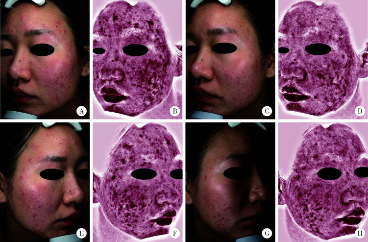
第2、4、6周治疗侧与对照侧4分红斑评分较治疗前相比差异均无统计学意义。治疗前治疗侧与对照侧的4分红斑严重程度评分比较差异无统计学意义(Z=1.41,P=0.16),治疗侧第8周与治疗前评分比较差异有统计学意义(Z=4.24, P < 0.01,Bonferroni校正P=0.01),对照侧第8周与治疗前评分比较差异无统计学意义(Z=1.73,P=0.08,Bonferroni校正P=0.01),第8周双侧面部4分红斑严重程度评分比较差异有统计学意义(Z=3.61, P < 0.01),结果显示585 nm Q开关激光治疗对于改善PIE有一定效果(表 2)。患者治疗侧第8周PIE程度较治疗前有明显改善,非治疗侧PIE前后差异无统计学意义(图 1)。
表 2.
585 nm Q开关激光治疗双侧治疗前后研究者红斑主观评分比较(n=22)
Comparison of investigator's subjective erythema score on the 585 nm Q-switched laser treatment side and the control side(n=22)
| Groups | Treatment side | Control side | Z 2 | P 2 | |||||||
| 0 | 1 | 2 | 3 | 0 | 1 | 2 | 3 | ||||
| Wilcoxon rank sum test, Z1, comparison of investigator’ s subjective erythema score prior treatment and on week 8 of treatment; Z2, comparison of Investigator’s subjective erythema score on the treatment side and on the control side. | |||||||||||
| Prior treatment, n | 0 | 5 | 14 | 3 | 0 | 6 | 14 | 2 | 1.41 | 0.16 | |
| Week 8 of treatment, n | 1 | 18 | 3 | 0 | 0 | 8 | 13 | 1 | 3.61 | < 0.01 | |
| Z 1 | 4.24 | 1.73 | |||||||||
| P 1 | < 0.01 | 0.08 | |||||||||
图 1.
患者治疗前后痤疮炎症性皮损和炎症后红斑变化
Changes of inflammatory skin lesions and erythema in a patient with acne before and after treatment
A and B show the photos of the treatment side before treatment, while C and D show the photos of week 8 of treatment. E and F show photos of the control side before treatment, while G and H show photos of the control side on week 8. A, C, E, G show photos in the ordinary light source while B, D, F, H show photos of the erythema area by VISIA. The inflammatory skin lesions on both sides were improved on week 8, the degree of postinflammatory erythema of the treatment side was significantly improved on week 8 compared with that before treatment, while it showed no significantly improvement on the control side.
2.4. 分光光度计测定红斑指数
患者不同时间治疗侧及对照侧红斑指数中位数及范围见表 3。多个样本Friedman检验结果提示,治疗侧χ2=17.844,P < 0.01,治疗前、第2、4、6、8周5个时间点间红斑指数差异具有统计学意义,两两比较检验结果提示第8周与治疗前比较差异具有统计学意义。对照侧χ2=7.590,P=0.11,5个时间点间红斑指数差异无统计学意义,结果显示585 nm Q开关激光治疗对于改善PIE有一定效果。
表 3.
585 nm Q开关激光治疗双侧红斑指数情况[M(Min, Max)]
Erythema index on the 585 nm Q-switched laser treatment side and the control side [M(Min, Max)]
| Groups | Prior treatment | Week 2 | Week 4 | Week 6 | Week 8 |
| Treatment side | 364.0(246.0, 564.0) | 356.5(250.0, 477.0) | 337.5(247.0, 503.0) | 339.5(224.0, 471.0) | 327.5(215.0, 464.0) |
| Control side | 344.5(251.0, 549.0) | 355.5(244.0, 488.0) | 348.5(234.0, 501.0) | 342.5(219.0, 497.0) | 336.0(233.0, 489.0) |
2.5. 患者满意度评价
22例患者均于末次评价进行满意度评分,其中9例患者评价为非常满意(40.9%),7例患者评价为满意(31.8%),6例患者评价为一般(27.3%)。
2.6. 安全性评价
全部22例患者均于治疗后即刻出现轻度红斑,均于3 d内消退。共17例患者于治疗后即刻出现轻度水肿,均于1 d内消退。共19例患者于治疗过程中出现轻度疼痛,3例患者出现中度疼痛,均无需药物治疗自行于1 d内缓解,未观察到瘙痒、渗出、紫癜、色素沉着等不良反应。
3. 讨论
痤疮是皮肤科的常见疾病,痤疮严重的炎症反应可导致PIE、PIH甚至瘢痕。痤疮PIE形成机制为炎症因子释放导致血管扩张[6],痤疮的炎症刺激也可引起血管增生,从而导致更为持久的痤疮PIE[6]。痤疮PIE一般会随时间逐渐好转,但速度较慢,当出现血管增生时在一些患者中甚至无法完全消退[7],对于患者的生活质量有巨大的影响。痤疮炎症若得不到及时处理,炎症刺激会引起黑素细胞活性增加、增生、体积增大,黑素转移到邻近的角质形成细胞,造成PIH[8],更难以消退。痤疮患者不仅追求皮损的早日消退,也希望能减少痤疮的后遗症如PIE、PIH等的发生,因此,寻找一种可以兼具治疗痤疮炎症性皮损和控制PIE的安全有效的治疗方法十分必要。
目前针对痤疮炎症后红斑虽然已经有多种治疗方法报告有效[5-7, 9-10],但仍无标准的治疗方案推荐。波长585 nm及595 nm的PDL对于扩张血管的治疗效果显著。小鼠模型研究证明,波长595 nm的PDL通过选择性光热作用,可造成血管凝固、血管收缩管径减小、血管消失[11]。PDL目前也被用于治疗炎症性皮肤病及某些非血管性皮肤病[12-13]。Seaton等[14]2003年的研究发现,PDL单次治疗12周后可见炎症性痤疮皮损明显改善,且没有严重副作用。痤疮PIE的组织病理表现为毛细血管扩张及新生血管[6],PDL治疗可使扩张血管凝固、收缩、管径缩小[11],从而达到治疗PIE的目的。2008年Yoon等[5]使用595 nm PDL治疗痤疮炎症性皮损和PIE,发现痤疮炎症性皮损数量和红斑指数均有明显改善。Park等[10]针对595 nm PDL及1 550 nm非剥脱点阵激光治疗PIE的疗效进行研究,发现两种激光均对PIE治疗有效,而Lekwuttikarn等[15]的临床研究发现595 nm PDL对于痤疮炎症性皮损以及PIE均未见显著疗效。Chalermsuwiwattanakan等[16]对比595 nm PDL及1 064 nm Nd: YAG激光治疗痤疮的疗效,发现两种激光对于炎症性皮损以及PIE均有显著疗效。PDL治疗的最主要副作用为紫癜形成,尤其当使用短脉宽治疗时较为常见,如果在治疗PIE时由于造成血管破裂及出血形成紫癜,可能会增加PIH的风险[1]。
585 nm Q开关激光为纳秒级脉冲激光,其脉宽短于微血管的热弛豫时间,且作为Q开关激光,可以产生光热作用及光声作用,使扩张血管凝固[1]。Panchaprateep等[1]应用585 nm Q开关激光对痤疮炎症性皮损和PIE进行治疗,在为期6周共3次的治疗后评估疗效,发现炎性皮损和红斑数量都有减少,红斑指数降低,且无严重不适感。本研究采用自身配对随机对照研究585 nm Q开关激光治疗痤疮炎症性皮损和PIE,结果显示经3次治疗后8周评价时,两侧面部IGA评分均较治疗前有显著性降低,但组间差异无统计学意义; 治疗侧较治疗前研究者红斑主观评分及红斑指数均有显著性下降,而对照侧红斑无改善,组间比较显示585 nm Q开关激光对于PIE有一定疗效。患者满意度较高,9例患者评价为非常满意,7例患者评价为满意,与上述文献报道的结果一致。
585 nm Q开关激光治疗的安全性较好,仅有治疗后即刻出现的轻度红斑、水肿,冰敷后可改善且1~3 d内自行消退; 患者疼痛感为轻至中度,可耐受; 该参数治疗后未见PIH及紫癜形成。Panchaprateep等[1]利用此激光治疗痤疮PIE,均未发现患者治疗后出现紫癜及PIH,与本研究结果相一致。Lekwuttikarn等[15]以及Chalermsuwiwattanakan等[16]使用PDL治疗痤疮及PIE均发现治疗后有少数患者出现PIH,于6~8周内恢复。综合上述文献报道,此治疗较PDL治疗出现紫癜及PIH可能性相对较低,且成本相对较低,可以考虑用于治疗痤疮PIE。
有研究显示595 nm PDL对于痤疮炎症性皮损有效[5, 14, 16-17]。本研究发现双侧面部IGA评分于8周均有降低,但两侧面部IGA评分差异无统计学意义,可能的原因包括:本研究中仅有4例患者没有联合使用系统与外用药物治疗,其余患者均有用药,因此双侧面部IGA评分均有显著降低,可能影响观察激光对于炎症性皮损的疗效; 痤疮本身容易复发及病情波动,部分患者在治疗期间有皮损波动,影响IGA评分。
本研究仍存在一定的不足之处,样本量相对较小可能造成一定的统计偏倚,且部分受试者有较多粉刺,对于评分可能有一定的影响,在以后的研究中应增加样本量,并完善患者研究前的洗脱期治疗。
综上所述,585 nm Q开关激光可以考虑用于痤疮PIE治疗,对于痤疮PIE治疗较为安全且有一定效果,但本研究样本量较小、治疗次数较少、评价时间较短,还需要进一步进行完善研究,希望本研究可以为痤疮PIE的激光治疗的选择上提供一定的帮助。
References
- 1.Panchaprateep R, Munavalli G. Low-fluence 585 nm Q-switched Nd: YAG laser: a novel laser treatment for post-acne erythema. Lasers Surg Med. 2015;47(2):148–155. doi: 10.1002/lsm.22321. [DOI] [PubMed] [Google Scholar]
- 2.Samuels DV, Rosenthal R, Lin R, et al. Acne vulgaris and risk of depression and anxiety: a meta-analytic review. J Am Acad Dermatol. 2020;83(2):532–541. doi: 10.1016/j.jaad.2020.02.040. [DOI] [PubMed] [Google Scholar]
- 3.鞠 强. 中国痤疮治疗指南(2019修订版) 临床皮肤科杂志. 2019;48(9):583–588. [Google Scholar]
- 4.Food and Drug Administration. Guidance for industry acne vulgaris: developing drugs for treatment[EB/OL]. (2016-10-16)[2021-02-26]. www.fda.gov/downloads/Drugs/Guidances/UCM071292.pdf.
- 5.Yoon HJ, Lee DH, Kim SO, et al. Acne erythema improvement by long-pulsed 595 nm pulsed-dye laser treatment: a pilot study. J Dermatolog Treat. 2008;19(1):38–44. doi: 10.1080/09546630701646164. [DOI] [PubMed] [Google Scholar]
- 6.Afra TP, Razmi TM, De D. Topical timolol for postacne erythema. J Am Acad Dermatol. 2020;84(6):e255–e256. doi: 10.1016/j.jaad.2020.04.144. [DOI] [PubMed] [Google Scholar]
- 7.Min S, Park SY, Yoon JY, et al. Fractional microneedling radiofrequency treatment for acne-related post-inflammatory erythema. Acta Derm Venereol. 2016;96(1):87–91. doi: 10.2340/00015555-2164. [DOI] [PubMed] [Google Scholar]
- 8.Chaowattanapanit S, Silpa-Archa N, Kohli I, et al. Postinflammatory hyperpigmentation: a comprehensive overview: treatment options and prevention. J Am Acad Dermatol. 2017;77(4):607–621. doi: 10.1016/j.jaad.2017.01.036. [DOI] [PubMed] [Google Scholar]
- 9.Mathew ML, Karthik R, Mallikarjun M, et al. Intense pulsed light therapy for acne-induced post-inflammatory erythema. Indian Dermatol Online J. 2018;9(3):159–164. doi: 10.4103/idoj.IDOJ_306_17. [DOI] [PMC free article] [PubMed] [Google Scholar]
- 10.Park KY, Ko EJ, Seo SJ, et al. Comparison of fractional, nonablative, 1 550 nm laser and 595 nm pulsed dye laser for the treatment of facial erythema resulting from acne: a split-face, evaluator-blinded, randomized pilot study. J Cosmet Laser Ther. 2014;16(3):120–123. doi: 10.3109/14764172.2013.854626. [DOI] [PubMed] [Google Scholar]
- 11.Li D, Chen B, Wu W J, et al. Experimental study on the vascular thermal response to visible laser pulses. Lasers Med Sci. 2015;30(1):135–145. doi: 10.1007/s10103-014-1631-3. [DOI] [PubMed] [Google Scholar]
- 12.Erceg A, de Jong EM, van de Kerkhof PC, et al. The efficacy of pulsed dye laser treatment for inflammatory skin diseases: a systematic review. J Am Acad Dermatol. 2013;69(4):609–615. doi: 10.1016/j.jaad.2013.03.029. [DOI] [PubMed] [Google Scholar]
- 13.Forbat E, Al-Niaimi F. Nonvascular uses of pulsed dye laser in clinical dermatology. J Cosmet Dermatol. 2019;18(5):1186–1201. doi: 10.1111/jocd.12924. [DOI] [PubMed] [Google Scholar]
- 14.Seaton ED, Charakida A, Mouser PE, et al. Pulsed-dye laser treatment for inflammatory acne vulgaris: randomised controlled trial. Lancet. 2003;362(9393):1347–1352. doi: 10.1016/S0140-6736(03)14629-6. [DOI] [PubMed] [Google Scholar]
- 15.Lekwuttikarn R, Tempark T, Chatproedprai S, et al. Rando-mized, controlled trial split-faced study of 595 nm pulsed dye laser in the treatment of acne vulgaris and acne erythema in adolescents and early adulthood. Int J Dermatol. 2017;56(8):884–888. doi: 10.1111/ijd.13631. [DOI] [PubMed] [Google Scholar]
- 16.Chalermsuwiwattanakan N, Rojhirunsakool S, Kamanamool N, et al. The comparative study of efficacy between 1 064 nm long-pulsed Nd: YAG laser and 595 nm pulsed dye laser for the treatment of acne vulgaris. J Cosmet Dermatol. 2020;20(7):2108–2115. doi: 10.1111/jocd.13832. [DOI] [PubMed] [Google Scholar]
- 17.Choi YS, Suh HS, Yoon MY, et al. Intense pulsed light vs. pulsed-dye laser in the treatment of facial acne: a randomized split-face tria. J Eur Acad Dermatol Venereol. 2010;24(7):773–780. doi: 10.1111/j.1468-3083.2009.03525.x. [DOI] [PubMed] [Google Scholar]


