Abstract
The rumen is a complex ecosystem that plays a critical role in our efforts to improve feed efficiency of cattle and reduce their environmental impacts. Sequencing of the 16S rRNA gene provides a powerful tool to survey the bacterial and some archaeal. Oral stomach tubing a cow to collect a rumen sample is a rapid, cost-effective alternative to rumen cannulation for acquiring rumen samples. In this study, we determined how sampling method (oral stomach tubing vs cannulated grab sample), as well as rumen fraction type (liquid vs solid), bias the bacterial and archaeal communities observed. Liquid samples were further divided into liquid strained through cheesecloth and unstrained. Fecal samples were also collected to determine how these differed from the rumen sample types. The abundance of major archaeal communities was not different at the family level in samples acquired via rumen cannula or stomach tube. In contrast to the stable archaeal communities across sample type, the bacterial order WCHB1-41 (phylum Kiritimatiellaeota) was enriched in both liquid strained and unstrained samples as well as the family Prevotellaceae as compared to grab samples. However, these liquid samples had significantly lower abundance of Lachnospiraceae compared with grab samples. Solid samples strained of rumen liquid most closely resembled the grab samples containing both rumen liquid and solid particles obtained directly from the rumen cannula; therefore, inclusion of particulate matter is important for an accurate representation of the rumen bacteria. Stomach tube samples were the most variable and were most representative of the liquid phase. In comparison with a grab sample, stomach tube samples had significantly lower abundance of Lachnospiraceae, Fibrobacter and Treponema. Fecal samples did not reflect the community composition of the rumen, as fecal samples had significantly higher relative abundance of Ruminococcaceae and significantly lower relative abundance of Lachnospiraceae compared with grab samples.
Introduction
The ruminant stomach consists of four chambers the reticulum, rumen, omasum, and abomasum. The rumen, which is the largest of the four compartments, is a complex pregastric anaerobic fermentation chamber that harbors a diverse microbial community of bacteria, archaea, protozoa, and fungi [1]. The diverse bacterial communities of the rumen comprise nearly 95% of the total microbial community as determined by 16S rRNA gene sequencing [2]. It is important to define the bacterial communities within the rumen because many genera have been linked to feed efficiency, milk yield, and milk composition in dairy cattle [3, 4]. In addition, factors such as age [5], breed [6–8], health status, and season [9] all contribute to variation in the microbiota of the rumen. However, the primary factor affecting the taxa present in the rumen, as well as the richness of those taxa, is the dietary composition; with the ratio of forage-to-concentrate in the diet being most important [10–12].
Next generation sequencing of 16S rRNA gene amplicons have been a successful tool for characterizing the diversity of the bacterial communities within the rumen [13, 14]. This technology is advantageous in that it allows the identification of a broader array of rumen bacterial taxa, than the small fraction of species that can be successfully cultured [15]. However, the most appropriate method of obtaining a representative rumen sample is still widely debated [16]. It is well known that the bacterial communities in the solid and liquid portions of the rumen digesta differ in composition, suggesting that the sampling method used will affect the characterization of the community [17–22]. Thus, identifying sampling methods that accurately represent both the liquid and solid fractions of the rumen digesta are necessary.
Much of the existing research describing the rumen microbiome was performed on animals surgically fitted with rumen cannula, which offer the accuracy and convenience of sampling both liquid and solid rumen digesta directly from the rumen chamber. However, the surgical fistulation procedure is invasive, and the costs associated with the procedure as well as the ongoing animal care limit the number of animals that can feasibly be used in an experiment. Importantly, if microbial biomarkers of health or disease are eventually identified for on-farm testing, retrieving rumen fluid through a cannula is not a practical approach on commercial dairy and livestock farms. Alternatively, many studies have used an oral stomach tube to collect rumen fluid without the need for a rumen fistula [8, 16, 23]. Oral stomach tubes are a cheaper, less invasive approach to rumen sampling that can be performed on as many cows as necessary, thus economically increasing the experimental sample size. In terms of bacterial community composition and diversity, rumen fluid extracted via the fistula was comparable to fluid extracted via the oral stomach tube [8, 24]. Some of the disadvantages of using an oral stomach tube include possible contamination by saliva (which affects the pH of the sample), inconsistent sampling region within the rumen, stress to the animal, skilled labor associated with use, and limited representation of particulate matter in samples; though the importance of these concerns to the bacterial community composition of a sample are widely debated among researchers [16, 25, 26].
The collection of fecal material from cattle is another non-invasive, simple, and inexpensive technique that is not as commonly regarded as a viable tool for collecting samples representative of the rumen microbiota. Although fecal sampling requires minimal equipment, is low-cost, and can be performed easily on any animal, bacterial communities of the feces were found to not reflect the rumen digesta [27, 28]. However, in these studies, the fecal microbiome was not compared with the liquid and solid fractions of the rumen digesta individually. If the feces reflect the bacterial communities in the solid fraction, fecal samples might be useful in evaluating the bacterial taxa involved in fiber digestion. Conversely, if fecal samples represent the liquid fraction, lactate-producing bacteria that contribute to ruminal acidosis could be diagnosed in a less invasive manner.
The aim of this study was to identify and compare the bacterial, and to a limited extent the archaeal community, present in samples collected using three methods–an oral stomach tube, fecal samples, and grab sample through a rumen fistula. Additionally, grab samples were separated into their solid and liquid components by straining through cheesecloth. Lastly, an unstrained liquid sample was taken from the rumen fistula for a total of six sample types. As grab samples containing both rumen fluid and particulate matter are considered the gold standard for surveying bacterial and archaeal communities in the rumen all sample types were compared to grab samples as a baseline. We hypothesized that the bacterial and archaeal communities observed would differ between sampling techniques. To the authors’ knowledge, no studies have considered this variety of sampling methods on a comparative basis using next generation sequencing. Our results will be useful in helping investigators design experiments that capture their bacterial or archaeal communities of interest.
Materials and methods
Animals
The experimental protocol and all procedures used in this study were approved by the UC Davis Institutional Animal Care and Use Committee. Four non-lactating Holstein (3) and Jersey (1) cows, each ruminally fistulated prior to the study, were used for the collection of samples. For the two-week duration of the study, cattle were housed individually with ad libitum access to water and offered the same maintenance total mixed ration (TMR) twice daily at approximately 08:00 and 16:00. Dietary composition of TMR was analyzed for protein, fiber, mineral, and energy content (Cumberland Valley Analytical Services, Hagerstown, MD; Table 1).
Table 1. Dietary composition of total mixed ration.
| Itema | Dietary Compositionb |
|---|---|
| Dry matter | 88.9% |
| Crude protein | 10.7% DM |
| Soluble protein | 28.6% CP, 3.1% DM |
| ADF | 78.5% NDF, 36.7% DM |
| aNDF | 46.8% DM |
| Ash | 9.62% DM |
| Calcium | 0.33% DM |
| Phosphorus | 0.22% DM |
| Magnesium | 0.25% DM |
| Potassium | 1.91% DM |
| Sodium | 0.08% DM |
| Iron | 449 ppm |
| Zinc | 54 ppm |
| Copper | 8 ppm |
| TDN | 57.5% DM |
| Net energy lactation c | 1.12 Mcal/kg |
| Net energy maintenance c | 1.07 Mcal/kg |
| Net energy gain c | 0.57 Mcal/kg |
| Non fiber carbohydrates | 32.9% DM |
Chemical composition of the total mixed-ration (TMR) fed to the rumen-fistulated dry cows. Dietary analysis conducted by Cumberland Valley Analytical Services (Hagerstown, MD) completed 12/01/2016. Ingredient composition of TMR on an as is a basis was 50% wheat hay, 25% alfalfa hay, 21.4% almond hulls and 3.6% mineral supplement.
a Acid detergent fiber (ADF); Ash free Neutral Detergent Fiber (aNDF); Total Digestible Nutrients (TDN).
b Dry Matter (DM); Crude Protein (CP); Neutral Detergent Fiber (NDF).
c Calculations for net energy metrics were estimated as described in Linn [29].
Sampling
Cows were given a one-week period for environmental adaption prior to sampling. This adaptation period was necessary to allow them to acclimate to an individual (rather than group) feeding approach and to reduce sorting of the feed. All cows were fed their normal maintenance TMR diet (Table 1) prior to and throughout the study, as was typical of all cows in their milking string. Sampling of fecal and ruminal contents occurred on days 7, 9, and 11 of the experiment, and took place approximately 4 hours after morning feeding. Fecal samples were collected from the rectum with sterile polyethylene gloves and stored in plastic bags. Grab samples (containing both liquid and particulate matter) from the fistula were collected from the medioventral region of the rumen and stored in plastic bags.
To obtain strained liquid and solid samples about 250 ml of rumen contents taken from the fistula was squeezed through 4 layers of cheesecloth to separate large particles, as previously done by others [17, 30, 31]. Solid rumen digesta contents remaining in the cheesecloth became the solid samples while the liquid that passed through the cheesecloth was the strained liquid sample. Solid samples were then stored in plastic bags until DNA extraction. Additionally on days 9 and 11, a liquid unstrained sample was taken by collecting rumen fluid with particulates small enough to fit in the tubing from the fistula using a PVC pipe, Tygon® tubing, and a large syringe. As with the other samples this liquid unstained sample was stored in 240 ml sterile plastic vials.
Lastly, to obtain the stomach tube sample, enough rumen liquid to fill a 240 ml sterile plastic vial was collected via an oral stomach tube using an oral speculum, Tygon® tubing (1.5 cm O.D. and 0.9 cm I.D.) and a vacuum pump. The first 100 ml of rumen fluid were discarded to avoid saliva contamination. A fresh Tygon® tube was used for each cow and was thoroughly rinsed and bleached between sampling days to avoid cross-contamination of samples. The pH of each of the liquid-containing samples was measured with a portable pH meter to ensure the pH was below 7 (Milwaukee Instruments, Rocky Mount, NC). Liquid samples ranged between a pH of 6.6 and 6.9. All samples were held on ice during transport and stored in triplicate 60 ml vials at -20°C for DNA extraction and dry matter analysis. In total there was 6 sample types taken: feces, stomach tube, grab sample from cannula, unstrained liquid from the rumen cannula, and separate solid and liquid fractions obtained by straining.
DNA extraction and PCR amplification
DNA extraction was performed using a ZR Fecal DNA MiniPrep™ kit (Zymo Research Corp., Irvine, CA), with slight modifications to the manufacturer’s instructions. Samples were thawed at room temperature, and 200 mg of each sample were used for DNA extraction, which included a bead bashing step to facilitate the mechanical lysis of cell walls. As the last step in the procedure, DNA was eluted from the column with elution buffer, and the resulting DNA was evaluated for concentration and purity on a NanoDrop 2000 spectrophotometer (Thermo Scientific, Waltham, MA, USA) and stored at -20° C. The V4 region of the bacterial 16S rRNA gene was amplified from each sample using forward primer F515 containing a unique 8 bp barcode (N) and linker region (GT) (5’-NNNNNNNNGTGTGCCAGCMGCCGCGGTAA-3’) and the reverse primer R806 (5’-GGACTACHVGGGTWTCTAAT-3’). The amplification was carried out in triplicate using GoTaq® Green Master Mix (Promega, Madison, WI) as previously described [32]. In brief, PCR conditions were set at initial denaturation for 94° C for 3 min; followed by 35 cycles of 94° C for 45 seconds, 50° C for 1 min, 72° C for 90 seconds with final extension step at 72° C for 10 min. Triplicates were combined in equal concentrations and amplicons were evaluated for off target bands by gel electrophoresis, pooled and then purified using a QIAquick PCR Purification Kit (QIAGEN, Hilden, Germany). A 50 μl aliquot of the final pooled PCR product was sequenced at the UC Davis Genome Center DNA Technologies Core via the Illumina MiSeq PE250 platform (Illumina, CA).
Amplicon library processing
Raw paired end reads were screened to remove phiX, human, and host contamination using Kneaddata v0.6.1 by aligning reads to the phiX174 (NCBI ACC: NC_001422.1), bovine (ARS-UCD1.2) and human (GRCh38) reference genomes [33]. Reads were demultiplexed followed by trimming of primers and barcodes with Cutadapt v1.18 [34]. Ends of reads were trimmed for quality, any read smaller than 150 bp was discarded and a max expected error of 2 was used as a quality filter using the filterAndTrim function from DADA2 v1.8.0 [35]. Sequences were merged, denoised, chimeras were removed, and exact amplicon sequence variants (ASVs) were identified using DADA2. Taxonomy was assigned using the RDP native Bayesian classifier algorithm in the DADA2 assignTaxonomy function with the SILVA reference database v.132 training set. A phylogenetic tree of unique ASVs was made using FastTree with default options in QIIME v.1.9.1 [36]. The ASV table, sequences and tree produced by DADA2 were imported into the R package Phyloseq v.1.24.2 for further analysis [37].
Bacterial and archaeal community analyses and statistics
First, unsupervised exploratory analysis was conducted with double principal coordinates analysis (DPCoA), which was calculated and graphed with the phyloseq R package [37, 38]. Both modeling and hypothesis testing of differentially abundant ASVs between sample types was determined using the Corncob R package [39]. All genera-level and ASV-level relative abundances were modeled using a beta-binomial regression with a logit-link for mean and dispersion as described by Martin et al. [39]. Differential relative abundances were modeled as a linear function of sample type, cow, and day. Significant differentially abundant ASVs were determined with the parametric Wald test with bootstrapping (n = 1000) as described by Martin et al. [39]. Within the Corncob algorithm the Benjamini-Hochberg (BH) adjustment for multiple comparisons was used to calculate adjusted p-values. An adjusted p-value ≤ 0.05 was considered significant. This model has the benefits of accommodating the absence of a taxon in samples without zero-inflation or pseudocounts, accounts for differences in library sizes, and gives valid inference even with small samples sizes [39]. Richness of sample types was estimated with the R package breakaway and evenness (Shannon diversity) was calculated using the R package DivNet, which accounts for the structure of the communities [40, 41]. Hypothesis testing of alpha diversity (richness and evenness) metrics was done using the betta() function using sample type, cow, and day as fixed effects in the breakaway R package [42]. Beta diversity was calculated by using weighted and unweighted UniFrac distances and graphed by PCA clustering in the Phyloseq R package [37]. The number of clusters in the data was determined with the gap statistic using the gapstat_ord() function in Phyloseq [43].
Results
Sequence processing of rumen and fecal samples
After filtering with Kneaddata and demultiplexing the single run of MiSeq yielded 747,961 250 bp raw paired-end reads that entered the DADA2 pipeline. After the quality trimming, initial filtering, and chimera removal, the library size ranged from 2,189 to 24,624 reads, with a median library size of 7,197 and an average size of 8,110 reads. The median read length of quality filtered merged reads was 257 bp. A total of 5,607 AVSs were identified, of which 94 were not assigned to a phylum and thus were removed for analysis along with 12 ASVs assigned to chloroplasts and mitochondria. The 94 unassigned taxa were found in all sample types with solid samples having the most reads of unknown taxa. This suggests there are still a diverse group of microbes attached to solid particles that have yet to be identified. The final feature table had 5,485 ASVs across 68 samples. Only 44 ASVs were assigned to Archaea and the remaining 5,441 ASVs were determined to be Bacteria.
Community composition of all sample types
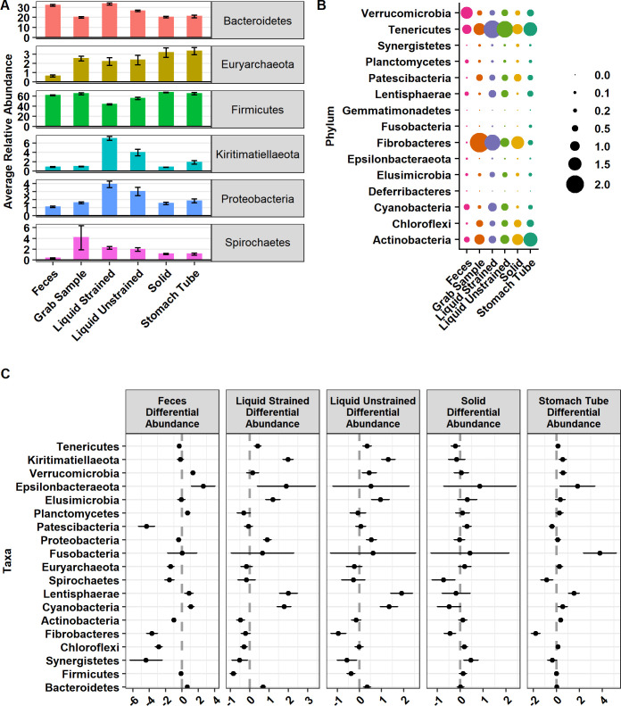
The 5,485 ASVs were assigned to 21 phyla, 78 orders, 117 families, and 293 genera. Here we define major phyla as those with a mean relative abundance in at least one sample type of greater than 3%. Major phyla were Firmicutes, Bacteroidetes, Kiritimatiellaeota, Proteobacteria, Euryarchaeota and Spirochaetes (Fig 1A). Of these major phyla, Firmicutes was lower in relative abundance (P ≤ 0.0001; Fig 1C) in both liquid strained and unstrained sample types compared to grab samples. In contrast, Bacteroidetes had higher relative abundance in feces, liquid strained, and liquid unstrained samples as compared with grab samples (P ≤ 0.0001; Fig 1C). Proteobacteria had higher relative abundance in liquid strained and unstrained samples and lower relative abundance in feces as compared to grab samples (P ≤ 0.002; Fig 1C). In addition, Kiritimatiellaeota was higher in relative abundance in stomach tube and liquid samples compared to grab samples (P ≤ 0.001; Fig 1C). Spirochaetes was lower in relative abundance in feces, stomach tube, and solid samples compared with grab samples (P ≤ 0.003; Fig 1C). While Euryarchaeota had lower relative abundance in feces, it had higher relative abundance in stomach tube samples compared with grab samples (P = 3.24x10⁻⁸). Minor phyla were those with less than 3% relative abundance in all samples (Fig 1B). For further data exploration an interactive version of Fig 1B with mean and standard deviations for each phyla is available at https://doi.org/10.5281/zenodo.4026849 with the title “Minor_phyla_plotly.html”. The phylum Gemmatimonadetes was only found in stomach tube samples and Deferribacteres was only found in fecal samples (Fig 1B). For the minor phyla in feces Tenericutes, Patescibacteria, Actinobacteria, Fibrobacteres, Chloroflexi and Synergistetes were lower in relative abundance and Verrucomicrobia, Epsilonbacteraeota, Cyanobacteria, Planctomycetes and Lentisphaerae were higher in relative abundance compared with grab samples (P ≤ 0.001; Fig 1C). Samples acquired with the oral stomach tube had significantly lower relative abundance of Patescibacteria and Fibrobacteres and significantly higher relative abundance of Verrucomicrobia, Epsilonbacteraeota, and Fusobacteria compared with grab samples. Only 1.68% of ASVs were assigned a species, but 67.3% were able to be assigned to a genus.
Fig 1.
Relative abundance of (A) major and (B) minor phyla and (C) their differential abundances. (A) Relative abundance of major phyla defined as those phyla found at greater than 3% relative abundance and graphed as relative abundance ± SE. (B) Minor phyla defined as those found below 3% relative abundance present in sample types. (C) Phyla that are significantly differentially abundant compared with grab samples. Graphed as coefficients with a 95% confidence interval from the corncob model. Families with negative coefficients for a sample type are expected to have a lower relative abundance when compared to the grab samples while positive coefficients suggest a higher relative abundance in that sample type compared to grab samples.
Liquid unstrained and fecal samples were the least variable samples as they shared 510 and 441 ASVs, respectively, with samples of their same type. On the other hand, stomach tube and liquid strained samples were the most variable as these sample types only shared 225 and 307 ASVs, respectively, with samples of their same type. Moderately variable sample types were grab and solid samples, which shared 319 and 405 ASVs, respectively, with samples of their same type.
Diversity
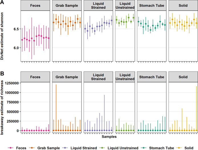
The evenness of fecal samples was lower than all rumen sample types (P ≤ 0.001; Fig 2A). Fecal, stomach tube, and liquid strained samples had lower evenness than grab samples (P ≤ 0.001; Fig 2A). Solid and liquid unstrained samples did not have significantly different evenness compared with grab samples (P ≥ 0.05; Fig 2A). Both the individual cow sampled, and day of sampling affected the evenness of a sample (P ≤ 0.05; Fig 2A).
Fig 2. Differences in estimated alpha diversity among sample types.
(A) DivNet estimate of Shannon diversity plotted as mean with 95% confidence intervals and (B) mean breakaway estimate of species richness with 95% confidence intervals. The x-axis ticks represent samples from each cow on different days. Both the richness and evenness of fecal samples were lower than all other rumen sample types (P ≤ 0.001). Stomach tube and liquid strained samples had lower evenness than grab samples (P ≤ 0.001). Solid and stomach tube samples were estimated to have fewer species than grab samples (P = 0.02 and P ≤ 0.001, respectively).
The richness of samples from the rumen were estimated to be higher than that of fecal samples (P ≤ 0.001; Fig 2B). Fecal samples were estimated to have a mean of 2,021 species, which was lower than the grab samples estimated mean of 4,119 species (P ≤ 0.001; Fig 2B). Liquid strained and unstrained samples did not have a significantly different mean number of estimated species compared with grab samples (P ≥ 0.05; Fig 2B). However, solid and stomach tube samples were estimated to contain a lower number of species compared with grab samples, an estimated 286 and 506 less species, respectively (P = 0.02, P ≤ 0.001; Fig 2B). Neither the day sampled, nor individual cow significantly affected the estimated number of species in a sample (P ≥ 0.05; Fig 2B).
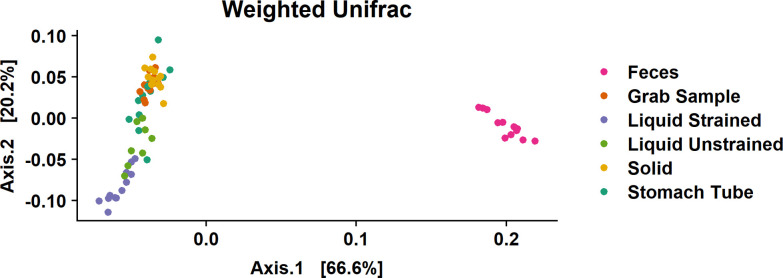
Weighted UniFrac distances were calculated to estimate beta diversity. Calculations of eigenvalues showed that 86.8% of the variance between samples was contained in the first two principal components, thus a two-dimensional visualization was deemed appropriate (Fig 3). Two distinct groups were present with fecal samples clustering away from all rumen sample types (Fig 3). Grab and solid samples exhibited low variability and overlapped each other, forming one group. Liquid samples were further down the second axis, which might indicate that there were distinct phylogenetic differences between these samples and grab samples. Stomach tube samples were the most variable with some of these samples found within the grab and solid sample cluster, while other stomach tube samples were more closely associated with liquid samples. The gap statistic of the weighted UniFrac distance indicated there were at least 3–5 clusters in the data. As there are six sample types in the dataset, this suggests that grab and solid samples are likely one cluster as these samples overlap the most (Fig 3). The unweighted UniFrac showed a similar pattern, with less variation explained in axis one and two, 45.8% and 6.9%, respectively (S1 Fig).
Fig 3. Beta diversity as weighted UniFrac distances between samples.
To faithfully reflect the variance in the coordinates, the height-to-width ratio was based on the ratio between the corresponding eigenvalues.
Overall differences between sample types
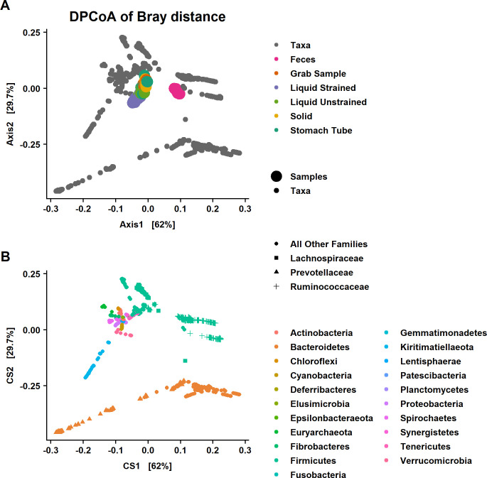
As an exploratory first step, DPCoA was performed (Fig 4A). An interactive version of this graph with taxon identification is available at https://doi.org/10.5281/zenodo.4026849 as interactive Fig 2 –with the title “DPCoA.html”. Additionally, since Firmicutes and Bacteroidetes dominated a majority of the graph, an interactive version without these phyla was created with the aim to allow a better visualization of minor phyla and is available at https://doi.org/10.5281/zenodo.4026849, titled “DPCoA_NoFrimBact.html. This phylogenetic ordination method provides a biplot representation of both samples and taxonomic categories. The DPCoA was used to visualize the underlying structure of these data and identify taxa that could be contributing to differences between sample types that will be specifically examined with differential abundance testing.
Fig 4. Double principal coordiant analysis (DPCoA) of the Bray-Curtis distances among samples.
DPCoA is a phylogenetic ordination method and that provides a biplot representation of both (A) samples and (B) taxonomic categories. Note that while the biplots are a square shape to fit the page the CS1 explains roughly twice the variation of CS2 similar to what is seen in Fig 1. The 1st axis discrimates fecal from rumen samples while the 2nd axis separates liquid strained samples from other rumen sample types. Samples that have larger scores on CS1 have a subset of taxa from Bacteroidetes and Firmicutes that is different than rumen samples. Liquid strained samples have lower values on CS2 suggesting they are distinguished from other rumen sample types by taxa in the phylum Kiritimatiellaeota and family Prevotellaceae. The DPCoA predictes that fecal samples will have a lower abundance of both Lachnospiraceae and Prevotellaceae and greater abundance of Ruminococcaceae compared to samples from the rumen.
Fecal samples clustered away from samples that were collected from the rumen, which was primarily driven by differences in the relative abundance of a subset of Firmicutes in the families Ruminococcaceae, Lachnospiraceae and Christensenellaceae and a subset of families in the phylum Bacteroidetes, mainly Rikenellaceae and Prevotellaceae on the 1st axis (Fig 4B). Additionally, fecal samples separate from samples from the rumen based on having more taxa from the family Akkermansiaceae and phylum Tenericutes and fewer from the families Fibrobacteraceae, and Spirochaetaceae (Fig 4B and S1 Fig). Liquid samples were found lower on the 2nd axis of the DPCoA, indicating these samples had more taxa from the phylum Kiritimatiellaeota and a subset of Bacteroidetes—most of them in the family Prevotellaceae (Fig 4B). Also, the separation of liquid strained samples away from other rumen samples was due to fewer taxa from the phylum Euryarchaeota and the family Eggerthellaceae that is within the phylum Actinobacteria (S1 Fig).
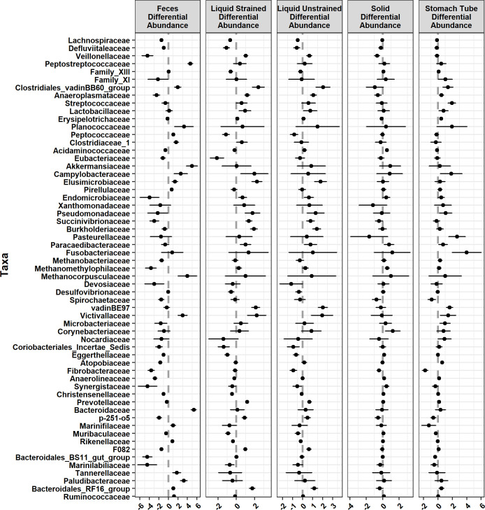
To test the significance of these differences, differential abundance testing was performed with Corncob using the gold standard grab samples as the baseline for all other sample types. The relative abundance of Prevotellaceae was lower in feces and was higher in liquid samples compared with grab samples (P ≤ 0.0004; Figs 5A and 6). Solid samples (P = 0.77) were not significantly different and stomach tube samples (P = 0.06) trended toward significance in the relative abundance of Prevotellaceae compared with grab samples. The relative abundance of Prevotellaceae was highest in liquid strained samples compared with other sample types (Fig 5A). In comparison to grab samples, the relative abundance of Ruminococcaceae was higher in feces (P ≤ 0.001; Fig 5B) and solid (P = 0.003; Fig 5B) samples while liquid strained samples had lower relative abundance (P ≤ 0.001; Fig 5B). Neither stomach tube nor liquid unstrained samples had significantly different relative abundance of Ruminococcaceae compared with grab samples. Fecal samples were lower in relative abundance of Lachnospiraceae compared with all other samples (P ≤ 6.96x10⁻¹⁵), while the relative abundance was higher for grab samples compared with all other sample types (P ≤ 0.03; Fig 5C). Neither day of sampling nor individual animal significantly affected the relative abundance of Prevotellaceae (P ≥ 0.05). In contrast, the relative abundance of Ruminococcaceae and Lachnospiraceae was significantly affected by individual animal (P ≤ 0.03), but not day of sampling.
Fig 5. Significant differences in the relative abundance of specific bacterial families.
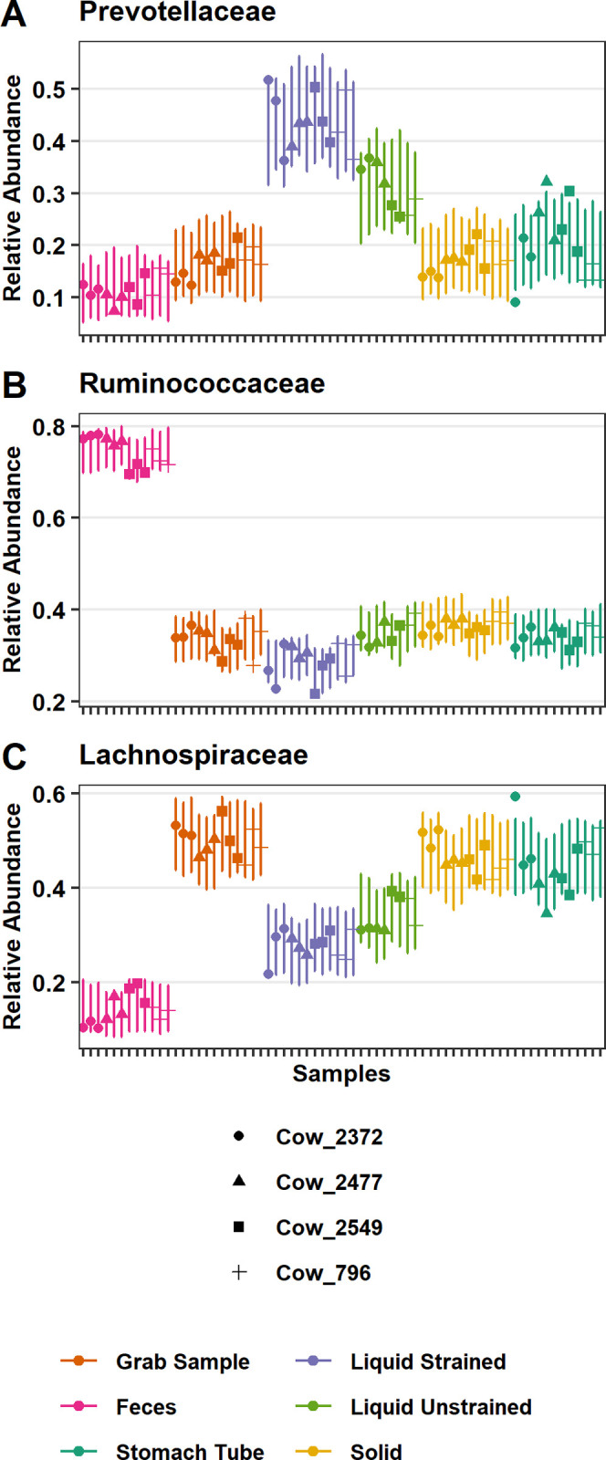
Relative abundance of (A) Prevotellaceae (B) Ruminococcaceae and (C) Lachnospiraceae as modeled by corncob. Points are the estimated relative abundance and bars are a 95% prediction interval for each cow on different days of sampling.
Fig 6. Families that were significantly differentially abundant across sample type compared with grab samples.
Graphed as coefficients with a 95% confidence interval calculated from the corncob model. Families with negative coefficients for a sample type are expected to have a lower relative abundance when compared to the grab samples while positive coefficients suggest a higher relative abundance in that sample type compared to grab samples.
Specific community differences between grab and fecal samples
To further distinguish what taxa were contributing to the separation of fecal samples from rumen grab samples on the DPCoA, we identified taxa that were found in one sample type and not the other. Within the phyla Firmicutes and Bacteroidetes, families Barnesiellaceae, Chitinophagaceae, p-2534-18B5_gut_group, GZKB124, and Hymenobacteraceae were found in fecal samples, but were not found in grab samples. Conversely, Leuconostocaceae, Carnobacteriaceae, Aerococcaceae, Syntrophomoadaceae, Bacteroidetes_DB2-2, PeH15, M2PB4-65_termite_group, COB_P4-1_termite_group, Spirosomaceae, and Porphyromonadaceae were found in grab samples, but were not found in fecal samples.
Next, we identified ASVs, genera and families that differed in relative abundance between sample types. There were 657 significant differentially abundant ASVs in fecal samples compared with grab samples, as well as 114 differentially abundant genera (P ≤ 0.05; S2 Fig). At the genera level, 131 ASVs were unable to be fit to the Corncob model for differential abundance testing. Primarily, this was due to either limited or lack of reads in one of the sample types. Of these genera that did not fit the model, Acetatifactor, Shuttleworthia, Succinivibrio, Veillonellaceae UCG-001, and Lachnospiraceae UCG-006 were found in all grab samples with greater than or equal to 50 reads across all samples, but were absent in fecal samples. Similarly, there were 11 genera found in all fecal samples with 50 or more reads, but these were not found in any grab samples including Coprococcus 3, Cellulosilytium, Clostridioides, Paeniclostridium, Parasutterella, Aeriscardovia, Odoribacter, Harryflintia, Negativibacillus, Pygmaiobacter, and Ruminococcaceae UCG-011.
The most common families with differentially abundant ASVs were Lachnospiraceae, Ruminococcaceae, Christensenellaceae, Family XIII, Rikenellaceae, and Prevotellaceae. These families are in the phyla Firmicutes and Bacteroidetes, which had the most significant differentially abundant ASVs. However, as a percent of total ASVs these phyla only had 4.9% and 16.3% significant differentially abundant ASVs, respectively. In contrast, 25.6% of the ASVs assigned to Chloroflexi and 29.5% of ASVs assigned to Euryarchaeota were significantly different between grab and fecal samples. The significant ASVs in Chloroflexi were all assigned to the genus Flexilinea. In addition to the significantly lower relative abundance of some Chloroflexi ASVs in fecal samples compared with grab samples, another 51.3% of the ASVs in the phyla were not found in any fecal samples (S2 Fig). In the phylum Euryarchaeota, feces had significantly lower relative abundance of Methanobrevibacter, Methanosphaera, and were almost devoid of Methanomethylophilaceae.
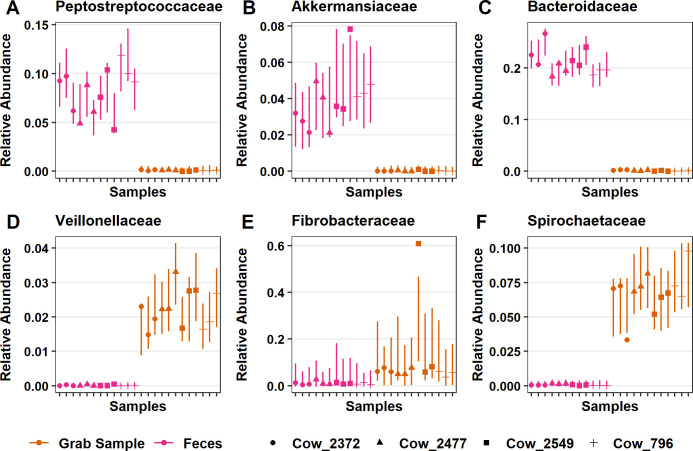
There were 30 families that had significantly lower relative abundance while 18 families had higher relative abundance in feces and compared to grab samples (Fig 6). Families that had the strongest positive relationship with fecal samples were Peptostreptococcaceae (P = 1.76x10⁻⁷; Fig 7A), Akkermansiaceae (P = 6.95x10⁻⁵; Fig 7B), and Bacteroidaceae (P = 7.87x10⁻¹²; Fig 7C), which were significantly higher in relative abundance compared with grab samples. Conversely, the families with largest negative relationship between fecal samples and that had lower relative abundance in comparison to grab samples were Veillonellaceae (P = 1.66x10⁻¹¹; Fig 7D) and Bacteroidales_BS11_gut_group (P = 6.20x10⁻¹¹; Fig 7E). Additionally, fecal samples separated from rumen samples on the DPCoA (S1 Fig) due in part to differences in the families Spirochaetaceae and Fibrobacteraceae both of which had lower relative abundance than grab samples (P = 7.88x10⁻⁹ and P = 4.73x10⁻⁸, respectively; Fig 7F).
Fig 7. Significant differences in the relative abundance of specific bacterial families between fecal and grab samples.
Fecal samples had significantly higher relative abundance of (A) Peptostreptococcaceae, (B) Akkermansiaceae, (C) Bacteroidaceae, compared to grab samples. Also, there was significantly lower relative abundance of (D) Veillonellaceae, (E) Bacteroidales_BS11_gut_group and (F) Spirochaetaceae compared to grab samples. Points are the estimated relative abundance and bars are a 95% prediction interval for each cow on different days of sampling.
Based on the DPCoA findings, the phyla Spirochaetes and Actinobacteria also played an important role in distinguishing feces from grab samples (Figs 1A and 4B). In the phylum Spirochaetes, there were 10 ASVs, all of which were from the genera Treponema_2, that had significantly lower relative abundance in fecal samples compared with grab samples. Within the phylum Actinobacteria, there were 4 ASVs in the genera Olsenella, 5 ASVs in Atopobium, 7 ASVs in the genera DNF00809, and 1 ASV assigned to Raoultibacter, which were all significantly lower in relative abundance compared with grab samples.
Specific community differences between grab and stomach tube samples
Oral stomach tube samples were composed of 20 phyla, 65 orders, 98 families, and 236 genera. There were 255 ASVs found in grab samples that were not found in the stomach tube samples. Likewise, 404 ASVs in stomach tube samples were not present in the grab samples. There were 3,615 ASVs that were in common between stomach tube and grab samples. Three families Rhodobacteraceae, Bacteriovoracaeae, and Spirosomaceae were found in grab samples, but were not present in stomach tube samples. The 5 families found in stomach tube samples, but not in grab samples were Cellvibrionaceae, Neisseriaceae, Bifidobacteriaceae, Micrococcaceae, and Solirubrobaceraceae.
In addition to the taxa not found in a particular sample type, there were 13 families, 43 genera, and 199 ASVs that showed significant differences in their relative abundance between stomach tube and grab samples. Lachnospiraceae, Ruminococcaceae, Prevotellaceae, and Erysipelotrichaceae were the most common families to have significant differentially abundant ASVs in stomach tube versus grab samples. The relative abundance of 39 ASVs in the family Lachnospiraceae were significantly lower while 15 were significantly higher in comparison to grab samples. At the genus level, 15 genera in the family Lachnospiraceae were significantly lower in relative abundance, while Blautia, Acetitomaculum and Howardella were the only genera that had higher relative abundance (S2 Fig) compared to grab samples. While Ruminococcaceae in stomach tube samples was not significantly different from grab samples at the family level (Fig 5B), eight genera in this family were significantly higher in relative abundance in samples taken with a stomach tube rather than a grab sample. Prevotellaceae in stomach tube samples only trended toward a significant increase in relative abundance compared to grab samples at the family level (P = 0.055; Fig 5A) However, at the genus level, two genera were significantly lower and one genus significantly higher in stomach tube samples compared to grab samples. Three genera in the family Erysipelotrichaceae, Catenisphaera, Erysipelotrichaceae _UCG-009, and Erysipelotrichaceae _UCG-004 were all significantly higher in stomach tube samples compared with grab samples. The only assigned genera in the family Fibrobacteraceae, Fibrobacter, was significantly lower in relative abundance in stomach tube samples compared to grab samples (S1 Fig). The genus Streptococcus had significantly higher relative abundance compared with grab samples (Fig 6).
The only genus in the phylum Euryarchaeota that had significant differences in relative abundance in samples from stomach tubing as compared with those collected from the rumen was Methanobrevibacter. This genus was significantly higher in stomach tube samples. At a finer resolution, there were only four ASVs assigned to Methanobrevibacter and one ASV assigned to Methanomethylophilaceae that were significantly higher in relative abundance in stomach tube samples compared with grab samples. However, at the family level three methanogenic families, Methanomethylophilaceae, Methanobacteriaceae and Methanocorpusculaceae, were not significantly different between the two sample types.
Comparing sub-fractions of the grab sample
Grab samples of rumen contents were placed in cheesecloth and squeezed to create the sub-fractions liquid strained and solid. There were 283 ASVs found in the grab samples that were not identified in the liquid strained samples. Conversely, there were 3,587 ASVs found in common between grab samples and liquid strained samples.
Based on the DPCoA, separation of strained liquid samples from other rumen sample types was driven in part by taxa from the phylum Kiritimatiellaeota (Fig 4). ASVs in this phylum were only assigned down to the order level with all ASVs assigned to WCHB1-41. Seventeen ASVs from Kiritimatiellaeota were significantly higher in liquid strained samples compared with grab samples while these ASVs were not significantly different in solid samples versus grab samples.
In addition to Kiritimatiellaeota, the DPCoA suggested that the families Lachnospiraceae and Prevotellaceae were also a major cause of differences between strained liquid and grab samples (Fig 4). Differential abundance testing found that indeed Lachnospiraceae, Ruminococcaceae, and Prevotellaceae were the most common families to have significant differences in the relative abundance of genera in liquid strained versus grab samples. Lachnospiraceae was lower in liquid samples compared with grab samples (P < 2.0x10⁻¹⁶; Fig 5C). Of all the rumen samples, liquid strained samples had the most genera that had significant differences in their relative abundance compared to grab samples. There were 22 that had lower relative abundance in liquid strained samples compared with grab samples and three with higher. One of these genera with significantly higher relative abundance was Howardella, which was also higher in relative abundance in the stomach tube samples compared to grab samples. Liquid unstrained and liquid strained samples had significantly higher relative abundance in Prevotellaceae than grab samples (Fig 5A). Within that family there was higher relative abundance of the genera Prevotella_1, Prevotellaceae_UCG-003, Prevotellaceae_UCG-001 and lower relative abundance of Prevotellaceae_NK3B31_group (P ≤ 0.01; S2 Fig). In the family Ruminococcaceae, there were 7 genera with significantly lower relative abundance compared to grab samples.
Liquid strained samples were also differentiated from grab samples by a significantly lower relative abundance of Actinobacteria, specifically the family Eggerthellaceae, and significantly higher relative abundance of Lentisphaerae and Cyanobacteria (Fig 1C). ASVs in the phylum Cyanobacteria were all within the order Gastranerophilales and were not classified any lower. Likewise, ASVs in the phylum Lentisphaerae were only assigned to the family Victivallaceae which were significantly higher in relative abundance in liquid samples compared with grab samples (Fig 6).
In liquid strained samples there were roughly an equal number of ASVs assigned to the genus Methanobrevibacter that were significantly higher and lower in relative abundance compared with grab samples (Fig 6). Therefore, at the genus level there was not a significant difference observed in the relative abundance of the genus Methanobrevibacter. Also, in the same phylum Euryarchaeota, there was significantly lower relative abundance of Methanosphaera in liquid strained samples when compared with grab samples.
Discussion
While other studies looked at differences in the rumen bacterial and archaeal communities due to rumen sampling method, they usually involved different diets and did not include all the sampling methods presented in the current study. As diet is the most determining factor that affects the rumen microbiome, we choose to keep the diet constant during the study to fully investigate the differences between sampling methods. To the authors’ knowledge, only one other study compares rumen sampling methods utilizing ASVs rather than OTUs [44]. Therefore, both this study and de Assis Lage et al. [44] have the advantage of identifying ASVs that are comparable across studies, which will improve the reproducibility of sequencing studies of the rumen [45]. However, de Assis Lage et al. [44] only examined differences between samples taken with a stomach tube and strained rumen contents from a cannula, both of which were strained through two layers of cheesecloth.
Diversity
In the present study, fecal samples had lower richness when compared to grab samples. This is in agreement with a study that used Faith’s Phylogenetic Diversity to compare samples from esophogeal tubing or feces of beef calves [46]. The same result was found using the number of ASVs present in fecal compared to rumen contents after slaughter [47]. Similar to fecal samples, we found that samples collected via the esophageal tube had lower richness than grab samples. Such a finding was expected as microbes that adhere to particles would be in low proportion or excluded in the stomach tube samples, even though the tube used did not have a screen. Using a stomach tube without a screen allowed the collection of small size particulates only, whereas the grab samples included small to large particulate sizes. Our finding contradicts Paz et al. [8] who reported no difference in richness between a rumen sample collected from a rumen cannula compared with a sample collected via esophageal tube. However, in Paz et al. [8], solid particles that adhered to the metal strainer of the esophageal tube were recovered and added to the esophageal sample to create a sample that was “more adequately representative of the rumen content”, which suggests the authors acknowledged that a sample collected by an esophageal tube that did not contain particles would not represent rumen contents. However, the present research differs in that particulate matter was not added to stomach tube samples.
Our work also differed from that of Ji et al. [48] who reported that the diversity of the bacterial community was not affected by sample type. The sample types in their study included rumen contents collected from a cannula as well as a fractioned grab sample from the cannula that was squeezed through cheesecloth to create liquid and solid samples. However, we determined that both liquid sample types did not have significant differences in the number of estimated species compared to grab samples, while solid samples had significantly lower estimated species than grab samples. The work of Weimer et al. [31] used a sample cup to collect 100 ml of digesta from the medio-ventral region of the rumen followed by squeezing through cheesecloth to create a liquid and a solid sample. While the study by Weimer et al. [31] found that community diversity and richness were greater in solids than in liquid, our data showed the opposite. Greater richness in liquid samples could potentially be explained by the greater relative abundance of Prevotellaceae, the most abundant species in the rumen, compared with the estimated number of species in solid samples. This is supported by Jewel et al. [49] who found liquid samples to have higher richness than solid samples in agreement with our data.
Some of the discrepancies in the estimates of which sample type had greater richness are in part due to differences in the metric used to estimate richness. All the previously mentioned studies reported Chao1 as a measure of richness, but the current study used breakaway to estimate richness. Many alpha diversity estimates that are ubiquitous in the literature are highly biased and require statistical adjustments to address this bias, which Chao1 does not [40, 41]. Furthermore, the strong negative bias of Chao1 is even further increased by the use of rarefying as a means of normalization in the previous studies [50]. It is true that Chao [51] proposed that Chao1 could be a useful metric for datasets that skewed toward low-abundance classes (in this case taxa) as microbiome data does; however, these low abundance counts are not reliable due to sequencing platform and PCR errors. Breakaway addresses some short comings of Chao1 by providing an estimate of the variance in its’ richness estimates used for hypothesis testing, estimating the number of missing taxa, and adjusting the richness estimate accordingly (bias correction) to provide a more accurate estimate of richness [41]. While this approach produces large error bars, the breakaway estimate provides a more accurate reflection of the uncertainly associated with estimating a true value that can never be known (Fig 2B).
Lastly, a recent study found rumen contents that were strained through cheesecloth to retain the liquid fraction had lower richness, as determined by the number of ASVs observed, than a liquid sample strained through cheesecloth obtained via a stomach tube [44]. This is the reverse of the conclusion made from our data. The disparity in our findings is in part due to the differences in the region amplified by the primers used and database used for taxonomic assignment. The V1–V2 region of the 16S rRNA gene was amplified and the Greengene database was used for taxonomic classification in de Assis Lage et al. while our methods amplified the V4 region of this gene and utilized the SILVA database [44, 52, 53]. Although the Greengenes database is still included in many packages it has not been updated since 2013, thus a reduction in the number of ASVs able to be assigned with this database as compare with regularly updated SILVA and RDP are expected. Additionally, utilization of TestPrime (v1.0) on the SILVA database relieved the V4 primers used in this work had a coverage of 82.9% of the Bacteria in the SILVA database while the V1–V2 primers used by de Assis Lage et al. only had a coverage of 59.6% [54]. Although it is likely de Assis Lage et al. used the Genegene database in combination with V1–V2 primers to maintain consistency with their previous work, the resulting estimation of alpha diversity will be limited by these methods.
Bacterial communities
Rumen samples
Based on the exploratory analysis with the DPCoA, differences between rumen liquid strained samples and other rumen sample types were driven mainly by variation in the relative abundance of ASVs in Lachnospiraceae, Prevotellaceae and Kiritimatiellaeota. Lachnospiraceae was significantly lower in liquid samples and Prevotellaceae had significantly higher relative abundance compared with grab samples (Fig 5A and 5C). Other studies that examined differences between the bacterial communities in liquid and solid phases have reported contradictory findings for both Lachnospiraceae and Ruminococcaceae, with some studies showing them in higher and some showing them in lower abundance in the liquid samples compared with the solid [17, 55]. These conflicting results could be due to the different diets used in these studies. Animals on all forage diets had higher abundances of both families in liquid phase, while cattle on a diet with a forage to concentrate ratio of 70:30 had lower abundances of these families in the solid phase [17, 55]. Lower resolution of the taxa might lend clues as to the cause of these differing results.
In agreement with our study, others have found that Prevotellaceae were most abundant in liquid phase compared with solid phase and that they are the dominant family in the liquid fraction [17, 19, 20]. Prevotella sp. are capable of degrading a wide variety of substrates including pectin, hemicellulose, protein, fatty acids, and starch [56]. Readily fermentable carbohydrates including sugars and soluble fiber in the liquid fraction likely support the presence of Prevotella. Thus, the lower abundance of Prevotella in samples with more particles, including grab samples and solid strained samples was expected.
Our data show that ASVs from Kiritimatiellaeota had significantly higher relative abundances in liquid strained samples, but these ASVs did not have significant differences in relative abundance in solid versus grab samples (Fig 1A and 1C). These data are in agreement with a study that found Kiritimatiellaeota in higher proportion in the liquid compared with the solid phase of a yak rumen [57]. Additionally, an order in this phyla, WCHB1-41, was identified to be part of the “core microbiome” in liquid samples from the rumen [58]. Kiritimatiellaeota was found in higher relative abundance in rumen samples of higher methane producers making it a potentially important microbial clade to understand in order to possibly reduce methane emissions [59]. Bioinformatic analysis has hypothesized that this phyla uses sodium for a coupling ion to generate the electrochemical gradient for ATP production rather than the typical H+ [60]. The role of this rumen microbe has yet to be understood and our data demonstrates that for investigators interested in elucidating the role of this microbe in the rumen ecosystem, samples can be enriched with Kiritimatiellaeota by filtering rumen samples through cheesecloth.
Stomach tube samples
In a previous study, when sampling was done by either rumen cannula or esophageal tube Prevotellaceae, Lachnospiraceae and Ruminococcaceae were the predominating families regardless of the sampling method [8]. Importantly, these authors made a point to include particles attached to the strainer to capture a representative sample in the rumen. Similarly, in the present study Prevotellaceae and Ruminococcaceae (Fig 5A and 5B) were not significantly different at the family level, while Lachnospiraceae was significantly lower in stomach tube samples compared to grab samples (Fig 5C). The lower relative abundance of Lachnospiraceae, specifically the genera Butyrivibrio and Coprococcus (S2 Fig), in samples collected by esophageal tube rather than through a rumen fistula was also determined in another study [61]. However, at a finer resolution our data showed that these three families had the most significant differentially abundant ASVs when comparing the stomach tube and grab samples.
In agreement with De Menezes et al. [62] who found Fibrobacter and Spirochaetes in the solid fraction, the only assigned genera in the family Fibrobacteraceae, Fibrobacter, was significantly lower in relative abundance in stomach tube samples compared with grab samples (Figs 1C, 6 and S2 Fig) as was the family Spirochaetaceae (Figs 1C and 6) due to a lower relative abundance of the genus Treponema (S2 Fig). Initially, we expected the lower relative abundance of Fibrobacter species in stomach tube samples would largely be driven by the exclusion of fibrous particles in the sample as Fibrobacter facilitates cellulose degradation in the rumen [63–65]. However, significantly lower relative abundances of the family Fibrobacteraceae and Fibrobacter at the genus level were seen in solid and liquid unstrained samples compared to grab samples (Fig 6 and S2 Fig). Alternatively, the differences could be attributed to location of rumen sampling as the stomach tube extracted samples from the cranial region of the rumen while solid and liquid unstrained samples were taken from the rumen cannula.
Another fiber adherent bacterium Ruminococcus flavefaciens (belonging to genus Ruminococcus_1) did follow the expected pattern of significantly lower relative abundance in stomach tube and liquid samples and significantly higher relative abundance in solid samples compared with grab samples (S2 Fig). The different distribution of these two cellulolytic species could be reflective of their differential preferences for particular plant tissues [66]. Thus, for studies that are interested in fibrolytic bacteria such as Fibrobacter, straining the liquid out of the sample does not enrich for these bacteria, but rather seems to disrupt these communities. Therefore, examination of fibrolytic bacteria using stomach tubing as the sampling method should be viewed with caution as our data suggest it will not accurately represent this population. An important phylum in defining stomach tube samples was Fusobacteria, which was significantly higher in relative abundance in stomach tube samples compared with grab samples (Fig 1C). This difference was driven by the genus Fusobacterium (S2 Fig) and to the authors’ knowledge this difference between stomach tube and rumen sampling methods has not been previously reported. Fusobacterium necrophorum is an important target species for improving rumen efficiency as it degrades lysine, a common limiting amino acid in diets linked to milk production [67, 68]. In addition, F. necrophorum was reported to be an opportunistic pathogen that causes liver abscesses in feedlot cattle [69, 70]. Thus, our data have identified a previously unreported difference between rumen and stomach tube samples. Further studies are needed to evaluate if shifts in abundance of F. necrophorum detected in stomach tube samples correlates to abscess formation and/or changes in milk production. If such studies confirm this correlation, monitoring this important genus with stomach tube sampling and could have implications for both dairy and beef cattle.
Stomach tube samples more closely reflected liquid samples, but stomach tube samples were highly variable (Fig 3 and S1 Fig). This high variability in the community could reflect the fact that the stomach tube did not have a screen, therefore the solid contribution to the stomach tube sample was also highly variable. Despite the high variability of this sample type a recent report corroborates our finding of significantly lower relative abundance of Ruminococcaceae and Lachnospiraceae as well as significantly higher relative abundance of Prevotellaceae in stomach tube samples as compared to liquid strained samples; demonstrating the durability of this conclusion (Fig 5) [44].
There were 3,615 ASVs that were in common between stomach tube and grab samples. Two families, Rhodobacteraceae and Spirosomaceae were found in grab, liquid strained and liquid unstrained samples, but were not present in stomach tube samples. However, Solirubrobacteraceae was found only in stomach tube samples. These differences could reflect differences in the location of the tube placement (cranial ventral) compared with the sampling the rumen from the cannula (central rumen).
Taken together, these data suggest that stomach tube samples could be reflective of rumen samples provided some solid particulates are included and attempts are made to place the tube at a consistent depth. Despite following these precautions, researchers should expect these samples to be more variable than grab samples and increase their sample size accordingly.
Feces vs rumen
In the current study, as anticipated, fecal samples were not representative of the bacterial community of the rumen [27, 28, 46]. The differences between fecal and rumen samples were driven by differences in two Firmicute families: Ruminococcaceae and Lachnospiraceae (Fig 4). Indeed, it was found that there was significantly higher relative abundance of Ruminococcaceae (Fig 5B) and significantly lower relative abundance of Lachnospiraceae in feces compared to grab samples (Fig 5C). Similarly, Noel et al. [71] found the abundance of Ruminococcaceae to be much higher in feces compared with rumen samples. However, they found no difference in the abundance of Lachnospiraceae. These data show that Ruminococcaceae is typically found in higher abundance in feces, while fecal Lachnospiraceae will have lower abundance than the rumen.
Both Lachnospiraceae and Ruminococcaceae are also members of the human gastrointestinal tract and have multiple glycoside hydrolases (GH) and carbohydrate-binding modules (CBM) that allow utilization of complex plant material, and transport degradation products of various sizes and compositions [72]. Their differences in abundance between the rumen and fecal samples was likely a reflection of their specialization in degrading the various types of substrates present in these two niches [72]. As both families contain butyrate producers, the shift in these families could represent a change in the major sources of butyrate in the rumen compared with the lower colon [73–75]. The reader should note that there are discrepancies in the literature as to the taxonomy of genera in Lachnospiraceae [76]. Of note is a prominent butyrate producer Eubacterium rectale that is cited as belonging to both Eubacteriaceae and Lachnospiraceae, despite its placement on a 16S rRNA gene tree near recognized members of Lachnospiraceae [77]. These inconsistencies can make appropriate comparisons at the level of family across studies difficult.
In addition to the families that drove the major differences between rumen and feces, other families were also found to be differentially abundant between these two sample types. There was significantly higher relative abundance of Akkermansiaceae in feces compared with grab samples (Fig 6B). Until 2016, Akkermansiaceae only contained the species Akkermansia muciniphila, when a novel strain, Akkermansia glycaniphila, was isolated from the feces of a reticulated python [78]. Muciniphila means “mucin-loving” in Latin and as its name suggests A. muciniphila is a mucin-degrader, which produces acetate and propionate from mucin fermentation [79]. This species is known to be one of the most abundant in the human colon making up 0.5–5% of the total bacteria, which was in agreement with the relative abundance we observed (Fig 7B) [80, 81]. Other studies have also noted the higher abundances of Akkermansia in feces compared with rumen samples [27, 82]. In humans, A. muciniphilia had a protective effect against obesity and played a role in both glucose and lipid metabolism [83, 84]. Akkermansia also had anti-inflammatory effects that were in part mediated through a membrane specific protein that interacted with the toll-like receptor-2 and improved gut-barrier function when given orally [85]. Due to the hypothesized role of A. muciniphilia in regulating intestinal inflammation and fat deposition, elucidating its niche in ruminant’s gastrointestinal tract is of interest.
Taken together, fecal samples were not an accurate representation of rumen samples as they have differences in the abundance of predominant families in the phyla Firmicutes and Bacteroidetes. Fecal samples differed from those taken from the rumen as they had significantly lower relative abundance of Lachnospiraceae, Christensenellaceae, Prevotellaceae, Fibrobacter and Treponema (Figs 5A and 5C, 6, 7 and S2 Fig). Also, fecal samples had significantly higher relative abundance of Ruminococcaceae, Rikenellaceae and Akkermansia compared with grab samples (Figs 5B, 6, 7 and S2 Fig).
Archaeal communities
Feces vs rumen
Methanogens are an important functional group within the rumen as their use of H2 to reduce CO2 to methane (CH4) removes H2 from the rumen that is generated during fermentation of carbohydrates [86, 87]. Methane has a global warming potential 28–34 fold higher than CO2 over 100 years, and therefore its mitigation is important to reducing the environmental impact of animal agriculture. Additionally, methane production is energy inefficient, resulting in a 2–12% loss in gross energy intake in cattle [88]. There is very limited data on differences between the archaeal communities in the rumen compared with the feces, as a majority of studies solely focus on the rumen community.
One study that has examined both the rumen and fecal communities of archaea of Nelore cattle was conducted by Andrade et al. [47]. Like this present study, Andrade et al. [47] also utilized DADA2 to identify ASVs and assigned taxa with the SILVA database v132; however, they used different primers that are specific for archaea and bacteria rather than universal primers, and classified archaeal sequences using the Rumen and Intestinal Methanogen database (RIM-DB). Together these choices allowed Andrade et al. [47] to classify archaeal ASVs down to the species level, which contrasted with this present study where methanogenic ASVs were only classified down to the genus level. Other than Methanobrevibacter and Methanosphaera, the other archaeal genera that this present study and Andrade et al. [47] identified were different. Our data contained Methanocorpusculum, Methanimicrococcus and Candidatus Methanomethylophilus while Andrade et al. [47] observed Methanomicrobium. Both studies found that Methanobrevibacter and Methanosphaera were found in both the rumen and feces; however, there were differences in the relative abundances of the main genera. In contrast to Andrade et al. [47] we found significantly lower relative abundance of Methanobrevibacter in fecal samples compared with samples from the rumen. Despite using similar methods there is not clear agreement as to the differences in abundance of genera and which genera are present in the two communities. These differences can be attributed in part to the fact that the bulls in the study by Andrade et al. had monensin in their diet, which causes shits in the abundance of methanogen species [89].
As an alternative to 16S rRNA gene sequencing, the mcrA gene can be sequenced to study methanogens [90, 91]. The mcrA gene encodes the α-subunit of the methyl coenzyme M reductase, which catalyzes the last step of methanogenesis and is conserved among all methanogens [92]. A study that used mcrA amplicon sequencing found that the most abundant genera in manure was Methanocorpusculum while in the rumen it was Methanobrevibacter [93]. Although we found Methanocorpusculum in our fecal samples, it was a minor genus, and the discrepancy is most likely explained by differences in the gene amplicon target. Taken together these data suggest Methanobrevibacter is a dominant archaeal genus in the rumen, and it remains unclear if Methanocorpusculum is a major or minor genera in fecal samples. The lack of data comparing the rumen and fecal communities indicates that further research is required to understand the archaeal community.
Rumen samples
In the present study relative abundance of archaeal families was similar across rumen samples, both liquid and solid phases, with wide variation in the relative abundance of Methanocorpusculaceae (Fig 6). In contrast, Bowen et al. [17] found methanogens to be overall more abundant in the solid phase. Our data more closely agree with De Mulder et al. [55] who found similar absolute abundance of methanogens in samples including solid, “crude” rumen liquid (similar to our unstrained liquid sample type), and liquid strained through cheesecloth. While the overall relative abundance of Methanobrevibacter was the not significantly different between the liquid and solid sample types, both our study and that of De Mulder et al. [55] found a more nuanced result at the species and ASV level with some of them being found at higher and some lower relative abundance; which led to the finding of overall no difference in abundance at the higher taxonomy. When we further examined the archaeal ASVs in our data at the genus level, Methanosphera was significantly lower in relative abundance in liquid samples compared with grab samples. This is in agreement with previous studies that found Methanosphera was more abundant in the solid phase, rather than the liquid phase [17, 55]. As a whole these data suggest that the collective abundance of methanogens was similar between solid and liquid phases, but that Methanosphera are found at lower abundance in the rumen liquid. Studies evaluating feed additives or diet alterations to modulate methanogen communities in the rumen should consider including the solid particles to capture changes in the abundance of Methanosphera.
At the family level three methanogenic families, Methanomethylophilaceae, Methanobacteriaceae and Methanocorpusculaceae, were not significantly different between the grab sample and samples acquired via a stomach tube (Fig 6). However, there were 4 ASVs assigned to Methanobrevibacter that were found to be significantly higher in relative abundance in stomach tube samples. As the coefficient for the difference in relative abundance of Methanobrevibacter is low (0.1–0.5), we believe that in practice with higher numbers of animals this difference would be negligible.
Many of the differences described thus far have focused on the major genera Methanobrevibacter and Methanosphaera, which are hydrogenotrophic methanogens. While the hydrogenotrophic pathway for methane production is the most common there are two alternative pathways: methylotrophic and acetoclastic that utilize methylated compounds and acetate, respectively. Thus far, only taxa within the order Methanosarcinales have been identified to be capable of acetoclastic methanogenesis [94, 95]. An acetoclastic methanogen in our data, Methanimicrococcus, was only present in two liquid samples with a mean relative abundance of 0.06%. The minuscule abundance of Methanimicrococcus in our data is in agreement with an in vivo study of acetoclastic methanogenesis in the rumen, which used an infusion of radiolabeled 14C-acetate to show that only 2–5% of methane was derived from acetate [96]. However, the order Methanosarcinales has been found in higher abundance in the developing rumen of calves as compared to adult cattle. Therefore, it has been proposed that acetoclastic methanogens might play a greater role in H2 consumption than hydrogenotrophic methanogens in early stages of rumen development [97]. As there was not a strong pattern as to the phase in which Methanimicrococcus may be found, and as very deep sequencing would be required to determine shifts in its abundance, targeted qRT-PCR would be a better choice to study its abundance in future research.
The other minor contributor to methane production are the methylotrophic methanogens that oxidizes methyl groups from methanol or methylamines and it is estimated that 22% of archaea in the rumen use this pathway [98]. There was one ASV assigned to Methanomethylophilaceae, a methylotrophic archaeon, that was significantly higher in relative abundance in stomach tube samples compared with grab samples, although at higher taxonomic levels no differences were found for the family Methanomethylophilaceae.
Taken together these results demonstrate that stomach tubing would likely provide a representative community of major populations of methanogens, Methanosphaera and Methanobrevibacter, compared with grab samples. For minor populations accurate surveys would require more targeted techniques, such as qRT-PCR or mcrA sequencing. While this study added to an understanding of how sampling methods will potentially impact archaea communities observed, it should not be considered a comprehensive evaluation of the archaeal communities. Specific archaeal primers and qRT-PCR could be used to clarify discrepancies between this study and past work. However, for those evaluating archaeal communities with 16S rRNA gene sequencing, this study can serve as a guide to help in study design to improve the chances of capturing an accurate picture of the taxa of interest.
Conclusion
In conclusion, sample types were mainly distinguished by significant differences in three bacterial families—Lachnospiraceae, Ruminococcaceae and Prevotellaceae. Fecal samples had significantly lower relative abundance of Lachnospiraceae and Prevotellaceae and significantly higher relative abundance of Ruminococcaceae compared to grab samples. Fecal samples did not accurately reflect any sub-fraction of the rumen making it an unreliable proxy for shifts in the rumen microbiota. Liquid strained samples were distinguished from stomach tube and grab samples by having significantly lower relative abundance of Lachnospiraceae and Ruminococcaceae and significantly higher relative abundance of Kiritimatiellaeota and Prevotellaceae. Samples acquired with a stomach tube were the most variable sample type and this should be taken into account in experiental designs.
Supporting information
To faithfully reflect the variance in the coordinates, the height-to-width ratio was based on the ratio between the corresponding eigenvalues.
(TIF)
DPCoA is a phylogenetic ordination method and that provides a biplot representation of both (A) samples and (B) taxonomic categories. The 1st axis separtes liquid strained samples from other rumen sample types while the 2nd axis discrimates fecal from rumen samples. Samples that have larger scores on the 1st axis have more taxa from the phylum Kiritimatiellaeota and less taxa from the phylum Euryarchaeota. Likewise, samples with higher scores on the 2nd axis have more taxa from the family Akkermansiaceae and less taxa from the families Fibrobacteraceae and Spirochaetaceae. To faithfully reflect the variance in the coordinates, the height-to-width ratio was based on the ratio between the corresponding eigenvalues.
(TIF)
Taxa with negative coefficients for a sample type are expected to have a lower relative abundance when compared to the grab samples while positive coefficients suggest a higher relative abundance in that sample type compared to grab samples. Taxa are presented with phylum, family, genus and species to the lowest assigned level.
(TIF)
Acknowledgments
We thank the UC Davis DNA Technologies Core for sequencing services. We appreciate the work of the UC Davis Dairy manager Douglas Gisi and Carlyn Peterson that helped during sample collection.
Data Availability
Scripts for sequence processing and analysis, interactive graphs, R objects as well as an Rmarkdown file to reproduce figures in this paper can be found at https://doi.org/10.5281/zenodo.4026849. Raw sequencing files are available through the Sequence Read Archive under the study accession number PRJNA692782 (https://www.ncbi.nlm.nih.gov/sra/?term=PRJNA692782).
Funding Statement
This work was supported by a multistate project #NC2042 from EJD. JH was supported by the Leland Roy Saxon and Georgia Wood Saxon fellowship. The funders had no role in study design, data collection and analysis, decision to publish, or preparation of the manuscript.
References
- 1.Stewart CS, Flint HJ, Bryant MP. The Rumen Bacteria. 2nd ed. In: Hobson PN, Stewart CS, editors. The Rumen Microbial Ecosystem. 2nd ed. London: Blackie Academic & Professional; 1997. pp. 10–72. [Google Scholar]
- 2.Brulc JM, Antonopoulos DA, Berg Miller ME, Wilson MK, Yannarell AC, Dinsdale EA, et al. Gene-centric metagenomics of the fiber-adherent bovine rumen microbiome reveals forage specific glycoside hydrolases. Proc Natl Acad Sci. 2009;106. doi: 10.1073/pnas.0806191105 [DOI] [PMC free article] [PubMed] [Google Scholar]
- 3.Jami E, White BA, Mizrahi I. Potential role of the bovine rumen microbiome in modulating milk composition and feed efficiency. PLoS One. 2014;9. doi: 10.1371/journal.pone.0085423 [DOI] [PMC free article] [PubMed] [Google Scholar]
- 4.Hernandez-Sanabria E, Goonewardene LA, Wang Z, Durunna ON, Moore SS, Guan LL. Impact of feed efficiency and diet on adaptive variations in the bacterial community in the rumen fluid of cattle. Appl Environ Microbiol. 2012;78: 1203–1214. doi: 10.1128/AEM.05114-11 [DOI] [PMC free article] [PubMed] [Google Scholar]
- 5.Liu C, Meng Q, Chen Y, Xu M, Shen M, Gao R, et al. Role of age-related shifts in rumen bacteria and methanogens in methane production in cattle. Front Microbiol. 2017;8: 1–14. doi: 10.3389/fmicb.2017.00001 [DOI] [PMC free article] [PubMed] [Google Scholar]
- 6.Guan LL, Nkrumah JD, Basarab JA, Moore SS. Linkage of microbial ecology to phenotype: correlation of rumen microbial ecology to cattle’s feed efficiency. FEMS Microbiol Lett. 2008;288. doi: 10.1111/j.1574-6968.2008.01343.x [DOI] [PubMed] [Google Scholar]
- 7.Fan P, Bian B, Teng L, Nelson CD, Driver J, Elzo MA, et al. Host genetic effects upon the early gut microbiota in a bovine model with graduated spectrum of genetic variation. ISME J. 2020;14: 302–317. doi: 10.1038/s41396-019-0529-2 [DOI] [PMC free article] [PubMed] [Google Scholar]
- 8.Paz HA, Anderson CL, Muller MJ, Kononoff PJ, Fernando SC. Rumen bacterial community composition in Holstein and Jersey cows is different under same dietary condition and is not affected by sampling method. Front Microbiol. 2016;7: 1–9. doi: 10.3389/fmicb.2016.00001 [DOI] [PMC free article] [PubMed] [Google Scholar]
- 9.Noel SJ, Attwood GT, Rakonjac J, Moon CD, Waghorn GC, Janssen PH. Seasonal changes in the digesta-adherent rumen bacterial communities of dairy cattle grazing pasture. PLoS One. 2017;12: 1–18. doi: 10.1371/journal.pone.0173819 [DOI] [PMC free article] [PubMed] [Google Scholar]
- 10.Petri RM, Forster RJ, Yang W, McKinnon JJ, McAllister TA. Characterization of rumen bacterial diversity and fermentation parameters in concentrate fed cattle with and without forage. J Appl Microbiol. 2012;112: 1152–1162. doi: 10.1111/j.1365-2672.2012.05295.x [DOI] [PubMed] [Google Scholar]
- 11.Zhang J, Shi H, Wang Y, Li S, Cao Z, Ji S, et al. Effect of dietary forage to concentrate ratios on dynamic profile changes and interactions of ruminal microbiota and metabolites in holstein heifers. Front Microbiol. 2017;8: 1–18. doi: 10.3389/fmicb.2017.00001 [DOI] [PMC free article] [PubMed] [Google Scholar]
- 12.Dehority BA, Orpin CG. Development of, and natural fluctuations in, rumen microbial populations. In: Hobson PN, Stewart CS, editors. The Rumen Microbial Ecosystem. London, UK: Blackie Academic & Professional; 1997. pp. 196–245. doi: 10.1111/j.1550-7408.1997.tb05693.x [DOI] [Google Scholar]
- 13.Xue M, Sun H, Wu X, Guan LL, Liu J. Assessment of rumen microbiota from a large dairy cattle cohort reveals the pan and core bacteriomes contributing to varied phenotypes. Appl Environ Microbiol. 2018;84: 1–13. doi: 10.1128/AEM.00970-18 [DOI] [PMC free article] [PubMed] [Google Scholar]
- 14.Wu S, Baldwin RL, Li W, Li C, Connor EE, Li RW. The Bacterial Community Composition of the Bovine Rumen Detected Using Pyrosequencing of 16S rRNA Genes. Metagenomics. 2012;1: 1–11. doi: 10.4303/mg/235571 [DOI] [Google Scholar]
- 15.Creevey CJ, Kelly WJ, Henderson G, Leahy SC. Determining the culturability of the rumen bacterial microbiome. Microb Biotechnol. 2014;7: 467–479. doi: 10.1111/1751-7915.12141 [DOI] [PMC free article] [PubMed] [Google Scholar]
- 16.Steiner S, Neidl A, Linhart N, Tichy A, Gasteiner J, Gallob K, et al. Randomised prospective study compares efficacy of five different stomach tubes for rumen fluid sampling in dairy cows. Vet Rec. 2015;176: 50. doi: 10.1136/vr.102399 [DOI] [PubMed] [Google Scholar]
- 17.Bowen JM, McCabe MS, Lister SJ, Cormican P, Dewhurst RJ. Evaluation of microbial communities associated with the liquid and solid phases of the rumen of cattle offered a diet of perennial ryegrass or white clover. Front Microbiol. 2018;9: 1–8. doi: 10.3389/fmicb.2018.00001 [DOI] [PMC free article] [PubMed] [Google Scholar]
- 18.Cho SJ, Cho KM, Shin EC, Lim WJ, Hong SY, Choi BR, et al. 16S rDNA analysis of bacterial diversity in three fractions of cow rumen. J Microbiol Biotechnol. 2006;16: 92–101. [Google Scholar]
- 19.Pitta DW, Pinchak WE, Dowd SE, Osterstock J, Gontcharova V, Youn E, et al. Rumen bacterial diversity dynamics associated with changing from bermudagrass hay to grazed winter wheat diets. Microb Ecol. 2010;59: 511–522. doi: 10.1007/s00248-009-9609-6 [DOI] [PubMed] [Google Scholar]
- 20.Schären M, Kiri K, Riede S, Gardener M, Meyer U, Hummel J, et al. Alterations in the rumen liquid-, particle- and epithelium-associated microbiota of dairy cows during the transition from a silage- and concentrate-based ration to pasture in spring. Front Microbiol. 2017;8. doi: 10.3389/fmicb.2017.00744 [DOI] [PMC free article] [PubMed] [Google Scholar]
- 21.Lengowski MB, Witzig M, Möhring J, Seyfang GM, Rodehutscord M. Effects of corn silage and grass silage in ruminant rations on diurnal changes of microbial populations in the rumen of dairy cows. Anaerobe. 2016;42: 6–16. doi: 10.1016/j.anaerobe.2016.07.004 [DOI] [PubMed] [Google Scholar]
- 22.McCann JC, Luan S, Cardoso FC, Derakhshani H, Khafipour E, Loor JJ. Induction of subacute ruminal acidosis affects the ruminal microbiome and epithelium. Front Microbiol. 2016;7: 1–18. doi: 10.3389/fmicb.2016.00001 [DOI] [PMC free article] [PubMed] [Google Scholar]
- 23.Ramos-Morales E, Arco-Pérez A, Martín-García AI, Yáñez-Ruiz DR, Frutos P, Hervás G. Use of stomach tubing as an alternative to rumen cannulation to study ruminal fermentation and microbiota in sheep and goats. Anim Feed Sci Technol. 2014;198: 57–66. doi: 10.1016/j.anifeedsci.2014.09.016 [DOI] [Google Scholar]
- 24.Lodge-Ivey SL, Browne-Silva J, Horvath MB. Technical note: Bacterial diversity and fermentation end products in rumen fluid samples collected via oral lavage or rumen cannula. J Anim Sci. 2009;87: 2333–2337. doi: 10.2527/jas.2008-1472 [DOI] [PubMed] [Google Scholar]
- 25.Duffield T, Plaizier JC, Fairfield A, Bagg R, Vessie G, Dick P, et al. Comparison of techniques for measurement of rumen pH in lactating dairy cows. J Dairy Sci. 2004;87: 59–66. doi: 10.3168/jds.S0022-0302(04)73142-2 [DOI] [PubMed] [Google Scholar]
- 26.Shen JS, Chai Z, Song LJ, Liu JX, Wu YM. Insertion depth of oral stomach tubes may affect the fermentation parameters of ruminal fluid collected in dairy cows. J Dairy Sci. 2012;95: 5978–5984. doi: 10.3168/jds.2012-5499 [DOI] [PubMed] [Google Scholar]
- 27.Liu J, Zhang M, Zhang R, Zhu W, Mao S. Comparative studies of the composition of bacterial microbiota associated with the ruminal content, ruminal epithelium and in the faeces of lactating dairy cows. Microb Biotechnol. 2016;9: 257–268. doi: 10.1111/1751-7915.12345 [DOI] [PMC free article] [PubMed] [Google Scholar]
- 28.Mohammadzadeh H, Yáñez-Ruiz DR, Martínez-Fernandez G, Abecia L. Molecular comparative assessment of the microbial ecosystem in rumen and faeces of goats fed alfalfa hay alone or combined with oats. Anaerobe. 2014;29: 52–58. doi: 10.1016/j.anaerobe.2013.11.012 [DOI] [PubMed] [Google Scholar]
- 29.Linn JG. Energy in the 2001 Dairy NRC: Understanding the System. Retrieved from the University of Minnesota Digital Conservancy; 2003. Available: https://hdl.handle.net/11299/108988. [Google Scholar]
- 30.Derakhshani H, Tun HM, Cardoso FC, Plaizier JC, Khafipour E, Loor JJ. Linking Peripartal Dynamics of Ruminal Microbiota to Dietary Changes and Production Parameters. Front Microbiol. 2017;7: 1–13. doi: 10.3389/fmicb.2016.02143 [DOI] [PMC free article] [PubMed] [Google Scholar]
- 31.Weimer PJ, Cox MS, Vieira de Paula T, Lin M, Hall MB, Suen G. Transient changes in milk production efficiency and bacterial community composition resulting from near-total exchange of ruminal contents between high- and low-efficiency Holstein cows. J Dairy Sci. 2017;100: 7165–7182. doi: 10.3168/jds.2017-12746 [DOI] [PubMed] [Google Scholar]
- 32.Mon KKZ, Saelao P, Halstead MM, Chanthavixay G, Chang H-C, Garas L, et al. Salmonella enterica Serovars Enteritidis Infection Alters the Indigenous Microbiota Diversity in Young Layer Chicks. Front Vet Sci. 2015;2: 61. doi: 10.3389/fvets.2015.00061 [DOI] [PMC free article] [PubMed] [Google Scholar]
- 33.McIver LJ, Abu-Ali G, Franzosa EA, Schwager R, Morgan XC, Waldron L, et al. BioBakery: A meta’omic analysis environment. Bioinformatics. 2018;34: 1235–1237. doi: 10.1093/bioinformatics/btx754 [DOI] [PMC free article] [PubMed] [Google Scholar]
- 34.Martin M. Cutadapt removes adapter sequences from high-throughput sequencing reads. EMBnet.journal. 2011;17: 10. doi: 10.14806/ej.17.1.200 [DOI] [Google Scholar]
- 35.Callahan BJ, McMurdie PJ, Rosen MJ, Han AW, Johnson AJA, Holmes SP. DADA2: High-resolution sample inference from Illumina amplicon data. Nat Methods. 2016;13: 581–583. doi: 10.1038/nmeth.3869 [DOI] [PMC free article] [PubMed] [Google Scholar]
- 36.Caporaso JG, Kuczynski J, Stombaugh J, Bittinger K, Bushman FD, Costello EK, et al. QIIME allows analysis of high-throughput community sequencing data. Nat Methods. 2010;7: 335–336. doi: 10.1038/nmeth.f.303 [DOI] [PMC free article] [PubMed] [Google Scholar]
- 37.McMurdie PJ, Holmes S. Phyloseq: an R package for reproducible interactive analysis and graphics of microbiome census data. PLoS One. 2013;8: e61217. doi: 10.1371/journal.pone.0061217 [DOI] [PMC free article] [PubMed] [Google Scholar]
- 38.Pavoine S, Dufour AB, Chessel D. From dissimilarities among species to dissimilarities among communities: A double principal coordinate analysis. J Theor Biol. 2004;228: 523–537. doi: 10.1016/j.jtbi.2004.02.014 [DOI] [PubMed] [Google Scholar]
- 39.Martin BD, Witten D, Willis AD. Modeling microbial abundances and dysbiosis with beta-binomial regression. Ann Appl Stat. 2020;14: 94–115. doi: 10.1214/19-aoas1283 [DOI] [PMC free article] [PubMed] [Google Scholar]
- 40.Willis AD, Martin BD. Estimating diversity in networked ecological communities. Biostatistics. 2020; 1–16. doi: 10.1093/biostatistics/kxy026 [DOI] [PMC free article] [PubMed] [Google Scholar]
- 41.Willis A, Bunge J. Estimating diversity via frequency ratios. Biometrics. 2015;71: 1042–1049. doi: 10.1111/biom.12332 [DOI] [PubMed] [Google Scholar]
- 42.Willis A, Bunge J, Whitman T. Improved detection of changes in species richness in high diversity microbial communities. J R Stat Soc Ser C Appl Stat. 2017;66: 963–977. doi: 10.1111/rssc.12206 [DOI] [Google Scholar]
- 43.Tibshirani R, Walther G, Hastie T. Estimating the number of clusters in a data set via the gap statistic. J R Stat Soc Ser B Stat Methodol. 2001;63: 411–423. doi: 10.1111/1467-9868.00293 [DOI] [Google Scholar]
- 44.de Assis Lage CF, Räisänen SE, Melgar A, Nedelkov K, Chen X, Oh J, et al. Comparison of Two Sampling Techniques for Evaluating Ruminal Fermentation and Microbiota in the Planktonic Phase of Rumen Digesta in Dairy Cows. Front Microbiol. 2020;11: 1–11. doi: 10.3389/fmicb.2020.00001 [DOI] [PMC free article] [PubMed] [Google Scholar]
- 45.Callahan BJ, McMurdie PJ, Holmes SP. Exact sequence variants should replace operational taxonomic units in marker-gene data analysis. ISME J. 2017;11: 2639–2643. doi: 10.1038/ismej.2017.119 [DOI] [PMC free article] [PubMed] [Google Scholar]
- 46.Lourenco JM, Kieran TJ, Seidel DS, Glenn TC, Da Silveira MF, Callaway TR, et al. Comparison of the ruminal and fecal microbiotas in beef calves supplemented or not with concentrate. PLoS One. 2020;15: 1–17. doi: 10.1371/journal.pone.0231533 [DOI] [PMC free article] [PubMed] [Google Scholar]
- 47.Andrade BGN, Bressani FA, Cuadrat RRC, Tizioto PC, De Oliveira PSN, Mourão GB, et al. The structure of microbial populations in Nelore GIT reveals inter-dependency of methanogens in feces and rumen. J Anim Sci Biotechnol. 2020;11: 1–10. doi: 10.1186/s40104-019-0409-7 [DOI] [PMC free article] [PubMed] [Google Scholar]
- 48.Ji S, Zhang H, Yan H, Azarfar A, Shi H, Alugongo G, et al. Comparison of rumen bacteria distribution in original rumen digesta, rumen liquid and solid fractions in lactating Holstein cows. J Anim Sci Biotechnol. 2017;8: 1–7. doi: 10.1186/s40104-016-0130-8 [DOI] [PMC free article] [PubMed] [Google Scholar]
- 49.Jewell KA, McCormick CA, Odt CL, Weimer PJ, Suen G. Ruminal bacterial community composition in dairy cows is dynamic over the course of two lactations and correlates with feed efficiency. Appl Environ Microbiol. 2015;81: 4697–4710. doi: 10.1128/AEM.00720-15 [DOI] [PMC free article] [PubMed] [Google Scholar]
- 50.Willis AD. Rarefaction, alpha diversity, and statistics. Front Microbiol. 2019;10. doi: 10.3389/fmicb.2019.00010 [DOI] [PMC free article] [PubMed] [Google Scholar]
- 51.Chao A. Nonparametric estimation of the number of classes in a population. Scand J Stat. 1984;11: 265. [Google Scholar]
- 52.Caporaso JG, Lauber CL, Walters WA, Berg-Lyons D, Lozupone CA, Turnbaugh PJ, et al. Global patterns of 16S rRNA diversity at a depth of millions of sequences per sample. Proc Natl Acad Sci U S A. 2011;108: 4516–4522. doi: 10.1073/pnas.1000080107 [DOI] [PMC free article] [PubMed] [Google Scholar]
- 53.Quast C, Pruesse E, Yilmaz P, Gerken J, Schweer T, Yarza P, et al. The SILVA ribosomal RNA gene database project: Improved data processing and web-based tools. Nucleic Acids Res. 2013;41: 590–596. doi: 10.1093/nar/gks1219 [DOI] [PMC free article] [PubMed] [Google Scholar]
- 54.Klindworth A, Pruesse E, Schweer T, Peplies J, Quast C, Horn M, et al. Evaluation of general 16S ribosomal RNA gene PCR primers for classical and next-generation sequencing-based diversity studies. Nucleic Acids Res. 2013;41: 1–11. doi: 10.1093/nar/gks1039 [DOI] [PMC free article] [PubMed] [Google Scholar]
- 55.De Mulder T, Goossens K, Peiren N, Vandaele L, Haegeman A, De Tender C, et al. Exploring the methanogen and bacterial communities of rumen environments: solid adherent, fluid and epimural. FEMS Microbiol Ecol. 2017;93: 1–12. doi: 10.1093/femsec/fiw251 [DOI] [PubMed] [Google Scholar]
- 56.Russell JB. Rumen Microbiology and its Role in Ruminant Nutrition. Ithaca. New York: Dept. of Microbiology, Cornell University; 2002. [Google Scholar]
- 57.Ren Q, Si H, Yan X, Liu C, Ding L, Long R, et al. Bacterial communities in the solid, liquid, dorsal, and ventral epithelium fractions of yak (Bos grunniens) rumen. Microbiologyopen. 2020;9: 1–16. doi: 10.1002/mbo3.963 [DOI] [PMC free article] [PubMed] [Google Scholar]
- 58.Wallace JR, Sasson G, Garnsworthy PC, Tapio I, Gregson E, Bani P, et al. A heritable subset of the core rumen microbiome dictates dairy cow productivity and emissions. Sci Adv. 2019;5. doi: 10.1126/sciadv.aav8391 [DOI] [PMC free article] [PubMed] [Google Scholar]
- 59.Auffret MD, Stewart R, Dewhurst RJ, Duthie CA, Rooke JA, Wallace RJ, et al. Identification, comparison, and validation of robust rumen microbial biomarkers for methane emissions using diverse Bos Taurus breeds and basal diets. Front Microbiol. 2018;8: 1–15. doi: 10.3389/fmicb.2017.02642 [DOI] [PMC free article] [PubMed] [Google Scholar]
- 60.Spring S, Bunk B, Spröer C, Schumann P, Rohde M, Tindall BJ, et al. Characterization of the first cultured representative of Verrucomicrobia subdivision 5 indicates the proposal of a novel phylum. ISME J. 2016;10: 2801–2816. doi: 10.1038/ismej.2016.84 [DOI] [PMC free article] [PubMed] [Google Scholar]
- 61.Henderson G, Cox F, Kittelmann S, Miri VH, Zethof M, Noel SJ, et al. Effect of DNA extraction methods and sampling techniques on the apparent structure of cow and sheep rumen microbial communities. PLoS One. 2013;8: 1–14. doi: 10.1371/journal.pone.0074787 [DOI] [PMC free article] [PubMed] [Google Scholar]
- 62.De Menezes AB, Lewis E, O’Donovan M, O’Neill BF, Clipson N, Doyle EM. Microbiome analysis of dairy cows fed pasture or total mixed ration diets. FEMS Microbiol Ecol. 2011;78: 256–265. doi: 10.1111/j.1574-6941.2011.01151.x [DOI] [PubMed] [Google Scholar]
- 63.Flint HJ. The rumen microbial ecosystem—some recent developments. Trends Microbiol. 1997;5: 483–488. doi: 10.1016/S0966-842X(97)01159-1 [DOI] [PubMed] [Google Scholar]
- 64.Hungate RE. The Rumen Microbial Ecosystem. Annual Review of Ecology and Systematics. 1975. doi: 10.1146/annurev.es.06.110175.000351 [DOI] [Google Scholar]
- 65.Dehority BA, Scott HW. Extent of Cellulose and Hemicellulose Digestion in Various Forages by Pure Cultures of Rumen Bacteria. J Dairy Sci. 1967;50: 1136–1141. doi: 10.3168/jds.S0022-0302(67)87579-9 [DOI] [Google Scholar]
- 66.Shinkai T, Kobayashi Y. Localization of ruminal cellulolytic bacteria on plant fibrous materials as determined by fluorescence in situ hybridization and real-time PCR. Appl Environ Microbiol. 2007;73: 1646–1652. doi: 10.1128/AEM.01896-06 [DOI] [PMC free article] [PubMed] [Google Scholar]
- 67.Polan CE, Cummins KA, Sniffen CJ, Muscato TV, Vicini JL, Crooker BA, et al. Responses of dairy cows to supplemental rumen-protected forms of methionine and lysine. J Dairy Sci. 1991;74: 2997–3013. doi: 10.3168/jds.s0022-0302(91)78486-5 [DOI] [PubMed] [Google Scholar]
- 68.Russell JB. Factors affecting lysine degradation by ruminal fusobacteria. FEMS Microbiol Ecol. 2006;56: 18–24. doi: 10.1111/j.1574-6941.2006.00041.x [DOI] [PubMed] [Google Scholar]
- 69.Tadepalli S, Narayanan SK, Stewart GC, Chengappa MM, Nagaraja TG. Fusobacterium necrophorum: A ruminal bacterium that invades liver to cause abscesses in cattle. Anaerobe. 2009;15: 36–43. doi: 10.1016/j.anaerobe.2008.05.005 [DOI] [PubMed] [Google Scholar]
- 70.Nagaraja TG, Chengappa MM. Liver abscesses in feedlot cattle: a review. J Anim Sci. 1998;76: 287–298. doi: 10.2527/1998.761287x [DOI] [PubMed] [Google Scholar]
- 71.Noel SJ, Olijhoek DW, McLean F, Løvendahl P, Lund P, Højberg O. Rumen and fecal microbial community structure of holstein and Jersey dairy cows as affected by breed, diet, and residual feed intake. Animals. 2019;9. doi: 10.3390/ani9080498 [DOI] [PMC free article] [PubMed] [Google Scholar]
- 72.Biddle A, Stewart L, Blanchard J, Leschine S. Untangling the genetic basis of fibrolytic specialization by lachnospiraceae and ruminococcaceae in diverse gut communities. Diversity. 2013;5: 627–640. doi: 10.3390/d5030627 [DOI] [Google Scholar]
- 73.Louis P, Flint HJ. Formation of propionate and butyrate by the human colonic microbiota. Environ Microbiol. 2017;19: 29–41. doi: 10.1111/1462-2920.13589 [DOI] [PubMed] [Google Scholar]
- 74.Vital M, Howe AC, Tiedje JM. Revealing the bacterial butyrate synthesis pathways by analyzing (meta)genomic data. MBio. 2014;5: 1–11. doi: 10.1128/mBio.00889-14 [DOI] [PMC free article] [PubMed] [Google Scholar]
- 75.Vital M, Karch A, Pieper DH. Colonic Butyrate-Producing Communities in Humans: an Overview Using Omics Data. mSystems. 2017;2: 1–18. doi: 10.1128/mSystems.00130-17 [DOI] [PMC free article] [PubMed] [Google Scholar]
- 76.Haas KN. Expansion of and reclassification within the family Lachnospiraceae. University of Massachusetts Amherst. 2016. [Google Scholar]
- 77.Meehan CJ, Beiko RG. A phylogenomic view of ecological specialization in the lachnospiraceae, a family of digestive tract-associated bacteria. Genome Biol Evol. 2014;6: 703–713. doi: 10.1093/gbe/evu050 [DOI] [PMC free article] [PubMed] [Google Scholar]
- 78.Ouwerkerk JP, Aalvink S, Belzer C, de Vos WM. Akkermansia glycaniphila sp. nov., an anaerobic mucin-degrading bacterium isolated from reticulated python faeces. Int J Syst Evol Microbiol. 2016;66: 4614–4620. doi: 10.1099/ijsem.0.001399 [DOI] [PubMed] [Google Scholar]
- 79.Derrien M, Vaughan EE, Plugge CM, de Vos WM. Akkermansia municiphila gen. nov., sp. nov., a human intestinal mucin-degrading bacterium. Int J Syst Evol Microbiol. 2004;54: 1469–1476. doi: 10.1099/ijs.0.02873-0 [DOI] [PubMed] [Google Scholar]
- 80.Collado MC, Derrien M, Isolauri E, De Vos WM, Salminen S. Intestinal integrity and Akkermansia muciniphila, a mucin-degrading member of the intestinal microbiota present in infants, adults, and the elderly. Appl Environ Microbiol. 2007;73: 7767–7770. doi: 10.1128/AEM.01477-07 [DOI] [PMC free article] [PubMed] [Google Scholar]
- 81.Derrien M, Collado MC, Ben-Amor K, Salminen S, De Vos WM. The mucin degrader Akkermansia muciniphila is an abundant resident of the human intestinal tract. Appl Environ Microbiol. 2008;74: 1646–1648. doi: 10.1128/AEM.01226-07 [DOI] [PMC free article] [PubMed] [Google Scholar]
- 82.Callaway TR, Dowd SE, Edrington TS, Anderson RC, Krueger N, Bauer N, et al. Evaluation of bacterial diversity in the rumen and feces of cattle fed different levels of dried distillers grains plus solubles using bacterial tag-encoded FLX amplicon pyrosequencing. J Anim Sci. 2010;88: 3977–3983. doi: 10.2527/jas.2010-2900 [DOI] [PubMed] [Google Scholar]
- 83.Cani PD, de Vos WM. Next-generation beneficial microbes: The case of Akkermansia muciniphila. Front Microbiol. 2017;8: 1–8. doi: 10.3389/fmicb.2017.00001 [DOI] [PMC free article] [PubMed] [Google Scholar]
- 84.Xu Y, Wang N, Tan HY, Li S, Zhang C, Feng Y. Function of Akkermansia muciniphila in obesity: Interactions with lipid metabolism, immune response and gut systems. Front Microbiol. 2020;11: 1–12. doi: 10.3389/fmicb.2020.00001 [DOI] [PMC free article] [PubMed] [Google Scholar]
- 85.Plovier H, Everard A, Druart C, Depommier C, Van Hul M, Geurts L, et al. A purified membrane protein from Akkermansia muciniphila or the pasteurized bacterium improves metabolism in obese and diabetic mice. Nat Publ Gr. 2016;23. doi: 10.1038/nm.4236 [DOI] [PubMed] [Google Scholar]
- 86.Ungerfeld EM. Metabolic Hydrogen Flows in Rumen Fermentation: Principles and Possibilities of Interventions. Front Microbiol. 2020;11. doi: 10.3389/fmicb.2020.00589 [DOI] [PMC free article] [PubMed] [Google Scholar]
- 87.Janssen PH. Influence of hydrogen on rumen methane formation and fermentation balances through microbial growth kinetics and fermentation thermodynamics. Anim Feed Sci Technol. 2010;160: 1–22. doi: 10.1016/j.anifeedsci.2010.07.002 [DOI] [Google Scholar]
- 88.Johnson KA, Johnson DE. Methane emissions from cattle. J Anim Sci. 1995;73: 2483–2492. doi: 10.2527/1995.7382483x [DOI] [PubMed] [Google Scholar]
- 89.Patra A, Park T, Kim M, Yu Z. Rumen methanogens and mitigation of methane emission by anti-methanogenic compounds and substances. J Anim Sci Biotechnol. 2017;8: 1–18. doi: 10.1186/s40104-016-0130-8 [DOI] [PMC free article] [PubMed] [Google Scholar]
- 90.Aguinaga Casañas MA, Rangkasenee N, Krattenmacher N, Thaller G, Metges CC, Kuhla B. Methyl-coenzyme M reductase A as an indicator to estimate methane production from dairy cows. J Dairy Sci. 2015;98: 4074–4083. doi: 10.3168/jds.2015-9310 [DOI] [PubMed] [Google Scholar]
- 91.Luton PE, Wayne JM, Sharp RJ, Riley PW. The mcrA gene as an alternative to 16S rRNA in the phylogenetic analysis of methanogen populations in landfill. Microbiology. 2002;148: 3521–3530. doi: 10.1099/00221287-148-11-3521 [DOI] [PubMed] [Google Scholar]
- 92.Friedrich MW. Methyl-coenzyme M reductase genes: Unique functional markers for methanogenic and anaerobic methane-oxidizing Archaea. Methods Enzymol. 2005;397: 428–442. doi: 10.1016/S0076-6879(05)97026-2 [DOI] [PubMed] [Google Scholar]
- 93.Ozbayram E, Ince O, Ince B, Harms H, Kleinsteuber S. Comparison of rumen and manure microbiomes and implications for the inoculation of anaerobic digesters. Microorganisms. 2018;6: 15. doi: 10.3390/microorganisms6010015 [DOI] [PMC free article] [PubMed] [Google Scholar]
- 94.Lang K, Schuldes J, Klingl A, Poehlein A, Daniel R, Brune A. New mode of energy metabolism in the seventh order of methanogens as revealed by comparative genome analysis of “Candidatus Methanoplasma termitum.” Appl Environ Microbiol. 2015;81: 1338–1352. doi: 10.1128/AEM.03389-14 [DOI] [PMC free article] [PubMed] [Google Scholar]
- 95.Fournier GP, Gogarten JP. Evolution of acetoclastic methanogenesis in Methanosarcina via horizontal gene transfer from cellulolytic Clostridia. J Bacteriol. 2008;190: 1124–1127. doi: 10.1128/JB.01382-07 [DOI] [PMC free article] [PubMed] [Google Scholar]
- 96.Oppermann RA, Nelson WO, Brown RE. In vivo studies of methanogenesis in the bovine rumen: dissimilation of acetate. J Gen Microbiol. 1961;25: 103–111. doi: 10.1099/00221287-25-1-103 [DOI] [PubMed] [Google Scholar]
- 97.Friedman N, Jami E, Mizrahi I. Compositional and functional dynamics of the bovine rumen methanogenic community across different developmental stages. Environ Microbiol. 2017;19: 3365–3373. doi: 10.1111/1462-2920.13846 [DOI] [PMC free article] [PubMed] [Google Scholar]
- 98.Seshadri R, Leahy SC, Attwood GT, Teh KH, Lambie SC, Cookson AL, et al. Cultivation and sequencing of rumen microbiome members from the Hungate1000 Collection. Nat Biotechnol. 2018. doi: 10.1038/nbt.4110 [DOI] [PMC free article] [PubMed] [Google Scholar]
Associated Data
This section collects any data citations, data availability statements, or supplementary materials included in this article.
Supplementary Materials
To faithfully reflect the variance in the coordinates, the height-to-width ratio was based on the ratio between the corresponding eigenvalues.
(TIF)
DPCoA is a phylogenetic ordination method and that provides a biplot representation of both (A) samples and (B) taxonomic categories. The 1st axis separtes liquid strained samples from other rumen sample types while the 2nd axis discrimates fecal from rumen samples. Samples that have larger scores on the 1st axis have more taxa from the phylum Kiritimatiellaeota and less taxa from the phylum Euryarchaeota. Likewise, samples with higher scores on the 2nd axis have more taxa from the family Akkermansiaceae and less taxa from the families Fibrobacteraceae and Spirochaetaceae. To faithfully reflect the variance in the coordinates, the height-to-width ratio was based on the ratio between the corresponding eigenvalues.
(TIF)
Taxa with negative coefficients for a sample type are expected to have a lower relative abundance when compared to the grab samples while positive coefficients suggest a higher relative abundance in that sample type compared to grab samples. Taxa are presented with phylum, family, genus and species to the lowest assigned level.
(TIF)
Data Availability Statement
Scripts for sequence processing and analysis, interactive graphs, R objects as well as an Rmarkdown file to reproduce figures in this paper can be found at https://doi.org/10.5281/zenodo.4026849. Raw sequencing files are available through the Sequence Read Archive under the study accession number PRJNA692782 (https://www.ncbi.nlm.nih.gov/sra/?term=PRJNA692782).


