

Abstract
Our objective was to study the steady-state plasma and intrapulmonary orally administered ethambutol concentrations in healthy volunteers and subjects with AIDS. Ethambutol (15 mg/kg of body weight) was administered orally once daily to 10 men with AIDS, 10 healthy men, 10 women with AIDS, and 10 healthy women. The mean (±standard deviation [SD]) CD4 cell count for the 20 subjects with AIDS was (350 ± 169) × 106 cells per liter. Blood was obtained for drug assay 2 h after the last dose and at the completion of bronchoalveolar lavage, performed 4 h after the last dose. Standardized bronchoscopy was performed without systemic sedation. The volume of epithelial lining fluid (ELF) was calculated by the urea dilution method. The total number of alveolar cells (AC) was counted in a hemocytometer, and differential cell counting was performed after cytocentrifugation. Ethambutol was measured by a new, sensitive and specific liquid chromotography-mass spectrometry method. The presence of AIDS, as defined in this study, or gender was without significant effect on the concentrations of ethambutol in plasma at 2 or 4 h or in ELF at 4 h following the last dose. Plasma drug concentrations (mean ± SD) at 2 and 4 h were 2.1 ± 1.2 and 2.1 ± 0.8 μg/ml, respectively, and both values were not significantly different from the concentration of ethambutol in ELF at 4 h (2.2 ± 1.1 μg/ml). The concentration of ethambutol was significantly greater in AC in all four groups (range, 44.5 ± 15.6 to 82.0 ± 39.4 μg/ml) than in ELF or plasma and was approximately 30 to 240 times the reported MIC for ethambutol-susceptible strains of Mycobacterium tuberculosis. The AC ethambutol concentration (mean ± SD) in the smoking women (97.2 ± 32.1 μg/ml) was more than twice the concentration in all other nonsmoking subjects (45.2 ± 16.8 μg/ml) combined (P < 0.05). Two- and 4-h concentrations of ethambutol in plasma were not affected by AIDS status or gender. The high AC/plasma and AC/ELF concentration ratios suggest that substantial antimycobacterial activity resides in these cells. The data confirm earlier observations of active transport ex vivo of ethambutol into pulmonary macrophages.
Ethambutol is an important orally administered drug that is used for the treatment of tuberculosis. It is recommended as a fourth drug with isoniazid, rifampin, and pyrazinamide (PZA) until the susceptibility of the organism is determined (7). The usual dose is 15 to 25 mg/kg of body weight administered as a single daily dose. The elimination half-life in humans is approximately 12 h (19). After administration of a single 25-mg/kg dose under fasting conditions, the mean maximum concentration of ethambutol in serum has been reported to be 4.5 μg/ml, with a range of 1.8 to 6.9 μg/ml, and the mean time to maximum concentration was 2.5 h, with a range of 1.5 to 4 h (19). Previous studies have suggested that the absorption of antimycobacterial agents, including ethambutol, is impaired in patients with AIDS (15, 20, 23; S. E. Berning, G. A. Huitt, M. D. Iseman, and C. A. Peloquin, Letter, N. Engl. J. Med. 327:1817–1818, 1992; C. A. Peloquin, A. A. MacPhee, and S. E. Benning, Letter, N. Engl. J. Med. 329:1122–1123, 1993). However, other reports have not confirmed this observation (8, 11, 12, 18, 26). The effects of gender and AIDS on the steady-state plasma and pulmonary kinetics of ethambutol have not been reported.
Ethambutol is moderately active against tubercle bacilli growing within cultured human macrophages (21). At a concentration of 6 μg/ml, a 1-log decrease (90% killing) in viable organisms was observed after 7 days of incubation (24). Mycobacterium tuberculosis enters and replicates within macrophages, has the ability to evade macrophage-mediated intracellular bactericidal mechanisms, and ultimately kills the cell (14). It is likely, although not proven, that the intrapulmonary concentrations of ethambutol are relevant to the drug's effectiveness in the treatment of tuberculosis. The in vivo penetration of ethambutol into pulmonary macrophages and pulmonary epithelial lining fluid (ELF) in humans has not been reported.
We (9–11) and others (2–4) have developed techniques for the measurement in vivo of the concentration of drugs in pulmonary ELF and alveolar cells (AC). We have also developed a sensitive and specific liquid chromatographic-mass spectrometric (LC-MS) method for determining ethambutol concentrations in plasma, AC, and ELF (J. E. Conte, Jr., E. Lin, and E. Zurlinden, submitted for publication). The purpose of this study was to compare the steady-state plasma and intrapulmonary ethambutol concentrations in normal volunteers and men and women with AIDS.
MATERIALS AND METHODS
Study design and subjects.
This was a prospective, nonblinded, controlled study of the effects of gender and AIDS on the concentrations of ethambutol in plasma, AC, and ELF. All subjects gave written informed consent and were required to be 18 years of age or older and to be within 10% of acceptable weight for height according to the Metropolitan Life height/weight tables (1). The research complied with all relevant federal guidelines and institutional policies. The evaluation included a medical history, physical examination, and a purified protein derivative (PPD) skin test. Baseline laboratory testing included a complete blood count (CBC), including CD4 counts for subjects with AIDS, platelet count, blood urea nitrogen, serum creatinine, aspartate aminotransferase, alanine aminotransferase, alkaline phosphatase, and total bilirubin. Women were required to be not pregnant and not lactating. Subjects were excluded who had a history of asthma requiring daily therapy, tuberculosis, or a positive PPD skin test (greater than 10 mM induration for normal subjects and greater than 5 mM induration for subjects with AIDS); intolerance to ethambutol or lidocaine; presence of clinically significant organ dysfunction; were required to take chronic medications other than self-prescribed vitamins, birth control pills, or thyroid replacement therapy; abnormal serum creatinine level; or other screening laboratory values outside the normal range (greater than twice normal for subjects with AIDS). The diagnosis of AIDS was based upon the revised Centers for Disease Control and Prevention criteria (6). Subjects with AIDS were permitted to continue all prescribed medications for their care. Subjects with AIDS were required to have (i) less than four soft stools per day without hematochezia, (ii) no abdominal pain or cramping, (iii) no nausea or vomiting, (iv) no symptoms of acute respiratory infection, and (v) a negative chest X ray within 4 months of enrollment. If an X ray had not been done, it was performed as part of the study. Ten men with AIDS, 10 healthy men, 10 women with AIDS, and 10 healthy women were enrolled. The age (mean ± standard deviation [SD]) of the 40 volunteers was 36.4 ± 7.8 years. The subjects with AIDS (men and women) were older than the subjects without AIDS (40.8 ± 5.9 versus 32.0 ± 7.0 years) when the two groups were compared (P ≤ 0.0001).
Ethambutol was administered orally in a dose of 15 mg/kg of body weight once daily for a total of five doses. The first and last doses of study medication were administered under direct supervision in the General Clinical Research Center at the University of California—San Francisco. Subjects were observed for adverse effects for 30 min after the first dose. Subsequent doses were taken according to verbal and written instructions. Compliance with the instructions was documented by the subjects in a written diary.
Bronchoscopy and BAL.
Standardized bronchoscopy and bronchoalveolar lavage (BAL) (9–13) were performed 4 h after the administration of the last dose. Blood pressure, heart rate, and respiratory rate were recorded prior to and at the completion of bronchoscopy and as clinically indicated following the procedure. Nasal oxygen was administered throughout the procedure, and fingertip oximetry was monitored in all subjects.
In preparation for bronchoscopy, subjects used a solution of 4% topical lidocaine as a gargle that was then followed by a 4% topical lidocaine spray. Cotton swabs soaked with 4% topical lidocaine were then applied to each side of the posterior pharynx followed by the application of topical 1% lidocaine more distally. Systemic sedation was not used.
A fiber-optic bronchoscope (Pentax FB-19H) was inserted in the right middle lobe. Four 50-ml aliquots of normal saline were instilled, and each was immediately aspirated into a trap. The average duration of the bronchoscopy was 4 min. The specimens were kept on ice until they were frozen. Because the first aspirate can be contaminated with proximal airway cells, it was discarded (5). The second, third and fourth aspirates were pooled (pooled BAL). The volume of the pooled BAL was measured and recorded. Measured aliquots of the pooled BAL were sent to the clinical laboratory for cell count and differential. A known volume of the pooled BAL was immediately spun at 400 × g for 5 min in a refrigerated centrifuge. The supernatant and the cells were separated and frozen at −70°C until assay. A small aliquot of the supernatant was frozen separately for urea assay.
Specimen handling.
Blood samples were kept on ice until centrifuged. The plasma was separated and then frozen at −70°C until assay. The cells from the BAL were volumetrically resuspended in water to a 10-fold concentration of the lavage fluid, which was centrifuged to produce the cell pellet. The cell suspension was sonicated for 2 min in a Model 550 Sonic Dismembrator (Fisher Scientific, Santa Clara, Calif.). Samples were prepared by a deproteinization step with acetonitrile, which contained an internal standard.
Ethambutol assay.
Ethambutol was measured in plasma, BAL fluid, and AC by a new high-pressure column liquid chromatography-tandem mass spectrometric technique (Conte, Jr., et al., submitted). Briefly, the mobile phase containing 80% acetonitrile, 4 mM ammonium acetate, and 0.10% trifluoroacetic acid was run through a Hypersil silica column (4.6 mm [inside diameter] by 50 mm; particle size, 5 μm) at a flow rate of 0.8 ml/min with using a Shimadzu LC-10 AD pump (Shimadzu, Columbia, Md.). Extracts from samples were injected into the system with a Waters intelligent Sample Processor 717 Plus (Waters, Milford, Mass.). The retention time for ethambutol was 2 min. The retention times for neostigmine and propranolol (used as internal standards) were 1.4 and 1.1 min, respectively, with a total run time of 2.8 min. Peak detection and area determinations for some plasma and BAL were carried out with a PE Sciex API III (Perkin-Elmer, Foster City, Calif.). Mass spectrometry settings and conditions were as follows. (i) The multiple reaction monitor scanning mode was set at 205 to 116 m/z for ethambutol and 209 to 71 m/z for neostigmine. (ii) Atmospheric pressure chemical ionization (APCI)-positive ionization was used. (iii) The sample inlet used a heated nebulizer at 450°C. (iv) The discharge current was +3 μA. (v) The gas curtain flow was 1.2 liter/min (N2 = 99.999%). (vi) The nebulizer pressure was 551.4 kPa. (vii) The collision gas consisted of a 9.99% nitrogen–90.01% argon mixture (set at 250 × 1012 molecules/cm2). Peak detection for alveolar cells and some plasma and BAL specimens was carried out on a Micromass Quattro LC (Micromass Co., Manchester, England). The following mass spectrometry conditions were used. (i) The reaction channel was 205.35 to 116.10 m/z for ethambutol and 260.18 to 115.95 m/z for propranolol. (ii) Electrospray-positive ionization was used. (iii) The sample inlet utilized a heated nebulizer: the sample cone was set to 25 V for ethambutol and 35 V for propranolol. (iv) The energy collision was set to 15.0 eV for both ethambutol and propranolol. A Macintosh Quadra 800 computer was used for peak integration and analysis. The detection limits for ethambutol were 0.05 μg/ml for plasma and 0.005 μg/ml for BAL supernatants and alveolar cell suspensions. The mean (± SD) coefficients of variation and ranges of the assay for intraday and inter-day determinations together for plasma, BAL supernatants, and alveolar cells were 7.81% ± 2.02% (range, 3.9 to 10.14%), 6.46% ± 3.69% (range, 1.42 to 11.42%), and 12.67% ± 4.59% (range, 6.0 to 20.0%), respectively. The mean (± SD) recoveries and ranges of the assay for intraday and interday determinations together in plasma, BAL supernatants, and alveolar cells were 105.91% ± 7.73% (range, 93.3 to 119.0%), 95.94% ± 10.43% (range, 80.0 to 106.88%), and 105.48% ± 3.60% (range, 100 to 110%), respectively. The accuracy ranges for all determinations in plasma, BAL supernatants, and alveolar cells were −6.67 to 19.0%, −20.0 to 6.88%, and 0.0 to 10.0%, respectively.
Quantitation of volume of ELF and concentration of antibiotics in ELF and AC.
The amount of ELF recovered was calculated by the urea dilution method described by Rennard et al. (22) and as reported in our previous pharmacokinetic studies (9–12). The concentration of urea in serum was analyzed by the clinical laboratory at the University of California—San Francisco by using a coupled urease-glutamate dehydrogenase enzymatic method (25) modified by the Boehringer Mannheim Corporation (Indianapolis, Ind.). Measurements were made at a fixed time interval permitting automated analysis with a BM 747 Analyzer (Boehringer Mannheim). Urea was measured in BAL supernatant with a modified enzymatic assay (blood urea nitrogen kit UV-66; Sigma, St Louis, Mo.) as previously reported (9–13). Controls were included with every run, and if not within 10% of the known value, the standard curve, controls, and specimen assays were repeated.
The volume of ELF, antibiotic concentration in ELF, volume of AC, and antibiotic concentration in AC were derived as we have previously reported (9–13). The volume of ELF in BAL was calculated from the following relationships:
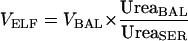 |
where VELF is the volume of ELF sampled by the BAL, VBAL is the volume of aspirated BAL fluid, UreaBAL is the concentration of urea in BAL fluid, and UreaSER is the concentration of urea in serum.
The concentration of antibiotic in the ELF (ABXELF), was calculated from the relationship
 |
where ABXBAL is the measured concentration of antibiotic in BAL.
The volume of AC collected in the pellet suspension was determined from the cell count in the BAL fluid. Cells were counted in a hemocytometer with a lower detection limit of 1.0 × 10⁶/liter. The number of cells in 1.0 ml of pellet suspension was calculated to be equal to the number of cells per liter of BAL fluid/100. Because of cell loss during centrifugation, the actual number of cells recovered may be lower than the number counted, and the antibiotic concentration may be approximately 20% greater than we calculated (28). Differential cell counting was performed after spinning the specimen in a cytocentrifuge. The volume of alveolar cells in the pellet suspension was determined with a mean macrophage cell volume of 2.42 μl/106 cells (4).
The concentration of antibiotic in alveolar cells, ABXAC, was calculated from the relationship
 |
where ABXPELLET is the antibiotic concentration in the 1-ml cell suspension, and VAC is the volume of AC in the 1-ml cell suspension
Statistical analysis.
PROPHET software, version 6.0 (Division of Research Resources, National Institutes of Health, Bethesda, Md., and Abtech Corporation, Charlottesville, Va.), was used to compute descriptive statistics and sample sizes and to perform the linear regression analyses. Analysis of variance (ANOVA) with a two-factor factorial model was used to assess the effects of gender and AIDS status on subject physical characteristics, clinical laboratory values, drug dosage, drug concentrations, AC recovery, ELF recovery, and AC/plasma and ELF/plasma ratios. The ethambutol concentrations in ELF and AC were compared for subjects with and without AIDS by one-way ANOVA. The two-sample equal-variances t test (two sided) was used to compare AC and ELF recovery and drug concentrations in plasma, AC, and ELF between the groups of women with AIDS who were smokers and nonsmokers. The equalities of variances of the smoking and nonsmoking groups were calculated by using the F test (Levene's test). The two-sample Mann-Whitney rank-sum test (two sided) was used to compare the daily doses in men and women, CD4 counts in men and women, and the serum creatinine determinations between healthy subjects and men or women with AIDS. The Shapiro-Wilk test was used to evaluate the normality of the distributions of the data sets prior to comparison. P < 0.05 was regarded as significant.
RESULTS
The CD4 counts (shown as mean ± SD, median, and range, respectively) for the 10 men with AIDS were 370 ± 208, 300, and 113 to 697 and for the 10 women with AIDS were 330 ± 127, 304, and 86 to 550 and were not significantly different (P > 0.05). All of the serum creatinine determinations were within normal limits; however, the values were greater for all 20 men (0.95 ± 0.25 mg/dl) than for all 20 women (0.71 ± 0.16 mg/dl) (P < 0.05). AIDS status was without effect (P > 0.05) on the serum creatinine determinations. Seven of the 10 female subjects with AIDS were cigarette smokers; the remainder of the subjects were nonsmokers.
Forty-eight subjects were recruited and signed informed consent for the study. Of the 48, 5 did not keep their study appointments and were dropped, 1 withdrew consent, 1 was discontinued due to intercurrent illness, and 1 refused bronchoscopy on day 5. The 40 remaining subjects successfully completed the bronchoscopy and BAL. One subject developed pneumonia postbronchoscopy and was successfully treated with antibiotics. There were no other major adverse events, and all of the subjects returned to their normal duties. One subject experienced mild, self-limited chest discomfort, and the temperature was transiently elevated in 1 subject. Mild cough or lightheadedness occurred in 4 subjects and 16 subjects, respectively; both of these symptoms were self-limited and did not require treatment.
The number (mean ± SD, median, and range) of AC recovered from BAL in the 40 subjects was 3.4 × 10⁸ ± 6.4 × 10⁸ cells/liter; AC recovery (Table 1) was not affected by gender or smoking history (P > 0.05), but was greater in subjects with AIDS (5.5 × 10⁸ cells/liter) than in the healthy subjects (1.2 × 10⁸ cells/liter) (P = 0.01). The percentage of cells in the monocyte/macrophage class was not affected by AIDS status or gender (P > 0.05) and was not significantly different when the smoking women (n = 7) were compared to the nonsmoking subjects (n = 33) (93% ± 9.9% versus 86% ± 14.9%). The volume (mean ± SD) of ELF recovered from the 20 subjects with AIDS (1.3 ± 0.6 ml) was greater than that recovered from healthy subjects (1.0 ± 0.4) (P = 0.03). Gender or smoking had no effect on the recovery of ELF (P > 0.05).
TABLE 1.
Recovery of cells from BAL in 40 subjects according to gender and AIDS stratification
| AIDS group (n) | Recovery from BAL (mean ± SD)a
|
|||||
|---|---|---|---|---|---|---|
| No. of cells (108/liter) (range) | % Polymorphonuclear leukocytes | % Lymphocytes | % Monocytes/macrophages | % Eosinophils | % Degenerated cells | |
| Men | ||||||
| With (10) | 6.5 ± 12.3 (0.47–40) | 1.7 ± 1.2 | 10.5 ± 8.3 | 83.7 ± 13.2 | 0.2 ± 0.4 | 3.9 ± 10.0 |
| Without (10) | 1.2 ± 0.80 (0.32–3.2) | 0.5 ± 0.5 | 6.7 ± 8.0 | 89.6 ± 11.0 | 0.4 ± 1.0 | 2.6 ± 6.0 |
| Women | ||||||
| With (10) | 4.6 ± 2.2 (1.3–8.0) | 2.1 ± 3.0 | 13.4 ± 17.8 | 83.6 ± 21.6 | 0.9 ± 1.4 | 0 |
| Without (10) | 1.2 ± 0.57 (0.41–2.4) | 3.0 ± 3.3 | 11.3 ± 8.8 | 82.2 ± 9.8 | 0.4 ± 0.7 | 3.1 ± 6.1 |
For all subjects with AIDS, the mean was higher than that for healthy subjects (P = 0.04). There was no effect of gender alone or interaction of gender and AIDS status on cell recovery (P > 0.05). Gender, AIDS status, and interaction between gender and AIDS had no significant effect on the percentages of polymorphonuclear leukocytes, lymphocytes, monocytes, or eosinophils in BAL (all P > 0.05).
Plasma ethambutol concentrations.
For all 40 subjects combined, there was no significant difference between the concentrations (mean ± SD) of ethambutol in plasma at 2 h (2.1 ± 1.2 μg/ml) or 4 h (2.1 ± 0.8 μg/ml) after the last dose (Table 2), and there was no correlation between the 2- and 4-h concentrations (r = 0.07, P = 0.65). There was no significant effect of gender or AIDS status on the concentrations of ethambutol in plasma at 2 h following the last dose. Plasma ethambutol concentrations at 4 h following the last dose were greater in the 20 men than in the 20 women (2.4 ± 0.9 versus 1.8 ± 0.6 μg/ml) (P < 0.05). There was no correlation between the weights of the subjects and the concentrations of ethambutol at 2 h (r = 0.27, P = 0.09) or 4 h (r = 0.27, P = 0.18).
TABLE 2.
Ethambutol concentration in plasma, ELF, and AC
| Samplea | Concn (μg/ml) in AIDS group (n = 10)b
|
|||
|---|---|---|---|---|
| Men
|
Women
|
|||
| With | Without | With | Without | |
| Plasma | ||||
| 2 hc | 2.5 ± 1.2 | 2.2 ± 1.1 | 1.9 ± 1.2 | 1.8 ± 1.5 |
| 4 hd | 2.4 ± 1.0 | 2.3 ± 0.7 | 1.7 ± 0.7 | 1.9 ± 0.6 |
| ELFe | 2.2 ± 1.0 (1.1–4.1) | 2.6 ± 1.7 (1.5–7.2) | 1.9 ± 0.5 (1.3–2.7) | 1.9 ± 0.6 (1.0–3.1) |
| ACf | 46.0 ± 17.0 (18.1–69.7) | 44.7 ± 14.2 (29.5–68.1) | 82.0 ± 39.4 (10.6–155.6) | 44.5 ± 15.6 (28.8–80.6) |
Within each group, the concentrations of ethambutol ELF were significantly greater than concentrations in plasma at 2 or 4 h or those in AC (P < 0.05).
Values are means ± SD. Ranges are given in parentheses.
There were no significant differences in plasma drug concentrations at 2 h among the four groups (P > 0.05).
There were no significant differences in plasma drug concentrations at 4 h among the four groups (P > 0.05). ANOVA confirmed a significant gender effect (P < 0.05), but no effect of AIDS status or interaction between gender and HIV status (P > 0.05) (see text).
There were no significant differences in ELF concentrations among the four groups (P > 0.05).
AC concentrations were significantly greater in women with AIDS than in any of the other three groups (P < 0.05). ANOVA confirmed a significant gender, AIDS status, and interaction effect (P < 0.05) (see text).
When smoking women with AIDS were compared to nonsmoking women with AIDS, plasma ethambutol concentrations (mean ± SD) at 2 h (1.54 ± 0.6 and 2.87 ± 1.9 μg/ml) and 4 h (1.68 ± .67 and 1.85 ± 0.6 μg/ml) were not significantly different (P > 0.05); however, the sample sizes were small, and a larger sample size may have detected a difference. CD4 counts in the subjects with AIDS (n = 20) were not correlated with the concentrations of ethambutol in plasma at 2 h (r = 0.0004, P = 1.0) or 4 h (r = −0.12, P = 0.6) or in AC (r = 0.004, P = 1.0) or ELF (r = −0.29, P = 0.2).
AC concentrations.
AC concentrations were greater in women with AIDS than in the other three groups (Table 2). The greater AC ethambutol concentrations in this group were associated with smoking. The AC ethambutol concentration (mean ± SD) in the smoking women (97.2 ± 32.1 μg/ml) with AIDS was more than twice the concentration in nonsmoking women (46.4 ± 34.5 μg/ml) with AIDS (P = 0.05) and more than twice the concentration in all other nonsmoking subjects (45.2 ± 16.8 μg/ml) combined (P < 0.05).
AC ethambutol concentrations were greater than plasma ethambutol concentrations at 2 and 4 h as well as ELF ethambutol concentrations for all four groups (Table 2). There was no correlation between the weights of the subjects and the concentrations of ethambutol in AC (r = −0.15, P = 0.4). The AC/plasma drug concentration ratios at 2 and 4 h were 18 and 19, 20 and 19, 43 and 48, and 25 and 23 for men with AIDS, men without AIDS, women with AIDS, and women without AIDS, respectively. For all 40 subjects, there was no correlation between the 2-h (r = −0.25, P =0.12) or the 4-h (r = 0.05, P = 0.77) plasma ethambutol concentrations and the AC ethambutol concentrations.
ELF concentrations.
There was no significant effect of gender or AIDS status on the concentrations of ethambutol in ELF (Table 2), although the volume of ELF recovered was approximately 30% greater in patients with AIDS (Table 1). When the male and female data were grouped to create a larger sample size (n = 20 in each group), the ELF concentrations (mean ± SD) of ethambutol in subjects with (2.1 + 0.8 μg/ml) and without (2.3 ± 1.3 μg/ml) AIDS were not significantly different (P > 0.05). For all 40 subjects, the ELF/plasma ratios at 2 and 4 h were 1.4 and 1.1 (P > 0.05). There was no correlation between the weights of the subjects and the concentrations of ethambutol in ELF (r = 0.27, P > 0.05). ELF ethambutol concentrations (mean ± SD) in smoking (1.8 ± 0.5 μg/ml) and nonsmoking (2.0 ± 0.6 μg/ml) women with AIDS were not significantly different (P > 0.05). Again, the sample sizes for the smoking and nonsmoking groups were small, and a larger sample size might have detected a difference. The 2-h plasma ethambutol concentrations were not correlated with the ELF ethambutol concentrations (r = 0.06, P = 0.7); however, there was a weak positive correlation between the 4-h plasma and ELF ethambutol concentrations (r = 0.52, P = 0.0006).
DISCUSSION
Concentrations of ethambutol in plasma at 2 and 4 h following the last dose and AC and ELF ethambutol concentrations were not affected by gender or the presence of AIDS, as defined in our subjects. Within the range of CD4 counts in our patients and within the limitations of the sample size of patients with AIDS (n = 20), CD4 cell counts were not correlated with ethambutol concentrations in plasma, AC, or ELF. Further investigation will be necessary to determine whether absorption of ethambutol would be more impaired in patients with greater degrees of immunosuppression (lower CD4 cell counts) than those included in this study. With all subjects combined, plasma drug concentrations at 2 h were not significantly different from the concentrations at 4 h. The time to maximum concentration of drug in serum (Tmax) for orally administered ethambutol has been reported to be a mean of 2.5 h with a range of 1.5 to 4.0 h (19), indicating considerable interpatient variability in this parameter. Since we drew blood specimens at 2 and 4 h after administration of the oral dose, it is likely that the actual Cmax was not detected in our subjects. This interpretation is supported by the lack of correlation, in our subjects, between the 2- and the 4-h measurements and by the wide range of Tmax that has been reported (19).
MICs of ethambutol for sensitive strains of Mycobacterium tuberculosis, tested by the BACTEC method, have been reported to be ≤2 μg/ml (21) and approximately 2 to 4 μg/ml (17). For clinical purposes, ≤2.5, 5.0, and ≥10.0 μg/ml, respectively, have been recommended as breakpoints for designating strains of M. tuberculosis as susceptible, intermediate, or resistant (21). It is of note that 27 (68%), 19 (48%), and 22 (55%) of the 40 subjects had 2-h plasma, 4-h plasma, and ELF drug concentrations below the MIC (2.0 μg/ml) that has been recommended as the laboratory breakpoint for susceptible strains of M. tuberculosis. A larger dose of ethambutol (e.g., 25 mg/kg), rather than the 15 mg/kg used in this study, likely would have resulted in greater plasma, AC, and ELF drug concentrations. More of the subjects would have had 2- and 4-h plasma and ELF drug concentrations that exceeded the published MICs of ethambutol for M. tuberculosis. The dose used in this study, 15 mg/kg once daily, was at the low end of the range recommended for clinical purposes (15 mg/kg to 25 mg/kg/day) and was chosen in order to minimize potential toxicity in volunteer subjects.
AC ethambutol concentrations in all 40 subjects were greater than 10 μg/ml. The high drug concentrations in AC relative to plasma and ELF were similar to those that we have described with macrolides (9, 10). Neither the 2-h nor the 4-h plasma concentrations were correlated with the AC ethambutol concentrations. The AC/plasma concentration ratio was 21 ± 13 at the time the bronchoscopy was performed (4 h following the last dose) in the 33 nonsmoking men and women. The AC/plasma concentration ratio at 4 h was 61 ± 30 in the seven smoking women (P < 0.05). This finding indicates that alveolar cells in smoking subjects concentrate ethambutol greater than in nonsmoking subjects and is consistent with the prior demonstration that the active transport ex vivo of ethambutol by alveolar macrophages is increased in smokers (16). Our ability to detect differences concentrations of ethambutol in plasma, AC, and ELF between the subgroups of smoking and nonsmoking women with AIDS or to measure effect of gender apart from smoking was limited by the small sample size of nonsmoking women. As with macrolides, considerable antibacterial activity resides in the AC. For example, for organisms with MICs of 0.25 to 2.0 μg/ml, inhibitory ratios between 30 to 1 and 240 to 1 would be present in AC.
High inhibitory and killing ratios are viewed as desirable in the treatment of infectious diseases. For concentration-dependent antibiotics such as fluoroquinolones, CMAX/MIC ratios of ≥10 in plasma have been associated with maximum efficacy and delay in emergence of resistance (24, 27). Whether these pharmacokinetic characteristics apply to ethambutol and the treatment of tuberculosis is unknown. However, it is likely, but not proven, that high intrapulmonary concentrations of antituberculous drugs would be related to their therapeutic efficacy.
The ELF/plasma ratio at the time of bronchoscopy (4 h after the last dose) was 1. ELF ethambutol concentrations were not affected by AIDS status or gender and were not significantly different from the plasma drug concentrations. These observations suggest that, at steady state, this compartment was in equilibrium with the intravascular compartment. The volume of ELF recovered in the AIDS patients was approximately 30% greater than that in the healthy subjects. This increased ELF recovery should not affect the calculated concentration of ethambutol in ELF, since the ELF concentration is also dependent upon the volume of BAL recovered and the concentration of ethambutol measured in BAL.
This study was not designed to detect interactions between ethambutol and other drugs taken by our AIDS patients. The clinical significance of the intrapulmonary ethambutol concentrations described in our study is unknown and requires further investigation.
ACKNOWLEDGMENTS
This work was carried out with funds provided by the NIH (grant AI36054) and NIH grant MO1RR00079 (General Clinical Research Center) at the University of California—San Francisco.
We acknowledge the assistance of Charles L. Daley, Margareta Andersson, and Ganfeng Wang for running the assays and Eve Benton and Maureen Morris for manuscript preparation.
REFERENCES
- 1.Anonymous. Metropolitan height and weight tables. 1983. Stat Bull. 1983;64:2–9. [PubMed] [Google Scholar]
- 2.Baldwin D R, Honeybourne D, Wise R. Pulmonary disposition of antimicrobial agents: in vivo observations and clinical relevance. Antimicrob Agents Chemother. 1992;36:1176–1180. doi: 10.1128/aac.36.6.1176. [DOI] [PMC free article] [PubMed] [Google Scholar]
- 3.Baldwin D R, Honeybourne D, Wise R. Pulmonary disposition of antimicrobial agents: methodological considerations. Antimicrob Agents Chemother. 1992;36:1171–1175. doi: 10.1128/aac.36.6.1171. [DOI] [PMC free article] [PubMed] [Google Scholar]
- 4.Baldwin D R, Wise R, Andrews J M, Ashby J P, Honeybourne D. Azithromycin concentrations at the sites of pulmonary infection. Eur Respir J. 1990;3:886–890. [PubMed] [Google Scholar]
- 5.Baldwin, D. R., R. Wise, J. M. Andrews, J. P. Ashby, and D. Honeyborne. The distribution of temafloxacin in bronchial epithelial lining fluid, alveolar macrophages, and bronchial mucosa. Eur. Respir. J. 5:471–476. [PubMed]
- 6.Centers for Disease Control and Prevention. 1993 revised classification system for HIV infection and expanded surveillance case definition for AIDS among adolescents and adults. JAMA. 1993;269:729–730. [PubMed] [Google Scholar]
- 7.Centers for Disease Control and Prevention. Prevention and treatment of tuberculosis among patients infected with human immunodeficiency virus: principles of therapy and revised recommendations. Morb Mortal Wkly Rep. 1998;47:1–58. [PubMed] [Google Scholar]
- 8.Choudhri S, Hawken H M, Gathua S, Minyiri G O, Watkins W, Sahai J, Sitar D S, Aoki F Y, Long R. Pharmacokinetics of antimycobacterial drugs in patients with tuberculosis, AIDS, and diarrhea. Clin Infect Dis. 1997;25:104–111. doi: 10.1086/514513. [DOI] [PubMed] [Google Scholar]
- 9.Conte J E, Jr, Golden J, Duncan S, McKenna E, Lin E, Zurlinden E. Single-dose intrapulmonary pharmacokinetics of azithromycin, clarithromycin, ciprofloxacin, and cefuroxime in volunteer subjects. Antimicrob Agents Chemother. 1996;40:1617–1622. doi: 10.1128/aac.40.7.1617. [DOI] [PMC free article] [PubMed] [Google Scholar]
- 10.Conte J E, Jr, Golden J A, Duncan S, McKenna E, Zurlinden E. Intrapulmonary pharmacokinetics of clarithromycin and of erythromycin. Antimicrob Agents Chemother. 1995;39:334–338. doi: 10.1128/aac.39.2.334. [DOI] [PMC free article] [PubMed] [Google Scholar]
- 11.Conte J E, Jr, Golden J A, Duncan S, McKenna E, Zurlinden E. Intrapulmonary concentrations of pyrazinamide. Antimicrob Agents Chemother. 1999;43:1329–1333. doi: 10.1128/aac.43.6.1329. [DOI] [PMC free article] [PubMed] [Google Scholar]
- 12.Conte J E, Jr, Golden J A, McQuitty M, Kipps J, Lin E T, Zurlinden E. Effects of AIDS and gender on steady-state plasma and intrapulmonary ethionamide concentrations. Antimicrob Agents Chemother. 2000;44:1337–1341. doi: 10.1128/aac.44.5.1337-1341.2000. [DOI] [PMC free article] [PubMed] [Google Scholar]
- 13.Conte J E, Jr, Golden J A, McQuitty M, Kipps J, Lin E T, Zurlinden E. Single-dose intrapulmonary pharmacokinetics of rifapentine in normal subjects. Antimicrob Agents Chemother. 2000;44:985–990. doi: 10.1128/aac.44.4.985-990.2000. [DOI] [PMC free article] [PubMed] [Google Scholar]
- 14.Fenton M J, Vermeulen M W. Immunopathology of tuberculosis: roles of macrophages and monocytes. Infect Immun. 1996;64:683–690. doi: 10.1128/iai.64.3.683-690.1996. [DOI] [PMC free article] [PubMed] [Google Scholar]
- 15.Gordon S M, Horsburgh C R, Jr, Peloquin C A, Havlik J A, Jr, Metchock B, Heifets L, McGowan J E, Jr, Thompson S E., III Low serum levels of oral antimycobacterial agents in patients with disseminated Mycobacterium avium complex disease. J Infect Dis. 1993;168:1559–1562. doi: 10.1093/infdis/168.6.1559. [DOI] [PubMed] [Google Scholar]
- 16.Hand W L, Boozer R M, King-Thompson N L. Antibiotic uptake by alveolar macrophages of smokers. Antimicrob Agents Chemother. 1985;27:42–45. doi: 10.1128/aac.27.1.42. [DOI] [PMC free article] [PubMed] [Google Scholar]
- 17.Heifets L B, Iseman M D, Lindholm-Levy P J. Ethambutol MICs and MBCs for Mycobacterium avium complex and Mycobacterium tuberculosis. Antimicrob Agents Chemother. 1986;30:927–932. doi: 10.1128/aac.30.6.927. [DOI] [PMC free article] [PubMed] [Google Scholar]
- 18.Jaruratanasirikul S. The pharmacokinetics of oral rifampicin in AIDS patients. J Med Assoc Thail. 1998;81:25–28. [PubMed] [Google Scholar]
- 19.Peloquin C A, Bulpitt A E, Jaresko G S, Jelliffe R W, Childs J M, Nix D E. Pharmacokinetics of ethambutol under fasting conditions, with food, and with antacids. Antimicrob Agents Chemother. 1999;43:568–572. doi: 10.1128/aac.43.3.568. [DOI] [PMC free article] [PubMed] [Google Scholar]
- 20.Peloquin C A, Nitta A T, Burman W J, Brudney K F, Miranda-Massari J R, McGuinness M E, Berning S E, Gerena G T. Low antituberculosis drug concentrations in patients with AIDS. Ann Pharmacother. 1996;30:919–925. doi: 10.1177/106002809603000901. [DOI] [PubMed] [Google Scholar]
- 21.Rastogi N, Labrousse V, Goh K S. In vitro activities of fourteen antimicrobial agents against drug susceptible and resistant clinical isolates of Mycobacterium tuberculosis and comparative intracellular activities against the virulent H37Rv strain in human macrophages. Curr Microbiol. 1996;33:167–175. doi: 10.1007/s002849900095. [DOI] [PubMed] [Google Scholar]
- 22.Rennard S, Basset I G, Lecossier D, O'Donnell K M, Pinkston P, Martin P G, Crystal R G. Estimation of volume of epithelial lining fluid recovered by lavage using urea as marker of dilution. J Appl Physiol. 1986;60:532–538. doi: 10.1152/jappl.1986.60.2.532. [DOI] [PubMed] [Google Scholar]
- 23.Sahai J, Gallicano K, Swick L, Tailor S, Garber G, Seguin I, Oliveras L, Walker S, Rachlis A, Cameron D W. Reduced plasma concentrations of antituberculosis drugs in patients with HIV infection. Ann Intern Med. 1997;127:289–293. doi: 10.7326/0003-4819-127-4-199708150-00006. [DOI] [PubMed] [Google Scholar]
- 24.Schentag J J. Antimicrobial action and pharmacokinetics/pharmacodynamics: the use of AUIC to improve efficacy and avoid resistance. J Chemother. 1999;11:426–439. doi: 10.1179/joc.1999.11.6.426. [DOI] [PubMed] [Google Scholar]
- 25.Talke H S. Enzymatische Harnstoffbestimmung im Blut und Serum im optischem Test nach Warburg. Klin Wochenschr. 1965;43:174. doi: 10.1007/BF01484513. [DOI] [PubMed] [Google Scholar]
- 26.Taylor B, Smith P J. Does AIDS impair the absorption of antituberculosis agents? Int J Tuberc Lung Dis. 1998;2:670–675. [PubMed] [Google Scholar]
- 27.Turnidge J. Pharmacokinetics and pharmacodynamics of fluoroquinolones. Drugs. 1999;58(Suppl. 2):29–36. doi: 10.2165/00003495-199958002-00006. [DOI] [PubMed] [Google Scholar]
- 28.Willcox M, Kervitsky A, Watters L C, King T E J. Quantification of cells recovered by bronchoalveolar lavage. Comparison of cytocentrifuge preparations with the filter method. Am Rev Respir Dis. 1988;138:74–80. doi: 10.1164/ajrccm/138.1.74. [DOI] [PubMed] [Google Scholar]