



Abstract
Background and Objectives:
Infective endocarditis (IE) involving the native pulmonary valve (PV) is extremely rare, with no data in Indian literature. The objective of this communication is to describe the clinical and diagnostic characteristics, underlying risk factors, microbiological features, and management of PVIE.
Methods:
This is a retrospective analysis of 8 cases of PVIE managed in a tertiary care center from 1992 to 2020.
Results:
PVIE was observed in 8 patients with underlying congenital cardiac malformation (Group A, 6 Patients) and in patients with central venous catheter (Group B, 2 patients). All the patients had prolonged febrile illness accompanied by right heart failure 4 (50%), septic pulmonary emboli 2 (25%), and pulmonary regurgitation 3 (37.5%). Trans-thoracic echocardiography demonstrated the vegetations, whereas computed tomography of chest diagnosed pulmonary emboli in 2 (25%), and pulmonary artery aneurysm in 1 (12.5%) patient. The early mortality was extremely high (5, 62.5%). Delayed diagnosis, fulminant septicemia, and multi-organ failure resulted in unfavorable outcomes.
Conclusions:
IE of the native PV is a rare and potentially lethal illness. Diagnosis should be considered in any febrile patient with an underlying congenital defect, central venous line, bacteremia, and comorbidities. Multi-modality imaging should be utilized to enhance the diagnostic yield and detect complications promptly.
Keywords: Infective endocarditis, mycotic pulmonary artery aneurysm, native pulmonary valve endocarditis, pulmonary valve
INTRODUCTION
Right heart infective endocarditis (IE) mostly involving tricuspid valve accounted for only 5%–10% of cases in the recent European Society of Cardiology data.[1] Endocarditis affecting the native pulmonary valve (PV) is extremely rare and ranges between 1% and 2%.[2,3] The infective process may be isolated to PV or concomitantly involve other valves.[4,5,6]
There are reports of PVIE from India in international journals.[7,8,9,10] The aim of this study is to report the clinical features, management, and outcome data of 8 patients of native PVIE seen over the last 28 years.
METHODS
The records of consecutive cases of PVIE seen in a tertiary care teaching institution form the material of this study. From October 1992 to October 2020, 8 cases of PVIE were diagnosed, treated, and followed up. Diagnosis of PVIE was guided by modified Dukes criteria.[11] Patients were categorized on the basis of predisposing factors. Group 1 consisted of patients with an underlying congenital cardiac malformation (CCM) (n = 6) and Group 2 consisted of IE related to an indwelling central venous catheter (CVC) (n = 2).
Clinical, investigative, management, and follow-up information was analyzed. Relevant laboratory investigations and blood cultures were studied. Electrocardiogram, Skiagram chest, echocardiography, high-resolution computed tomography (HRCT) of chest,[3] pulmonary angiography,[1] and management data were analyzed.
RESULTS
Table 1 summarizes the clinical, investigative, and outcome data. Group 1 includes 6 patients (all male) aged 4–22 years with an underlying CCM. All had prolonged (6–16 weeks) febrile illness and had received multiple antibiotics prior to referral to our center. Case 1 had features of congestive cardiac failure (CCF) and pulmonary regurgitation (PR). This patient was considered too sick for surgery and died due to septicemia. Patient 2 had staphylococcus endocarditis and responded favorably to antibiotics and underwent a successful balloon PV dilatation 6 months after the index episode. The clinical picture of patient 3 was complicated by septic pulmonary emboli (SPE). Two-dimensional echocardiography (2DE) in short axis view revealed vegetations attached to the PV. The vegetations were protruding into the pulmonary artery (PA) and suggested a flail valve [Figure 1a and b]. The family was unwilling for surgery due to guarded prognosis. Case 4 was transferred to the pediatric intensive care unit with febrile illness of 4 months duration unresponsive to anti-tubercular treatment and multiple antibiotics. Physical examination of this sick, cachexic child (weight 8 kg) revealed anemia, tachypnea, tachycardia, ejection click, and a grade 3/6 ejection systolic murmur along the left sternal border. Multiple blood cultures and fungal cultures were negative. Skiagram of the chest posterior-anterior view revealed a homogenous shadow occupying the right lung field [Figure 2a]. This shadow has remained unaltered in several X-rays. HRCT of the chest revealed a large enhancing vascular structure with surrounding low-density thrombus in relation to the right PA (RPA). This structure extended upon the right paracardiac region and measured approximately 6.1 cm × 5.0 cm [Figure 2b]. A 2DE with Doppler interrogation revealed thickened and mildly stenotic valve with a trans-PV gradient of 24 mm of Hg. Diagnosis of PVIE with mycotic aneurysm of PA was entertained. Pressure data (mm of Hg) at cardiac catheterization revealed: Aorta 100/60 mean 85, left ventricle 100/8, main, left, and RPA 20/8, right ventricle (RV) 50/6, trans PV gradient 30 and right atrium (RA) mean 6. Selective RPA angiography revealed a large round aneurysm with marked stasis of contrast occupying the entire right hemi-thorax. A narrow neck communicated between middle lobe branch of RPA and the aneurysm. Cardiothoracic surgery was refused due to moribund condition of the child. Partial percutaneous closure was achieved by multiple coils however, further attempts to close the residual aneurysm proved fatal.
Table 1.
Clinical, echocardiographic, microbiological data and outcome
| Age (year), sex | Underlying CCM/Risk factor | 2DECHO | Infecting organism | Outcome |
|---|---|---|---|---|
|
| ||||
| Group 1 | ||||
| 17, male | TOF | Mobile mass on PV, PR, TR | Polymicrobial organisms | Died of septicemia |
| 12, male | PVS | Mobile mass on PV | S. aureus | Improved on antibiotics, balloon dilation of PV after 6 months |
| 18, male | VSD and PVS | Mobile mass on PV, protruding in PA (flail PV), PR | Culture negative | Patient unwilling for surgery (died) |
| 4, male | PVS | Thickening of PV | Culture negative | Rupture of pseudoaneurysm and death |
| 7, male | VSD | Thickened PV, vegetations | Streptococcus viridans | Closure of VSD and pulmonary valvectomy (alive) |
| 22, male | TOF | Mobile mass on PV, mild PR | S. aureus | Intracardiac repair (VSD-patch and RVOT - patch repair), alive |
|
| ||||
| Group 2 | ||||
|
| ||||
| 56, female | CKD, DM, CVC | Mobile mass on PV, protruding in PA, free PR | Klebsiella, C. albicans | Considered too sick for surgery (died) |
| 45, female | RA, DM, CVC | Mobile vegetations on PV | Pseudomonas, Candida | Considered too sick for surgery (died) |
CCM: Congenital cardiac malformation, CVC: Central venous catheter, DM: Diabetes mellitus, PR: Pulmonary regurgitations, PV: Pulmonary valve, PVS: PV stenosis, RA: Rheumatoid arthritis, RVOT: Right ventricular outflow tract, TOF: Tetralogy of Fallot, TR: Tricuspid regurgitation, VSD: Ventricular septal defect, 2DECHO: Two-dimensional echocardiography, CKD: Chronic kidney disease, PA: Pulmonary artery, S. aureus: Staphylococcus aureus, C. albicans: Candida albicans, S. viridans: Streptococcus viridans
Figure 1.
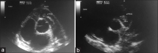
(a and b) Two-dimensional echocardiography short axis view shows vegetation (veg) on pulmonary valve. The excessive mobility into RVO and PA raise the possibility of a flail leaflet. AO: Aorta, RA: Right atrium, LA: Left atrium, TV: Tricuspid valve, RVO: Right ventricular outflow, PA: Pulmonary artery
Figure 2.
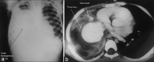
Skiagram chest pulmonary artery view shows homogenous shadow occupying large portion of right lung (Panel a). High resolution computed tomography of chest revealed a large (6.1 cm × 5.0 cm) enhancing vascular structure with surrounding low density thrombus in relation to right pulmonary artery interpreted as aneurysm (Panel b)
Cases 5 and 6 presented with a febrile illness with blood cultures showing streptococcus viridans and Staphylococcus aureus. Both underwent successful surgical procedures after medical stabilization.
Patient 2 was doing well up to 5 years follow up, patients 5 and 6 followed up only for 2 years.
Group 2 includes two female patients aged 45 and 56 years. These patients were extremely sick and displayed features of fulminant septicemia. Both had CVC inserted for different reasons. Additional risk factors included diabetes mellitus, chronic kidney disease, rheumatoid arthritis, and history of immunosuppressive therapy [Table 1]. Both the patients succumbed to multi-organ failure secondary to fungal and Gram-negative septicemia.
DISCUSSION
IE of native PV is rare. There are reports published by Indian authors in international journals.[7,8,9,10] Our experience of diagnosing and managing 8 cases of PVIE during the last 28 years is reported. There has been continued progress in the diagnostic abilities and the management of IE over the years. However, the recognition of this entity remains a challenge due to its rarity and varying clinical presentation. In an autopsy series from India, PV involvement was observed in 5.6% of cases.[10]
Symptoms and signs of infection are invariably present. Most had prolonged febrile illness and have received at least 2–3 weeks of antibiotics prior to referral to a tertiary care. The dominant etiology in this series is underlying CCM and included tetralogy of Fallot, ventricular septal defect, and PV stenosis [Table 1]. Almost all the variety of congenital lesions can be complicated by PVIE.[4,5,6,8] As reported previously, the diagnosis of PVIE should be entertained in the presence of fever, SPE, PR with or without CCF.[8] Heart failure in 4 (50%), SPE in 2 (25%) and murmur of PR in 3 (37.5%) supported the diagnosis. Signs of right-sided CCF depend on degree of PR which occurs as a sequel to the valve destruction. The most challenging presentation was of a 4-year-old moribund child (case 4) seen 2 decades back and was referred as persistent consolidation despite multiple rounds of antibiotics and a course of ATT. The pediatric resident recorded a murmur and sought a cardiac evaluation. Multiple chest X-rays showed a persistent homogenous opacity which was misinterpreted as nonresolving consolidation. Echocardiography revealed a stenotic PV with vegetation and HRCT of chest provided a clue to aneurysm of RPA. PA aneurysm (PAA) is a rare vascular anomaly which can occur due to a variety of conditions.[12] Mycotic PAA has been described after right heart endocarditis and is attributed to septic emboli or a jet hitting the wall of the PA as in patent ductus arteriosus.[13,14] The management is challenging as they are inherently unstable and can rupture. It is also important to achieve eradication of infection by intensive antibiotic therapy. Definitive removal or obliteration of the aneurysm is best achieved by surgical procedures such as aneurysmectomy, aneurysmorrhaphy, banding, or lobectomy.[13,14] These aneurysms have been treated by detachable coils[15] and stent-graft.[16] Percutaneous closure was preferred as surgery was considered very high risk; however, the child succumbed to rupture of the aneurysm. Modern-day hardware and experience certainly promise a better outcome.
During the last 5 years, we encountered two middle-aged patients presenting with fulminant septicemia. One had severe PR and other multiple SPE which were diagnosed as recurrent pneumonia. Both had CVC inserted for dialysis and pharmacotherapy. A wide spectrum of risk factors ranging from intravenous drug addiction, cardiac implantable electronic devices (CIED), central lines, immunosuppressive therapy, and several health care associated procedures have been associated with PVIE.[17,18,19] These cases highlight the need to recognize PV endocarditis in presence of CVC and extra-cardiac comorbidities. Infection of PV can occur even in individuals without any known illness.[20] There are fewer patients with CCM being encountered in recent PVIE series and this change is attributed to early surgical treatment of these defects.[18,19] On the contrary, there is an increase in conduit and prosthetic PV endocarditis.[19]
The Duke's criteria utilize clinical, echocardiographic, and blood culture findings for the diagnosis of IE.[11] The applicability of these criteria in PVIE is unproven. Anemia, leukocytosis, and elevation of inflammatory markers though nonspecific raise a suspicion of IE in the presence of underlying risk factors. The yield of blood cultures has improved due to refined techniques and organisms could be isolated in six out of eight cases (75%). A favorable outcome was observed in patients who grew either staphylococcus (25%), or streptococcus viridans (12.5%). The remaining with poly-microbial (12.5%), Gram-negative and fungal (25%), and culture-negative (25%) endocarditis did not survive. Fungal, poly-microbial, and culture-negative endocarditis pose diagnostic challenges and a poor prognosis.[21,22]
TTE remains a key modality for the diagnosis. Vegetations cause thickening of valve, shaggy/fluffy echoes, or mobile mass [Table 1 and Figure 1]. PV is extremely difficult to be visualized especially in adults, and it is quite likely that endocarditis involving this valve is missed in real-world scenario. M-mode and 2-DE identification of PV vegetations have been well reported.[7,23] Parasternal short-axis view is the most useful and along with parasternal long axis, RV inflow and subcostal views provide diagnosis in 87%–91%.[18] Better visualization of valve and Doppler assessment of PR is possible on TTE than trans-esophageal (TEE) with experienced operators. TEE of the anteriorly located PV can be challenging and is superior in evaluating CIED lead involvement.[18]
It is suggested that adjunctive imaging techniques are utilized to complement the information derived from the echocardiography. HRCT diagnosed SPE in two patients (25%) and PAA in one patient (12.5%) in this study. This modality diagnoses pulmonary complications (SPE, abscess, infarction, effusion) and large (>10 mm, mobile) vegetations which usually cause embolization.[24,25] CT angiography and cardiac magnetic resonance imaging provide high-resolution images and can help in the diagnosis of pseudo-aneurysm, fistulas, valve perforation, abscess and valvular dehiscence.[26] 18F-fluorodeoxyglucose positron emission tomography/(PET/CT) has been utilized for diagnosis of right-sided endocarditis.[27,28]
High mortality was reported in an earlier communication over 3 decades ago.[8] Unfortunately, the mortality trends remain unchanged in this study, despite better diagnostic and therapeutic facilities. This may be partly attributable to the inclusion of cases of group 2 who succumbed to septicemia. The cases referred to tertiary care are often too sick to be considered for surgery. Only three patients (37.5%) (2 with surgical and one with initial conservative treatment) survived. High mortality ranging from 20% to 66% has been observed.[19] Early surgery should be considered in the presence of highly mobile vegetations, recurrent SPE, and failure of intensive medical therapy.[29]
PVIE is a rare entity. A high index of clinical suspicion is needed for an early diagnosis. If the diagnosis is delayed, the outcome may be less than satisfactory.
Financial support and sponsorship
Nil.
Conflicts of interest
There are no conflicts of interest.
REFERENCES
- 1.Habib G, Lancellotti P, Antunes MJ, Bongiorni MG, Casalta JP, Del Zotti F, et al. 2015 ESC Guidelines for the management of infective endocarditis: The task force for the management of infective endocarditis of the European Society of Cardiology (ESC). Endorsed by: European Association for Cardio-Thoracic Surgery (EACTS), the European Association of Nuclear Medicine (EANM) Eur Heart J. 2015;36:3075–128. doi: 10.1093/eurheartj/ehv319. [DOI] [PubMed] [Google Scholar]
- 2.Murdoch DR, Corey GR, Hoen B, Miró JM, Fowler VG, Jr, Bayer AS, et al. Clinical presentation, etiology, and outcome of infective endocarditis in the 21st century: The International Collaboration on Endocarditis-Prospective Cohort Study. Arch Intern Med. 2009;169:463–73. doi: 10.1001/archinternmed.2008.603. [DOI] [PMC free article] [PubMed] [Google Scholar]
- 3.Habib G, Erba PA, Iung B, Donal E, Cosyns B, Laroche C, et al. Clinical presentation, aetiology and outcome of infective endocarditis. Results of the ESC-EORP EURO-ENDO (European infective endocarditis) registry: A prospective cohort study. Eur Heart J. 2019;40:3222–32. doi: 10.1093/eurheartj/ehz620. [DOI] [PubMed] [Google Scholar]
- 4.Cassling RS, Rogler WC, McManus BM. Isolated pulmonic valve infective endocarditis: A diagnostically elusive entity. Am Heart J. 1985;109:558–67. doi: 10.1016/0002-8703(85)90563-0. [DOI] [PubMed] [Google Scholar]
- 5.Cremieux AC, Witchitz S, Malergue MC, Wolff M, Vittecocq D, Vilde JL, et al. Clinical and echocardiographic observations in pulmonary valve endocarditis. Am J Cardiol. 1985;56:610–3. doi: 10.1016/0002-9149(85)91021-5. [DOI] [PubMed] [Google Scholar]
- 6.Nakamura K, Satomi G, Sakai T, Ando M, Hashimoto A, Koyanagi H, et al. Clinical and echocardiographic features of pulmonary valve endocarditis. Circulation. 1983;67:198–204. doi: 10.1161/01.cir.67.1.198. [DOI] [PubMed] [Google Scholar]
- 7.Sharma S, Katdare AD, Munsi SC, Kinare SG. M-mode echographic detection of pulmonic valve infective endocarditis. Am Heart J. 1981;102:131–2. doi: 10.1016/0002-8703(81)90426-9. [DOI] [PubMed] [Google Scholar]
- 8.Sharma S, Desai AG, Pillai MG, Kinare SG, Panday SR. Clinical and diagnostic features of pulmonary valve endocarditis in the setting of congenital cardiac malformations. Int J Cardiol. 1985;9:457–64. doi: 10.1016/0167-5273(85)90241-4. [DOI] [PubMed] [Google Scholar]
- 9.Ranjith MP, Rajesh KF, Rajesh G, Haridasan V, Bastian C, Sajeev CG, et al. Isolated pulmonary valve endocarditis: A case report and review of literature. J Cardiol Cases. 2013;8:161–3. doi: 10.1016/j.jccase.2013.07.007. [DOI] [PMC free article] [PubMed] [Google Scholar]
- 10.Vaideeswar P, Jawale RM, Tullu M. Isolated infective endocarditis of the pulmonary valve: An autopsy analysis of nine cases. Cardiovasc Pathol. 2009;18:231–5. doi: 10.1016/j.carpath.2008.06.011. [DOI] [PubMed] [Google Scholar]
- 11.Li JS, Sexton DJ, Mick N, Nettles R, Fowler VG, Jr, Ryan T, et al. Proposed modifications to the Duke criteria for the diagnosis of infective endocarditis. Clin Infect Dis. 2000;30:633–8. doi: 10.1086/313753. [DOI] [PubMed] [Google Scholar]
- 12.Kreibich M, Siepe M, Kroll J, Höhn R, Grohmann J, Beyersdorf F. Aneurysms of the pulmonary artery. Circulation. 2015;131:310–6. doi: 10.1161/CIRCULATIONAHA.114.012907. [DOI] [PubMed] [Google Scholar]
- 13.Bozkurt AK, Oztunç F, Akman C, Kurugoglu S, Eroğlu AG. Multiple pulmonary artery aneurysms due to infective endocarditis. Ann Thorac Surg. 2003;75:593–6. doi: 10.1016/s0003-4975(02)04303-5. [DOI] [PubMed] [Google Scholar]
- 14.Surgical Repair of Pulmonary Artery Aneurysm following Infective Endocarditis in a Patient with Persistent Ductus Arteriosus – Abstract-Europe PMC. [Last accessed on 2021 Jan 15]. Available from: https://europepmc.org/article/med/10721646 .
- 15.Wells IP, Roobottom CA, Morgan-Hughes G, Perrin D, Marshall AJ. Pulmonary mycotic aneurysm secondary to left-sided infective endocarditis treated with detachable coils. Clin Radiol Extra. 2004;59:37–9. [Google Scholar]
- 16.Chou MC, Liang HL, Pan HB, Yang CF. Percutaneous stent-graft repair of a mycotic pulmonary artery pseudoaneurysm. Cardiovasc Intervent Radiol. 2006;29:890–2. doi: 10.1007/s00270-005-0034-z. [DOI] [PubMed] [Google Scholar]
- 17.Bindra A, Iqbal R, Sapico F, Szlachcic Y. Isolated pulmonic valve endocarditis. Infect Dis Clin Pract. 2001;10:193–7. [Google Scholar]
- 18.Miranda WR, Connolly HM, DeSimone DC, Phillips SD, Wilson WR, Sohail MR, et al. Infective endocarditis involving the pulmonary valve. Am J Cardiol. 2015;116:1928–31. doi: 10.1016/j.amjcard.2015.09.038. [DOI] [PubMed] [Google Scholar]
- 19.Prieto-Arévalo R, Muñoz P, Cuerpo G, Marí-Hualde A, Castelo-Corral L, Navas-Elorza E, et al. Pulmonary infective endocarditis. J Am Coll Cardiol. 2019;73:2782–4. doi: 10.1016/j.jacc.2019.03.480. [DOI] [PubMed] [Google Scholar]
- 20.Simone J, Elizabeth S, Robert S, Rukmini K. Native pulmonary valve endocarditis in the absence of risk factors: A unique occurrence. J Am Coll Cardiol. 2019;73 9(Suppl 1):2998. [Google Scholar]
- 21.Bouabdallaoui N, Demondion P, Lebreton G, Leprince P. Fungal native pulmonary valve endocarditis: Facing both medical and surgical challenges. Eur J Cardiothorac Surg. 2017;51:184–5. doi: 10.1093/ejcts/ezw219. [DOI] [PubMed] [Google Scholar]
- 22.Bekker T, Govind A, Weber DM. Case of polymicrobial, gram-negative pulmonic valve endocarditis. Case Rep Infect Dis. 2019;2019:6439390. doi: 10.1155/2019/6439390. [DOI] [PMC free article] [PubMed] [Google Scholar]
- 23.Dander B, Righetti B, Poppi A. Echocardiographic diagnosis of isolated pulmonary valve endocarditis. Br Heart J. 1982;47:298–300. doi: 10.1136/hrt.47.3.298. [DOI] [PMC free article] [PubMed] [Google Scholar]
- 24.Feuchtner GM, Stolzmann P, Dichtl W, Schertler T, Bonatti J, Scheffel H, et al. Multislice computed tomography in infective endocarditis: Comparison with transesophageal echocardiography and intraoperative findings. J Am Coll Cardiol. 2009;53:436–44. doi: 10.1016/j.jacc.2008.01.077. [DOI] [PubMed] [Google Scholar]
- 25.Passen E, Feng Z. Cardiopulmonary manifestations of isolated pulmonary valve infective endocarditis demonstrated with cardiac CT. J Cardiovasc Comput Tomogr. 2015;9:399–405. doi: 10.1016/j.jcct.2015.03.013. [DOI] [PubMed] [Google Scholar]
- 26.Dursun M, Yılmaz S, Yılmaz E, Yılmaz R, Onur İ, Oflaz H, et al. The utility of cardiac MRI in diagnosis of infective endocarditis: Preliminary results. Diagn Interv Radiol. 2015;21:28–33. doi: 10.5152/dir.2014.14239. [DOI] [PMC free article] [PubMed] [Google Scholar]
- 27.Taylor B, Kahli Z, Kishan P. Diagnosis of pulmonic valve infective endocarditis with the use of positron emission tomography-computed tomography scan. J Am Coll Cardiol. 2016;67(Suppl 13):1031. [Google Scholar]
- 28.El Barzouhi A, Tanis W, van Gelder RE, Vriend JW. The pivotal role of cardiac computed tomography angiogram and 18F-fluorodeoxyglucose positron emission tomography-computed tomography in the diagnosis of right sided endocarditis: A case report. Eur Heart J Case Rep. 2019;3:yty165. doi: 10.1093/ehjcr/yty165. [DOI] [PMC free article] [PubMed] [Google Scholar]
- 29.Deng H, Ma Y, Zhai H, Miao Q. Surgical valve repair of isolated pulmonary valve endocarditis. Interact Cardiovasc Thorac Surg. 2013;16:384–6. doi: 10.1093/icvts/ivs509. [DOI] [PMC free article] [PubMed] [Google Scholar]