Correction to: Medical & Biological Engineering & Computing
10.1007/s11517-021-02490-z
The original article contained a mistake.
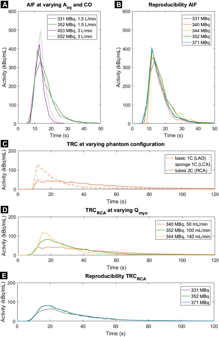
Figure 4 is not displayed correctly in the published paper. The correct Figure 4 is shown below.
Fig. 4.
A–E Time activity curves obtained using the myocardial perfusion phantom. Arterial input functions (AIFs) were acquired in the left ventricle at varying injected activity of 99mTc-tetrofosmin (Ainj)and cardiac output (CO). Resulting tissue response curves (TRCs) in the three myocardial segments were executed at varying myocardial fow rates (Qmyo) and tissue inlays (1 or 2 compartments). Each line colour denotes a single fow measurement (n=7). LAD=left anterior descending coronary artery, RCA=right coronary artery, LCX=left circumfex coronary artery
In addition, the caption was Fig. 4A-D and should have been Fig. 4A-E. The correct Figure caption is included.
The original article has been corrected.
Footnotes
The online version of the original article can be found at 10.1007/s11517-021-02490-z
Publisher’s note
Springer Nature remains neutral with regard to jurisdictional claims in published maps and institutional affiliations.


