Abstract
Bio-catalysis is an attractive alternative to replace chemical methods due to its high selectivity and mild reaction conditions. Furfural is an important bio-based platform compound generated from biomass. Herein, the bio-catalytic reduction of furfural (FAL) to furfuryl alcohol (FOL) was performed by using a furfural tolerant strain, Bacillus coagulans NL01. An efficient co-substrate was explored and a high conversion and selectivity of FAL to FOL was reported over this bio-catalytic system using glucose as co-substrate. As the bioconversion occurred over 42 mM FAL, 20 g L−1 glucose and 9 mg mL−1 at 50 °C, a high conversion and selectivity was obtained by 3 h reaction. This transformation rate of FAL was the highest compared with other reactions. Furthermore, about 98 mM FOL was produced from FAL within 24 h by a fed-batch strategy with a conversion of 92% and selectivity of 96%. These results indicate that this bio-catalytic reduction of FAL has high potential for application to upgrading of FAL and B. coagulans NL01 is a promising biocatalyst for the synthesis of FOL. In addition, this bio-catalytic reduction shows a high potential application for catalytic upgrading of FAL from biomass.
Bio-catalytic reduction of furfural to furfuryl alcohol was successfully carried out by Bacillus coagulans NL01 whole-cells in the presence of glucose.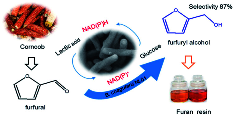
1. Introduction
Lignocellulosic biomass represents a vast renewable resource on earth, and is made of cellulose, hemicelluloses, and lignin. Recently, in order to realize sustainable development, plenty of government and industry attention has focussed on fundamental research and process development for the production of fuels and fine chemicals from biomass.1 FAL is an important industrial bio-based compound from lignocellulosic biomass. After treatment with mineral acid at high temperature, the hemicelluloses are readily hydrolyzed into pentose and the pentose is dehydrated to FAL.2 In this context, great effort has been devoted to the production and application of FAL and its derivatives.1 To date, FAL has been considered as one of the “Top 10 + 4” bio-based chemicals by the U.S. Department of Energy.3 It can be used as the starting material to produce FOL, furoic acid (FA), furan, tetrahydrofuran, 2-methyltetrahydrofuran and related resins.4
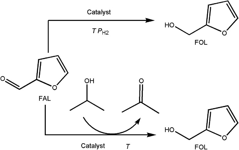
Among FAL derivatives, FOL is the hydrogenation product of FAL through the reduction of the C O bond in FAL. Recently it has gained an increase interest as a potential important building block for the production of various lysine, vitamin C, lubricants, dispersing agents and plasticizers.5 FOL is mainly produced by chemical reduction of FAL in either direct hydrogenation by H2 or catalytic transfer hydrogenation (CTH) processes (Scheme 1).6 By contrast, the direct hydrogenation of FAL by H2 has been widely commercialized using copper chromite catalysts. This method exhibited a high selectivity in the liquid phase process at high temperature (130–200 °C) and high H2 pressure.5 However, its main drawbacks are the toxicity of Cu–Cr catalysts. Although many other catalysts were designed and considered as alternatives for this method, an acceptable activity and selectivity of these catalysts still remains an issue.7 On the other hand, CTH of FAL into FOL is a recently developed method. It is mainly performed over metal catalysts and different hydrogen donors were used as alterative for H2.6 Due to avoid the use of explosive H2, this method seemed to be safer of FAL into FOL. But most reported CTH methods demonstrated a high metal loading and low selectivity.6 Recently, magnetic γ-Fe2O3@HAP catalyst was applied to promote this CTH with iso-propanol as the hydrogen donors. High FOL yield of 92% was achieved after 10 h at 180 °C.6 Thereafter, CTH showed a potential for the reduction of FAL. But its kinetic studies revealed that CTH of FAL was sensitive to the reaction temperature and a high temperature in the process.
Scheme 1. Chemical conversion of FAL into FOL (catalysts represent noble metal and non-noble metals; T = 130–200 °C; high H2 pressure about 2.0 MPa).
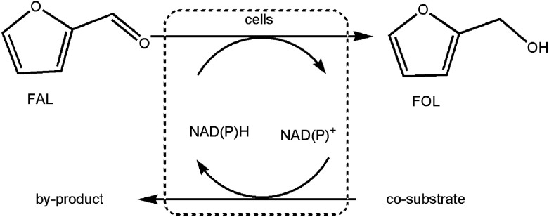
Biochemical catalysis for production of chemicals from biomass presents a promising alternative for conventional chemical routes. In contrast to chemical process, it has many advantages over chemical catalysis, such as mild reaction conditions, high productivity and environmental friendliness. Microbial degradation of FAL has been widely studied and reported as inhibitor produced during pre-treatment process of biomass.8 Some strains, including Pseudomonas putida, Saccharomyces cerevisiae, Amorphotheca resinae and so on, had been found that could metabolize FAL by reduction and oxidation reaction.2 Moreover, the reduction of furans to correspond furan alcohol has been demonstrated for S. cerevisiae, Pichia stipites, and B. coagulans under anaerobic condition.9 It provides the basis for the production of FOL by biotransformation (Scheme 2). Reduction of FAL into FOL with whole cells began to gained considerable interest. Liu et al. described that S. cerevisiae 307-12-F40 could convert 100% FAL into FOL at 30 mM.10 A methanogenic archaea, Methanococcus sp. was found that the end product of FAL metabolism was FOL in the bio-detoxification.11 He et al. reported chemical-enzymatic conversion of biomass-derived xylose to FOL by sequential acid catalyzed dehydration and bio-reduction.12 A recombination of E. coli CCZU-A13 harboring a NADH-dependent reductase (SsCR) was attempted to convert FAL to FOL.13 Although culturing cells of some microorganisms were reported to be FAL-tolerant, their transformation efficacies were low. Overall, the proof-of-concept of the bio-reduction is promising and further technical development and investigation would be desirable.
Scheme 2. Biotransformation of FAL to FOL.
Moderately thermophilic Bacillus coagulans are ideal organisms for the industrial manufacture of lactic acid from biomass. Our group ever found that Bacillus coagulans GKN316 could effectively convert several aldehyde inhibitors to the less toxic corresponding alcohols in situ.9 To continue our interest in biotransformation of FAL by this strain, herein we reported the whole-cell bio-reduction of FAL to FOL by employing B. coagulans NL01. Various parameters affecting bio-reduction reaction were investigated and the catalytic performances were evaluated in the FOL synthesis. Besides, a fed-batch strategy was applied for improving FOL production.
2. Experimental
2.1. Strains and materials
B. coagulans NL01 was isolated from soil samples in hot spring environments and used in this study.14
FAL (99%), FOL (99%) and FA (99%) were purchased from Sigma chemicals. The chemicals used in microbiological culture media were purchased from Sinochem or Fluka Chemical. Corn steep liquor was from Shandong Kangyuan Biotechnology Co. (Heze, China).
2.2. Fermentation experiments
B. coagulans NL01 were firstly pre-cultivated at 50 °C and 150 rpm for 12 h under aerobic condition. The used medium contained the following (in grams per liter): glucose 20; yeast extract 2; corn steep 2.5; NH4Cl 1; MgSO4 0.2 and CaCO3 10. After preculture, 10% seed culture was inoculated to the fresh above medium supplemented with FAL (0–52 mM) for fermentation. Fermentation experiments were performed in 150 mL shake flasks containing 50 mL of the culture mediums at 50 °C and 150 rpm for 60 h. During the fermentation, samples were taken periodically for analysis. All experiments were performed at least in duplicate and the values were expressed as the means ± standard deviations.
2.3. Cultivation of B. coagulans NL01 cells
B. coagulans NL01 were firstly pre-cultivated at 50 °C and 150 rpm for 12 h under aerobic condition. Then, 10% culture seed was inoculated into the fresh above medium. After incubation at 50 °C and 150 rpm for 12 h, the cells were harvested by centrifugation (8000g, 3 min and 4 °C) and washed twice with phosphate buffer (200 mM, pH 7.0).
2.4. General procedure for bio-reduction of FAL to FOL
Typically, 9 mg B. coagulans NL01 were added in 25 mL phosphate buffer (200 mM, pH 7.0) containing 42 mM FAL and 20 g L−1 co-substrate glucose. The bio-reduction experiments were conducted in a thermostatic shaker at 50 °C and 150 rpm and aliquots were withdrawn from the reaction mixtures at a specified time intervals for analysis. The conversion was defined as the percentage of the consumed FAL amount compared to the initial amount of FAL. The selectivity of FOL was defined as the ratio of FOL amount to the consumed FAL amount (in mmol). All of experiments were performed at least in duplicate and the values were expressed as the means ± standard deviations.
Various parameters (substrate, co-substrate, cell, temperature, time etc.) were investigated based on the above typical experiment of bio-reduction. To assess the impact of the co-substrate type, 10 g L−1 glucose, ethanol, maltose, sucrose or glycerol were added into reaction mixtures as co-substrate, respectively. The optimization of FAL concentration was carried out in 0–52 mM. The optimum cell concentration and co-substrate concentration was determined at 5–21 g L−1 cells and 5–50 g L−1 glucose respectively. After 24 h reaction, the conversion and selectivity were measured under the selected collocations and standard conditions. Additionally, the optimal temperature was determined with the temperature ranging from 35 to 55 °C for 24 h reaction. Fed-batch experiments were performed by adding the FAL at 3 h and 9 h.
2.5. Analytic methods
For monitoring bacterial growth, the turbidity measurements of cells were measured at 600 nm (OD600) by UV-visible spectrophotometer (Spectrum lab 752s). Cell amount was measured according to dry cellular weight. Cell samples were collected, washed by water twice, and dried at 105 °C for 6 h to gain the constant weight. FAL and FOL concentration were analyzed on a Zorbax Eclipse XDB-C18 column (4.6 mm × 250 mm, 5 μm) by using a reversed-phase HPLC (Agilent 1260 system) equipped with a DAD detector set at 220 nm. The column was set at 25 °C and the mobile phase was a mixture of acetonitrile/water (50 : 50, v/v) at flow rate of 1 mL min−1.
2.6. Statistical analysis
Statistical analyses were performed using SPSS statistics 20.0. The differences of the corresponding values between exposed groups were tested by one-way analysis of variance (ANOVA). p < 0.05 was regarded as statistically significant.
3. Results and discussion
3.1. Characteristic of B. coagulans strain NL01
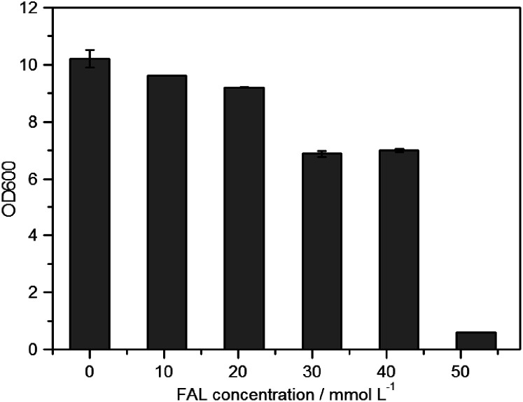
Recently, there is the growing interest in developing B. coagulans as an ideal platform microorganism for biological conversion of biomass.9 Prior reports have identified that some B. coagulans could detoxify some aldehyde inhibitors during fermentation in situ by aldehyde reduction.9,15 Thus in this study the tolerance of B. coagulans NL01 toward FAL were firstly investigated under anaerobic condition. As shown in Fig. 1, B. coagulans NL01 demonstrated a good growth in the presence of ≤21 mM of FAL. An obvious inhibitory effect was observed at 31 mM FAL and further increasing the FAL concentration resulted in complete cell death. According to previous reports, many microorganisms are inhibited by FAL at concentration of 1–12 mM.11Bacillus spp. IFA 119 showed 78% growth inhibition by 10 mM FAL.16 Delgenes et al. found that 21 mM FAL inhibited S. stipitis growth by 99%.17 The growth of E. coli LY01 was inhibited by 50% in the presence of 25 mM FAL.18 By contrast, B. coagulans NL01 revealed a higher FAL tolerance than most microorganisms in the anaerobic condition.
Fig. 1. Effect of FAL concentration on cell viability. Cell viability was measured by turbidity measurements at 600 nm (OD600) after 60 h fermentation.
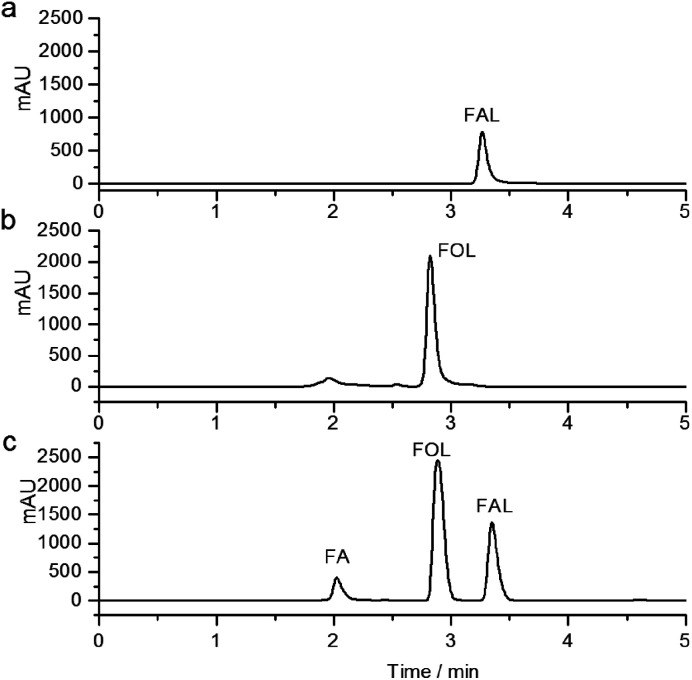
As previously reported, the tolerance mechanism of microorganisms for FAL mainly includes two kinds: completely metabolize or only transform into a less toxic form by reduction or oxidization.2 Therefore, the fate of the FAL by B. coagulans NL01 was investigated by HPLC analysis. The result showed that all of FAL was transformed primarily into most of FOL and a small portion FA after fermentation (Fig. 2). Table 1 presents the bioconversion of FAL into FOL during fermentation. It was found that FAL eliminated gradually and FOL appeared in the culture medium along with the time. When the initial FAL concentration is below 33 mM, the FAL could be completely disappeared by 24 h (Table 1, entry 3) fermentation. Moreover, a FOL concentration of 27 mM was achieved at the initial FAL concentration of 33 mM after 36 h fermentation. When the initial concentration of FAL was up to 52 mM, the FAL concentration remained throughout fermentation (Table 1 entry 5), indicating the death of the cells. It is consistent with the result of cell growth (Fig. 1). In our previous study, a similar reductive transformation of FAL was observed in B. coagulans GKN316. Our observations confirmed that B. coagulans NL01 might have a tolerance mechanism of FAL similar to some ethanologenic strains, either S. cerevisiae or Z. mobilis, which could reduce aldehydes to the less toxic corresponding alcohols.18–21 More importantly, its higher FAL tolerance and selectivity of FOL (highest 91%) suggest that B. coagulans NL01 may play a biocatalyst role in the FAL to FOL biotransformation.
Fig. 2. HPLC analysis of culture samples of B. coagulans in the presence of FAL after 60 h fermentation. (a) Samples for HPLC analysis was taken at the time of 0 h; (b) samples for HPLC analysis was taken at the time of 60 h fermentation; (c) FAL and its derivatives standard samples; FA (1.978 min); FOL (2.847 min); FAL (3.310 min).
Bioconversion of FAL during fermentation with B. coagulans NL01a.
| Entry | Initial FAL [mM] | t [h] | FAL [mM] | FOL [mM] | t [h] | FAL [mM] | FOL [mM] | t [h] | FAL [mM] | FOL [mM] | t [h] | FAL [mM] | FOL [mM] |
|---|---|---|---|---|---|---|---|---|---|---|---|---|---|
| 1 | 14.7 ± 0.1 | 12 | 1.8 ± 0.23 | 9.1 ± 0.04 | 24 | 0.0 | 11.1 ± 0.1 | 36 | 0.0 | 10.8 ± 0.1 | 60 | 0.0 | 10.6 ± 0.2 |
| 2 | 22.2 ± 0.1 | 12 | 3.2 ± 0.1 | 18.0 ± 0.2 | 24 | 0.0 | 19.7 ± 0.1 | 36 | 0.0 | 19.2 ± 0.1 | 60 | 0.0 | 19.1 ± 0.1 |
| 3 | 33.3 ± 0.1 | 12 | 31.5 ± 0.1 | 1.5 ± 0.1 | 24 | 4.1 ± 0.1 | 25.9 ± 0.1 | 36 | 0.0 | 27.2 ± 0.2 | 60 | 0.0 | 27.1 ± 0.2 |
| 4 | 40.6 ± 0.1 | 12 | 37.5 ± 0.1 | 1.4 ± 0.1 | 24 | 14.1 ± 0.3 | 24.0 ± 0.2 | 36 | 1.6 ± 0.1 | 35.9 ± 0.0 | 60 | 0.0 | 35.6 ± 0.2 |
| 5 | 52.1 ± 0.0 | 12 | 53.6 ± 0.1 | 0.0 | 24 | 53.6 ± 0.1 | 0.0 | 36 | 52.0 ± 0.2 | 0.0 | 60 | 49.8 ± 0.1 | 0.0 |
Reaction conditions: 10–52 mM FAL were added into 50 mL culture medium, 150 rpm, 50 °C; after microbial cells were anaerobic incubated for 60 h under the above conditions.
3.2. The co-substrate requirement of B. coagulans NL01
Bio-reduction of FAL to FOL is a typical redox reaction. Earlier, the reduction of furan in microorganisms has been found to be both NADPH- and NADH- coupled, which indicated that bio-reduction process must be cofactor dependent and hydrogen donor is needed.17 Likewise, the reported bio-catalytic reduction of furans also needed the addition of glucose as co-substrate to regenerate the enzyme cofactor.2 Therefore, in order to realize highly efficient bio-reduction, whole cell catalysis was considered for the FOL production and the requirement of co-substrate to promote bio-reduction as hydrogen donors was studied.
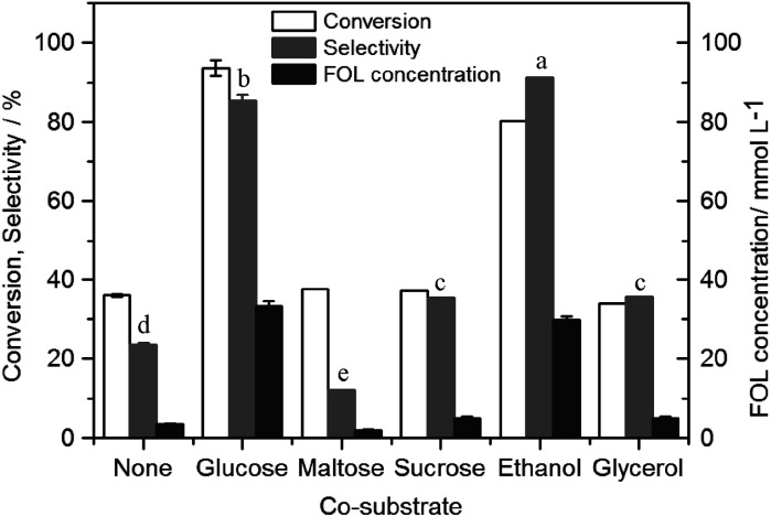
As shown in Fig. 3, FOL was produced with a low FAL conversion of 36% after 24 h without co-substrate. The result indicated that coupling the reduction of oxidized cofactors to the oxidation of a co-substrate was needed for redox cofactor recycle in cells. Thus some common sugars and alcohols were studied as co-substrate and the results were summarized in Fig. 3. It was observed that the conversion of FAL and the selectivity of FOL were greatly affected by the co-substrate type. The result of co-substrate consumption showed that both glucose and ethanol consumed quickly along with the degradation of FAL (data not shown). A maximal FAL conversion of 94% was achieved using glucose as co-substrate. Meanwhile, a high selectivity (85%) was simultaneously observed. In addition, despite that the conversion of FAL was only 80%, the highest selectivity (91%) was found using ethanol as co-substrate. The higher selectivity revealed that ethanol was more suitable for the reductase coenzyme regeneration. But to our understanding, glucose not only could regenerate efficiently reduced cofactors (e.g., NAD(P)H) and make the reduction reaction readily, but also could contribute to hold the cell status and activity as a broad-spectral carbon source. Meanwhile, the cells were easily inhibited by ethanol due to its polarity.22 Therefore, glucose was the best co-substrate for bio-reduction of FAL to FOL by B. coagulans NL01 and the highest FOL concentration (32 mM) was achieved using glucose as co-substrate. As for other sugars and alcohol, there was no obvious difference in conversion of FAL compared to the control. But the selectivity of FOL varied significantly. Both sucrose and glycerol had significant improvement for the selectivity of FOL, while the lowest selectivity was observed at 12% using maltose as co-substrate.
Fig. 3. Effect of co-substrate on reduction of furfural by whole cell catalytic of B. coagulans NL01. Conditions: 42 mM FAL, 10 g L−1 co-substrate, 9 mg (dry weight) per mL microbial cells, 25 mL phosphate buffer (200 mM pH 7.0), 150 rpm, 50 °C; anaerobic reaction for 24 h under the above conditions, with different letters representing significant differences between the treatment means (p < 0.05).
3.3. Effect of substrate and co-substrate concentration on reduction of FAL to FOL
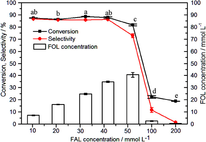
Compared with conventional chemical catalysts, the disadvantage of biocatalyst is lower substrate tolerance. As described above, FAL is an toxic compound toward the cell growth of B. coagulans NL01. Thus the influence of FAL on FOL biosynthesis was studied (Fig. 4). Although the cell growth was almost completely inhibited in the presence of 52 mM FAL (Fig. 1), a bio-reduction system seemed to demonstrate a better substrate tolerance than the fermentation process. Effect of initial concentration of FAL exerted no significant change on the conversion when FAL concentrations were less than 52 mM. But when the FAL concentration was over 52 mM, the conversion and selectivity of FAL began to decrease considerably, which suggested that FAL inhibition occurs at this substrate concentration. A conversion of 82% and selectivity of 73% was obtained at 52 mM FAL and a further increase in FAL concentration led to a quick decrease of FOL production. It meant that the related enzyme activity was completely inhibited. Therefore, considering the effect of FAL on the cell growth, 42 mM FAL was chosen for the next study.
Fig. 4. Effect of substrate concentration on reduction of furfural by whole cell catalytic of B. coagulans NL01. Conditions: 0–200 mM FAL, 10 g L−1 glucose, 9 mg (dry weight) per mL microbial cells, 25 mL phosphate buffer (200 mM pH 7.0), 150 rpm, 50 °C; anaerobic reaction for 24 h under the above conditions, with different letters representing significant differences between the treatment means (p < 0.05).
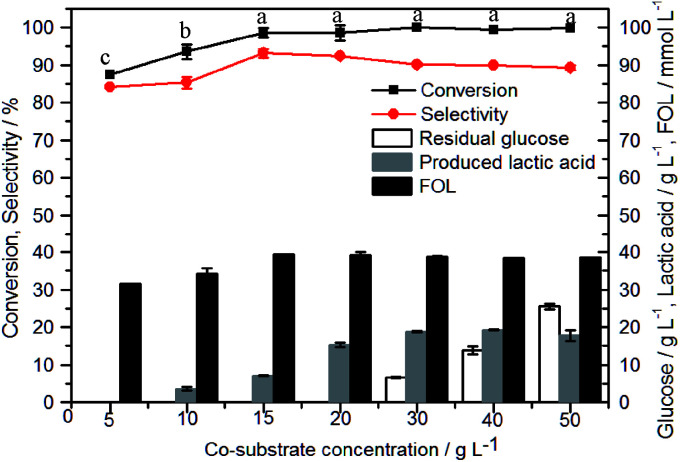
As described above, glucose is a preferred co-substrate toward bioreduction system. The amount of glucose is critical for efficient synthesis of FOL from FAL. As shown in Fig. 5, satisfactory conversions (>98%) and selectivities (>90%) were obtained when initial glucose concentration was over 15 g L−1. In the presence of 15 g L−1 glucose, the conversion and selectivity were 99% and 93% respectively. Meanwhile, it was found that glucose is completely consumed and transformed into lactic acid under the anaerobic condition. When the addition of glucose reached 30 g L−1, the accumulation of glucose occurred and the amount of lactic acid reached the maximum. It indicated that 15–20 g L−1 glucose might be a suitable amount for the bio-reduction of 42 mM FAL by B. coagulans NL01. In a previous study on HMF bio-reduction by Meyerozyma guilliermondii SC1103, an assumption was proposed that supplementation of excess co-substrate might favor to enhance HMF-tolerance of cells and a high glucose concentration could improve the bio-catalytic reduction of HMF.1 Therefore, the reduction of FAL at 40 mM and 100 mM were conducted in the presence of 10 and 30 g L−1 glucose (Fig. S1†). Unfortunately, when the concentration of FAL was 100 mM, poor results were obtained and no obvious improvements were observed at higher concentration of glucose.
Fig. 5. Effect of co-substrate concentration on reduction of furfural by whole cell catalytic of B. coagulans NL01. Conditions: 42 mM FAL, 5–50 g L−1 glucose, 9 mg (dry weight) per mL microbial cells, 25 mL phosphate buffer (200 mM pH 7.0), 150 rpm, 50 °C; anaerobic reaction for 24 h under the above conditions, with different letters representing significant differences between the treatment means (p < 0.05).
3.4. Effect of key reaction conditions on the bio-reduction of FAL
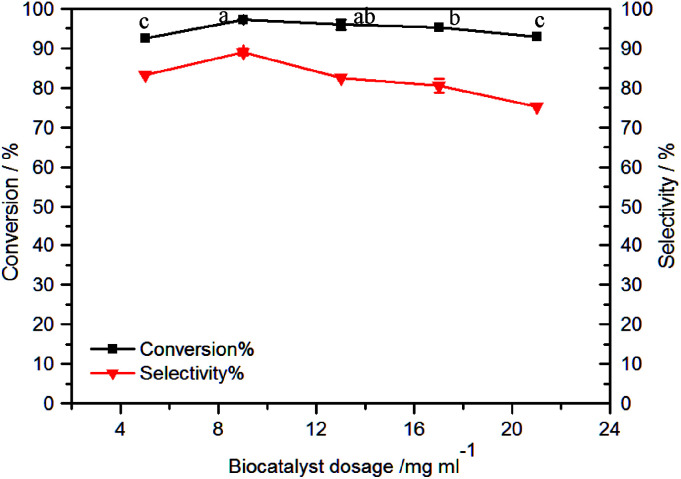
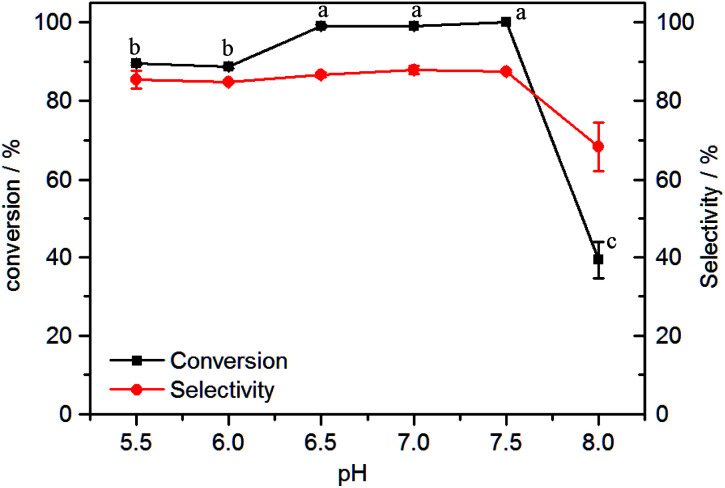
Fig. 6 showed the effect of the cell dosage on FOL synthesis. Despite that the higher cell loading could accelerate the reaction rate due to the presence of more enzymes, the cell dosage of 5–21 mg mL−1 exerted no significant effect on FAL conversion. The highest FAL conversion reached 97% after 24 h with 9 mg mL−1 of cell dosage. In addition, the cell loading showed obvious effect on the catalyst selectivity. 83% selectivity was observed with the use of 5 mg mL−1 of cell dosage and then increased to 90% with 9 mg mL−1 of cell dosage. Upon further increasing the cell loading, FOL selectivity began to decrease slowly. During most studies on biodegradation of furanic compounds, it was found that the oxidation or reduction of furanic aldehydes was quite commonly observed and the reduction reaction of FAL and HMF was often reversible.23 FOL could be oxidized to FAL again by alcohol dehydrogenase or aldehyde oxidase, and then to FA consequently by aldehyde oxidase from the cells.24 In this study, the decline of selectivity and more FA produced at higher cell loading suggested that B. coagulans NL01 might also have a metabolic pathway for FAL to FA. Therefore, we reasoned that the decrease of FOL selectivity might be owed to the proceeding oxidization of produced FOL to FA along the degradation pathway for FAL by B. coagulans NL01 as the cell loading was excess. Fig. 7 shows the effect of pH value on the reduction of FAL. B. coagulans NL01 cells exhibited good catalytic performances within the pH range 6.5–7.5. The maximal conversions of 99% were achieved after a reaction time of 24 h. In addition excellent selectivities (87%) were observed.
Fig. 6. Effect of cell concentration on reduction of furfural by whole cell catalytic of B. coagulans NL01. Conditions: 42 mM FAL, 20 g L−1 glucose, 5–21 mg (dry weight) per mL microbial cells, 25 mL phosphate buffer (200 mM pH 7.0), 150 rpm, 50 °C; anaerobic reaction for 24 h under the above conditions, with different letters representing significant differences between the treatment means (p < 0.05).
Fig. 7. Effect of pH on reduction of furfural by whole cell catalytic of B. coagulans NL01. Conditions: 42 mM FAL, 20 g L−1 glucose, 9 mg (dry weight) per mL microbial cells, 25 mL phosphate buffer (200 mM pH 5.5–8.0), 150 rpm, 50 °C; anaerobic reaction for 24 h under the above conditions, with different letters representing significant differences between the treatment means (p < 0.05).
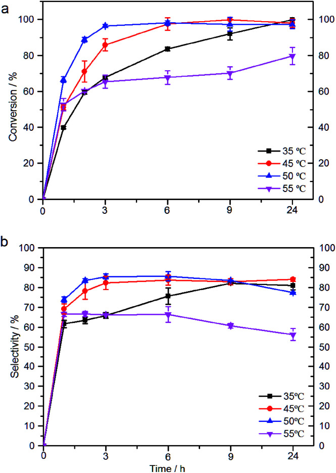
The effect of the reaction temperature on FOL synthesis was shown in Fig. 8. Fig. 8a showed the influence of the reaction temperature on the bio-reduction of FAL. It was found that FAL was consumed completely after 24 h reaction when the temperature varied from 35 to 50 °C and the decrease of conversion at a higher temperature might be due to the thermal inactivation of the cells. Moreover, the actual reaction time was reduced significantly by increasing the temperature at 35–50 °C (e.g., 24 h at 35 °C vs. 6 h at 45 °C or 3 h at 50 °C). A complete conversion was observed after 6 h reaction at 45 °C and the conversion of 96% was achieved after 3 h reaction at 50 °C. The effect of temperature on FOL selectivity was similar with the conversion (Fig. 8b). The high selectivity (>85%) were obtained within 3–6 h at 45 °C and 50 °C. Furthermore, although the conversion of FAL remained high, a longer time reaction resulted to the decrease of the FOL selectivity at 45 °C and 50 °C possible due to excess metabolism of FAL and by-product formed. Hence, a maximum selectivity of 87% was achieved at 50 °C and 6 h reaction. Recently, Mo-doped Co–B amorphous catalyst was reported that exhibited excellent activity and nearly 100% selectivity to FOL at 100 °C under 1.0 MPa.25 Compared with these metals and amorphous alloys catalysts,7 a similar high efficient conversion of FAL into FOL was obtained even under a mile temperature (50 °C) using whole cells of B. coagulans NL01 as bio-reduction catalyst. The results obtained in this work were compared with selected literature results. As shown in Table 2, B. coagulans NL01 gave the highest biotransformation rates of FAL ever reported. FAL was hydrogenated to FOL with high temperature and low reaction rate using SBA-15Cu as catalyst. The efficiency of CTH of FAL to FOL remained low although the method appeared to be environmentally. Although Saccharomyces cerevisiae culturing cells were reported to be FAL-tolerant, their transformation efficacies were very poor.
Fig. 8. Effect of temperature on reduction of furfural by whole cell catalytic of B. coagulans NL01. (a) The conversion of FAL; (b) the selectivity of FOL. Conditions: 42 mM FAL, 20 g L−1 glucose, 9 mg (dry weight) per mL microbial cells, 25 mL phosphate buffer (200 mM pH 7.0), 150 rpm, 35–55 °C; anaerobic reaction for 24 h under the above conditions.
FAL reduction catalyzed by various catalysts.
| Catalysts | Reaction temperature [°C] | Productivitya [g L−1 h−1] | References |
|---|---|---|---|
| SBA-15Cu | 170 | 0.22 (1 h) | 5 |
| γ-Fe2O3@HAP | 180 | 0.01 (10 h) | 6 |
| Zymomonas mobilis | 30 | 0.06 (48 h) | 15 |
| Clostridium acetobutylicum ATCC 824 | 35 | 0.13 (12 h) | 28 |
| Escherichia coli CCZU-K14 | 30 | 0.80 (24 h) | 29 |
| Escherichia coli KO11 | 37 | 0.10 (8 h) | 30 |
| S. cerevisiae 354 | 30 | 1.00 (6 h) | 31 |
| Issatchenkia occidentalis CCTCC M 206097 | 30 | 0.0003 (48 h) | 32 |
| B. coagulans NL01 | 50 | 1.29 (3 h) | This work |
The productivity is defined as the ratio of consumed FAL to the corresponding reaction time.
3.5. Production of FOL by a fed-batch strategy
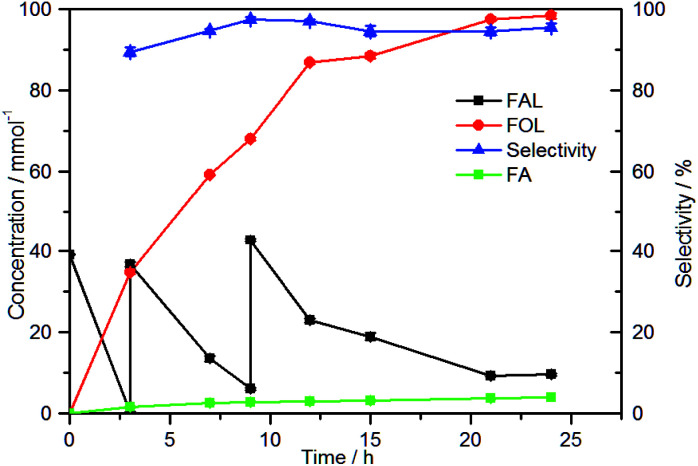
Under the established conditions above, in the presence of 20 g L−1 glucose, 42 mM FAL could be converted into 35 mM FOL at 50 °C with the cells of 9 mg mL−1 after 6 h. Meanwhile, FAL conversion and FOL selectivity reached 97% and 87% respectively. However, inhibition of FAL towards microbial cells still limits the level of FOL production. Fed-batch strategy is considered as an efficient approach for getting a high concentration of product. On the other hand, in addition to substrate inhibition and toxicity, the product also affects the biocatalytic reduction reactions.26 Therefore, fed-batch experiment was performed to evaluate the highest amount of FOL with the cells of 9 mg mL−1. Reaction was initiated with 40 mM FAL and FAL was added to 40 mM at 3 h and 9 h respectively when the FAL concentration reached almost zero (Fig. 9). During the first 12 h, reaction rate was almost linear and about 90 mM FAL was converted to about 87 mM FOL. But thereafter, the consumption rate of FAL decreased continuously until at 24 h. This decline in productivity might be attributed to FOL accumulation, active enzyme depletion, or a combination of these factors.27 Although the rate of FOL production decreased over time with the accumulation of FOL, the 24 h bio-reduction by B. coagulans cells still produced 98.4 mM FOL at 50 °C from 112 mM FAL. The conversion of FAL and the selectivity toward the desired product reached 92% and 96%, respectively.
Fig. 9. FOL production in a fed-batch culture of B. coagulans NL01. Conditions: 40 mM FAL, 20 g L−1 glucose, 9 mg (dry weight) per mL microbial cells, 25 mL phosphate buffer (200 mM pH 7.0), 150 rpm, 50 °C. After FAL was almost used up, 0.925 mmol FAL were supplemented.
4. Conclusions
Herein, we have demonstrated that FOL can be selectively produced from the bio-reduction of FAL by using highly FAL-tolerant B. coagulans NL01 cells. The presence of co-substrate glucose is required for the bio-reduction of FAL to FOL. Optimal conditions were 42 mM FAL, 15–20 g L−1 glucose, 9 mg mL−1, at 50 °C and 6 h. A satisfactory conversion of 97% and selectivity of 87% was obtained under optimized conditions. Fed-batch strategy may be a promising approach for improving FOL production. The cells were able to efficiently transform FAL to FOL by a fed-batch process with good selectivity and up to 98 mM FOL was synthesized in 24 h, which corresponding to 92% conversion and 96% selectivity. These results mean that B. coagulans NL01 can be considered as a promising biocatalyst for FOL production. Compared to other systems for FOL production, this developed bio-reduction system has three main advantages: low temperature condition, atmospheric pressure and no extra hydrogen. Thus it offers a potential strategy for ambient pressure hydrogenation of FAL to FOL and other catalytic reduction reaction.
Conflicts of interest
There are no conflicts to declare.
Supplementary Material
Acknowledgments
This study was supported by the National Natural Science Foundation of China (51776099) and the Major Program of the Natural Science Foundation of Jiangsu Higher Education of China (16KJA220004). We also kindly acknowledge partial support from the Priority Academic Program Development of Jiangsu Higher Education Institutions (PAPD).
Electronic supplementary information (ESI) available. See DOI: 10.1039/c8ra05098h
Notes and references
- Li Y. M. Zhang X. Y. Li N. Xu P. Lou W. Y. Zong M. H. Chemsuschem. 2016;10:372–378. doi: 10.1002/cssc.201601426. [DOI] [PubMed] [Google Scholar]
- Dominguez P. D. M. Guajardo N. V. Chemsuschem. 2017;10:4123–4134. doi: 10.1002/cssc.201701583. [DOI] [PubMed] [Google Scholar]
- Bozell J. J. Petersen G. R. Green Chem. 2010;12:539–554. doi: 10.1039/B922014C. [DOI] [Google Scholar]
- Tuck C. O. Pérez E. Horváth I. T. Sheldon R. A. Poliakoff M. Science. 2012;337:695–699. doi: 10.1126/science.1218930. [DOI] [PubMed] [Google Scholar]
- Vargas-Hernández D. Rubio-Caballero J. M. Santamaría-González J. Moreno-Tost R. Mérida-Robles J. M. Pérez-Cruz M. A. Jiménez-López A. Hernández-Huesca R. Maireles-Torres P. J. Mol. Catal. A: Chem. 2014;383–384:106–113. doi: 10.1016/j.molcata.2013.11.034. [DOI] [Google Scholar]
- Wang F. Zhang Z. ACS Sustainable Chem. Eng. 2016;5:942–947. doi: 10.1021/acssuschemeng.6b02272. [DOI] [Google Scholar]
- Audemar M. Ciotonea C. Royer S. Ungureanu A. Dragoi B. Dumitriu E. Jérôme F. Chemsuschem. 2015;8:1985. doi: 10.1002/cssc.201403398. [DOI] [PubMed] [Google Scholar]
- Zhang D. X. Yeeling O. Zhi L. Wu J. C. Biochem. Eng. J. 2013;72:77–82. doi: 10.1016/j.bej.2013.01.003. [DOI] [Google Scholar]
- Jiang T. Qiao H. Zheng Z. Chu Q. Li X. Qiang Y. Jia O. PLoS One. 2016;11:e0149101. doi: 10.1371/journal.pone.0149101. [DOI] [PMC free article] [PubMed] [Google Scholar]
- Liu Z. L. Slininger P. J. Gorsich S. W. Appl. Biochem. Biotechnol., Part A. 2005:451–460. doi: 10.1385/ABAB:121:1-3:0451. [DOI] [PubMed] [Google Scholar]
- Belay N. Boopathy R. Voskuilen G. Appl. Environ. Microbiol. 1997;63:2092–2094. doi: 10.1128/aem.63.5.2092-2094.1997. [DOI] [PMC free article] [PubMed] [Google Scholar]
- He Y. C. Jiang C. X. Chong G. G. Di J. H. Wu Y. F. Wang B. Q. Xue X. X. Ma C. L. Bioresour. Technol. 2017;245:841–849. doi: 10.1016/j.biortech.2017.08.219. [DOI] [PubMed] [Google Scholar]
- He Y. Ding Y. Ma C. Di J. Jiang C. Li A. Green Chem. 2017;19:3844–3850. doi: 10.1039/C7GC01256J. [DOI] [Google Scholar]
- Ouyang J. Ma R. Zheng Z. Cai C. Zhang M. Jiang T. Bioresour. Technol. 2013;135:475–480. doi: 10.1016/j.biortech.2012.09.096. [DOI] [PubMed] [Google Scholar]
- Franden M. A. Pilath H. M. Mohagheghi A. Pienkos P. T. Zhang M. Biotechnol. Biofuels. 2013;6:99. doi: 10.1186/1754-6834-6-99. [DOI] [PMC free article] [PubMed] [Google Scholar]
- Thomasser C. Danner H. Neureiter M. Saidi B. Braun R. Appl. Biochem. Biotechnol., Part A. 2002:765–773. doi: 10.1385/ABAB:98-100:1-9:765. [DOI] [PubMed] [Google Scholar]
- Hanly T. J. Henson M. A. Biotechnol. Bioeng. 2014;111:272–284. doi: 10.1002/bit.25101. [DOI] [PubMed] [Google Scholar]
- Kennethm B. Liu S. Stephenr H. Josepho R. Biotechnol. Lett. 2010;32:823. doi: 10.1007/s10529-010-0222-z. [DOI] [PubMed] [Google Scholar]
- Ou M. S. Ingram L. O. Shanmugam K. T. J. Ind. Microbiol. Biotechnol. 2011;38:599–605. doi: 10.1007/s10295-010-0796-4. [DOI] [PubMed] [Google Scholar]
- Mussatto S. I. Roberto I. C. Bioresour. Technol. 2004;93:1–10. doi: 10.1016/j.biortech.2003.10.005. [DOI] [PubMed] [Google Scholar]
- Jiang M. Wan Q. Liu R. Liang L. Chen X. Wu M. Zhang H. Chen K. Ma J. Wei P. J. Ind. Microbiol. Biotechnol. 2014;41:115–123. doi: 10.1007/s10295-013-1346-7. [DOI] [PubMed] [Google Scholar]
- Hunt J. R. Carter A. S. Murrell J. C. Dalton H. Hallinan K. O. Crout D. H. G. Holt R. A. Crosby J. Biocatal. Biotransform. 2009;12:159–178. doi: 10.3109/10242429508998160. [DOI] [Google Scholar]
- Wierckx N. Koopman F. Ruijssenaars H. J. de Winde J. H. Appl. Microbiol. Biotechnol. 2011;92:1095–1105. doi: 10.1007/s00253-011-3632-5. [DOI] [PMC free article] [PubMed] [Google Scholar]
- Ran H. Zhang J. Gao Q. Lin Z. Bao J. Biotechnol. Biofuels. 2014;7:1–12. doi: 10.1186/1754-6834-7-1. [DOI] [PMC free article] [PubMed] [Google Scholar]
- Chen X. Li H. Luo H. Qiao M. Appl. Catal., A. 2007;233:13–20. doi: 10.1016/S0926-860X(02)00127-8. [DOI] [Google Scholar]
- Ni Y. Xu J. H. Biotechnol. Adv. 2012;30:1279–1288. doi: 10.1016/j.biotechadv.2011.10.007. [DOI] [PubMed] [Google Scholar]
- Koopman F. Wierckx N. de Winde J. H. Ruijssenaars H. J. Bioresour. Technol. 2010;101:6291–6296. doi: 10.1016/j.biortech.2010.03.050. [DOI] [PubMed] [Google Scholar]
- Zhang Y. Han B. Ezeji T. C. New Biotechnol. 2012;29:345–351. doi: 10.1016/j.nbt.2011.09.001. [DOI] [PubMed] [Google Scholar]
- He Y. C. Jiang C. X. Jiang J. W. Di J. H. Liu F. Ding Y. Qing Q. Ma C. L. Bioresour. Technol. 2017;238:698–705. doi: 10.1016/j.biortech.2017.04.101. [DOI] [PubMed] [Google Scholar]
- Gutiérrez T. Buszko M. L. Ingram L. O. Preston J. F. Appl. Biochem. Biotechnol., Part A. 2002;98–100:327–340. doi: 10.1385/ABAB:98-100:1-9:327. [DOI] [PubMed] [Google Scholar]
- Villa G. P. Bartroli R. López R. Guerra M. Enrique M. Peñas M. Rodríquez E. Redondo D. Jglesias I. Díaz M. Acta Biotechnol. 1992;12:509–512. doi: 10.1002/abio.370120613. [DOI] [Google Scholar]
- Fonseca B. G. Moutta R. D. O. Ferraz F. D. O. Vieira E. R. Nogueira A. S. Baratella B. F. Rodrigues L. C. Zhang H. R. Da Silva S. S. J. Ind. Microbiol. Biotechnol. 2011;38:199–207. doi: 10.1007/s10295-010-0845-z. [DOI] [PubMed] [Google Scholar]
Associated Data
This section collects any data citations, data availability statements, or supplementary materials included in this article.


