Abstract
Background
Methanol (methyl alcohol) is a form of toxic alcohol that is found in illicit alcohol as well as household products such as solvents and paint removers. The most common cause of methanol poisoning is through ingestion of adulterated alcohol; however, other routes of poisoning may also occur including cutaneous exposure and, rarely, inhalation.
Methods/results
We are reporting a case of a young woman with vision loss, parkinsonism and widespread cerebral artery spasms due to methanol inhalation from domestically made perfume.
Conclusion
Our case highlights the increased need for awareness on the part of the public and health authorities with regard to the manufacturing and use of homemade perfumes produced with poorly processed alcohol having a high methyl alcohol content.
Keywords: CLINICAL NEUROLOGY, MOVEMENT DISORDERS
Key messages.
WHAT IS ALREADY KNOWN ON THIS TOPIC
Methanol poisoning through oral ingestion is the most common route of its toxicity, wit a variety of presentations including vision loss, metabolic alterations and irreversible neurological dysfunction, and it may even lead to mortality
WHAT THIS STUDY ADDS
Though methanol poisoning is usually caused by ingestion, other routes of poisoning may also occur including cutaneous exposure. Our study highlights the accidental exposure through inhalation that may lead do devastating outcome including blindness, parkinsonism, and cerebral vasculopathy.
HOW THIS STUDY MIGHT AFFECT RESEARCH, PRACTICE AND/OR POLICY
Our case is aimed to increase the public awareness about the accidental poisoning through the poor manufacturing of homemade perfumes and highlight the presence of cerebral vasospasms as a possible cause of multiple symptoms.
Introduction
Methanol is an alternative to ethanol because it is cheaper and easier to manufacture. Its illegal oral consumption is the most commonly reported route of toxicity in the literature.1 Methanol poisoning is thought to cause severe morbidities such as vision loss, metabolic alterations and irreversible neurological dysfunction, and it may even lead to mortality.2 We are reporting an unusual presentation of incidental methanol intoxication through the inhalation of homemade perfume during the manufacturing process.
Case report
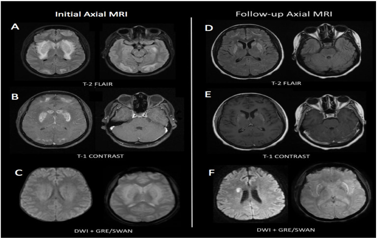
A woman in her 30s living in a rural area of Saudi Arabia was referred to our tertiary care facility for the management of sudden vision loss and parkinsonism. The patient makes a living by manufacturing local perfumes at home, and to compensate for increased demand for her product, she admitted to using adulterated alcohol from a different local vendor. One day after producing a large batch of perfume, she experienced sudden vision loss and became encephalopathic. Subsequently, she was admitted to her local hospital as a case of encephalopathy with optic neuritis, and she required intubation. Due to the lack of the aforementioned noted history, she was misdiagnosed as having autoimmune encephalitis, for which she received a course of pulse methylprednisolone followed by intravenous immunoglobulin G (IVIG). Her initial laboratory investigations (including cerebrospinal fluid analysis) were unremarkable, and the brain MRI showed bilateral and symmetrical areas of bright T2 signals involving the basal ganglia with surrounding oedema. The lentiform nuclei demonstrated blooming in T2-weighted images donating blood degeneration products, likely secondary to necrosis. The frontal and occipital lobes showed bilateral, symmetrical, subcortical, hyperintense T2 signals with heterogeneous contrast enhancements in addition to enhancement of the optic nerves bilaterally (figure 1).
Figure 1.
(A) Bilateral symmetrical areas of bright T2 signals involving the subcortical frontal, temporal and occipital areas involving the basal ganglia with surrounding oedema in the internal, external capsule and insula. (B) Heterogeneous contrast enhancements of the basal ganglia and the frontal and occipital lobes (left) and the optic nerve bilaterally (right). (C) the lentiform nuclei demonstrated high signals with susceptibility artefact. (D) Axial FLAIR MRI of the brain showing bilateral symmetrical T2 hyperintensity in caudate, peripheral putamen and external capsule, with associated cystic changes of the putamen (left) and symmetric subcortical curvilinear hyperintensity in bilateral frontal and occipital lobes sparing the U-fibres without corresponding diffusion restriction nor enhancement (right). (E) Corresponding putaminal enhancement (left) and mild thickening and enhancement of the bilateral optic nerves with patchy enhancement reaching optic chiasm (right). (F) DWI images showing diffusion restriction on the right centrum semiovale (left), central tiny dots of susceptibility artefact in SWAN sequence (right). DWI, diffusion-weighted imaging; FLAIR, fluid-attenuated inversion recovery; GRE, Gradient echo sequences; SWAN, susceptibility-weighted angiography.
On discharge from her local hospital, the patient showed improvement only in her level of consciousness with no significant change in her visual acuity. She became apathetic and bradykinetic, which led to her referral to our tertiary centre.
The patient was hypomemic, hypophonic and apathic with minimal verbal output. She had bilateral polyminimyoclonus in her hands. The higher mental functions and cranial nerves were intact, aside from bilateral optic atrophy and decreased visual acuity with appreciation of hand motions only. The power and deep tendon reflexes were normal. Significant rigidity and bradykinesia were noticed. A sensory examination was unremarkable to all modalities as well as cerebellar examination. Her gait revealed a decreased arm swing bilaterally with a shuffling gait and mild postural instability. The estimated unified Parkinson’s disease rating scale (UPDRS) III was 50.
Laboratory investigations in our hospital, including a haematological profile, renal profile and hepatic profile, were unremarkable. An antinuclear antibody screening, a pyruvate kinase test, lactate levels, and a Wilson workup were normal. A visual evoked potential test showed prolonged P100 bilaterally.
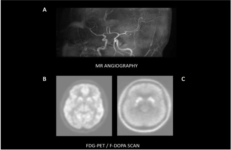
Repeated MRI brain scans showed evolution of the previously mentioned changes with the development of patchy cystic enhancement and microhemorrhages in the basal ganglia, as well as in the frontal and occipital lobes bilaterally. There was enhancement of the bilateral optic nerves and optic chiasm (figure 2). Brain magnetic resonance angiography demonstrated multiple instances of long segmented narrowing along the bilateral A2, A3 and right A1 segments of the anterior cerebral arteries and mild stenosis of the bilateral M2 segments of the middle cerebral arteries (figure 2). MRI spectroscopy showed a decrease of the N-acetylaspartate (NAA) peak at 2.2 parts per million (ppm) and the lactic acid peak at 1.3 ppm.
Figure 2.
(A) MR angiography of the brain demonstrate multiple long segmented moderate smooth narrowing along the bilateral A2, A3 and right A1, M2 as well as mild stenosis of left M2. (B) PET-CT of the brain FDG showing symmetrical FDG uptake within the caudate nucleus with marked symmetrical reduced FDG activity within the lentiform nucleus and mild hypometabolism seen in the occipital lobe bilaterally. (C) PET-CT of the brain and F18-DOPA scan showing reduced tracer activity involving the caudate nuclei bilaterally as well as the inhomogeneous, asymmetrical activity involving the putamen and globus pallidus bilaterally more pronounced on the left. FDG, fluorodeoxyglucose; F-DOPA, 6-[18F]-L-fluoro-L-3, 4-dihydroxyphenylalanine; MR, magnetic resonance; PET, positron emission tomography
A fluorodeoxyglucose (FDG)-positron emission tomography (PET) scan revealed symmetrical uptake within the caudate nuclei with marked reduced symmetrical FDG activity within the lentiform nucleus, and mild hypometabolism was seen in the occipital lobe bilaterally (figure 2B). A 6-[18F]-L-fluoro-L-3, 4-dihydroxyphenylalanine (F-DOPA) PET scan showed reduced tracer activity in the caudate nuclei bilaterally and asymmetrically reduced activity involving the lentiform nuclei bilaterally that was more pronounced on the left side (figure 2C).
The preliminary diagnosis was methanol toxicity due to the nature of her disease progression and typical radiological imaging. As the patient denied any history of alcohol intake, a sample of her domestically manufactured perfume was sent for governmental toxicology screening, and it was found to have a high concentration of methyl alcohol.
Symptomatic therapy consisting of carbidopa–levodopa and pramipexole was started with a gradual increase in the dosages.
During a follow-up visit a month later, the patient exhibited no change in her visual symptoms, but she reported an improvement in her activities of daily living which corresponded to her UPDRS improvement by 20%.
Discussion
This report represents an unusual presentation of incidental methanol intoxication through inadvertent inhalation while manufacturing homemade perfume. It also highlights an additional, yet unrecognised finding of cerebral artery spasms as a squeal of methanol poisoning.
Methanol intoxication can cause severe visual disturbances, metabolic alterations, gastrointestinal complaints and irreversible neurological dysfunction, and it may even be fatal.2 Symptoms may manifest after a latency period of a few hours after exposure (as seen with our patient) or may develop days later.2 The damage is due to alcohol dehydrogenase’s oxidisation of methanol, which causes its conversion to formaldehyde, leading to formic acid production—a toxic metabolite.2 Formic acid causes toxicity by alterations in the cytochrome c oxidase complex at the mitochondria’s terminal end of the respiratory chain.1–5 This change causes disruption of the mitochondria, which are represented heavily in the optic nerves, retina and putamen.1–5
MRI brain findings in methanol toxicity include haemorrhagic putaminal necrosis, diffuse white matter lesions, cerebellar or brainstem lesions that may enhance, and intracerebral or intraventricular haemorrhaging.6 In a case series following 58 patients with methanol intoxication, 33 cases suffered from severe visual disturbances, and brain imaging revealed bilateral optic nerve enhancement, while 45 cases revealed bilateral putaminal ischaemic changes or necrosis.7 All of these cases experienced extrapyramidal symptoms.7 Of note, there was no correlation between the degree of optic atrophy and putaminal involvement.7 Other findings mentioned included asymmetric putaminal involvement, caudate involvement and subcortical white matter oedema.7
What makes our case significant is the presence of multiple cerebral vessel stenosis, which has not been reported in the literature previously (figure 2A). The mechanism of methanol-induced vasospasm was studied in the late 1990s as a response to methanol exposure in canines.8 The study reported a similar finding to ethanol-induced vasospasm, hypothesised to be secondary to its direct action on the smooth muscles of the cerebral vasculature in opposition to alteration in metabolic demand.8 On autopsy, brain infarctions have been previously reported in humans in cases of methanol intoxication; however, vessel stenosis has not been highlighted.9
This is the second case reported to have presynaptic dopaminergic denervation as manifested by reduced tracer activity in the F-DOPA scan (figure 2C). The first case was a female presenting with delayed-onset parkinsonism–dystonia and hypophonia after methanol intoxication, as reported by Franquet et al.4 In our patient, [18F] fluoro-L-DOPA PET imaging confirmed dopaminergic nigrostriatal neuron functional impairment via reduced tracer activity involving the basal ganglia, which explains the parkinsonian features induced by methanol toxicity. These findings were concurrent with the patient’s MRI findings that were reported previously (figure 2C).
Parkinsonian symptoms resulting from methanol poisoning have been managed by the use of dopaminergic agents, as was the case with our patient who showed a fair response to levodopa–carbidopa and pramipexole; however, the literature showed conflicting evidence in response to treatment.9
Our case highlights the increased need for awareness on the part of the public and health authorities with regard to the manufacturing and use of homemade perfumes produced with poorly processed alcohol having a high methyl alcohol content.
Footnotes
Contributors: Writing—original draft: WBM and ST. Writing—review and editing: WBM, ST, SB.
Funding: The authors have not declared a specific grant for this research from any funding agency in the public, commercial or not-for-profit sectors.
Competing interests: None declared.
Provenance and peer review: Not commissioned; externally peer reviewed.
Data availability statement
Data sharing not applicable as no datasets were generated and/or analysed for this study.
Ethics statements
Patient consent for publication
Consent obtained directly from patient(s)
Ethics approval
Not applicable.
References
- 1.Cosentino C, Torres L, Apaza L. Methanol-Induced parkinsonism. Basal Ganglia 2016;6:23–4. 10.1016/j.baga.2015.11.003 [DOI] [Google Scholar]
- 2.Kumar P, Gogia A, Kakar A, et al. An interesting case of characteristic methanol toxicity through inhalational exposure. J Family Med Prim Care 2015;4:470. 10.4103/2249-4863.161359 [DOI] [PMC free article] [PubMed] [Google Scholar]
- 3.Mojica CV, Pasol EA, Dizon ML, et al. Chronic methanol toxicity through topical and inhalational routes presenting as vision loss and restricted diffusion of the optic nerves on MRI: a case report and literature review. eNeurologicalSci 2020;20:100258. 10.1016/j.ensci.2020.100258 [DOI] [PMC free article] [PubMed] [Google Scholar]
- 4.Franquet E, Salvadó-Figueres M, Lorenzo-Bosquet C, et al. Nigrostriatal pathway dysfunction in a methanol-induced delayed dystonia-parkinsonism. Mov Disord 2012;27:1220–1. 10.1002/mds.25049 [DOI] [PubMed] [Google Scholar]
- 5.Liesivuori J, Savolainen H. Methanol and formic acid toxicity: biochemical mechanisms. Pharmacol Toxicol 1991;69:157–63. 10.1111/j.1600-0773.1991.tb01290.x [DOI] [PubMed] [Google Scholar]
- 6.Blanco M, Casado R, Vázquez F, et al. Ct and MR imaging findings in methanol intoxication. AJNR Am J Neuroradiol 2006;27:452–4. [PMC free article] [PubMed] [Google Scholar]
- 7.Elkhamary SM, Fahmy DM, Galvez-Ruiz A, et al. Spectrum of MRI findings in 58 patients with methanol intoxication: long-term visual and neurological correlation. Egypt J Radiol Nucl Med 2016;47:1049–55. 10.1016/j.ejrnm.2016.06.011 [DOI] [Google Scholar]
- 8.Li W, Altura BT, Altura BM. Methanol-Induced contraction of canine cerebral artery and its possible mechanism of action. Toxicol Appl Pharmacol 1998;150:361–8. 10.1006/taap.1998.8425 [DOI] [PubMed] [Google Scholar]
- 9.McLean DR, Jacobs H, Mielke BW. Methanol poisoning: a clinical and pathological study. Ann Neurol 1980;8:161–7. 10.1002/ana.410080206 [DOI] [PubMed] [Google Scholar]
Associated Data
This section collects any data citations, data availability statements, or supplementary materials included in this article.
Data Availability Statement
Data sharing not applicable as no datasets were generated and/or analysed for this study.


