



Abstract
Background and Objectives:
The human papillomavirus (HPV) is associated with more than 70% of the cervical neoplasm. The current study aims to evaluate the distribution of HPV genotypes in suspected women cytological specimens from Tehran, Iran.
Materials and Methods:
In the current cross-sectional study, HPV genotype prevalence was investigated in 433 subject women. DNA extraction was performed by High Pure Viral Nucleic Acid kit. A semi-automatically hybriSpot 24™ (HS24) setting was used for HPV typing and data interpreted by hybriSoft™ software according to instructions.
Results:
Pathologic data showed 181 (41.8%) had non-malignant lesions, 212 (49%) had inflammation and 40 (9.2%) reported LSIL in primary Pap-smear result. HPV was found in 143 (33%) specimens and the most comment high-risk and low-risk HPV types were HPV-16 and −6, respectively. Also, 62 (43%) were co-infected with multiple genotypes includes, 34 (24%) cases had co-infection with two HPV types, 17 (12%) cases had co-infection with three HPV types, 6 (4%) cases had co-infection with four HPV types and 5 (3%) cases had co-infection with five HPV types. There was statistically different domination on high-risk genotype in most of the co-infected samples (p<0.01).
Conclusion:
Current study indicates that the lesion pathology assessment was significantly associated with the HPV infection (p<0.01). Furthermore, the age group assessment shows that most of the HPV positive cases were 21 to 40 (p<0.01). The HPV infection prevalence in the current study was 33% and the most frequently reported high-risk and low-risk HPV types were 16 and 6, respectively.
Keywords: Human papillomavirus (HPV), Papillomavirus infections, Uterine cervical neoplasms, In situ hybridization, Co-infection
INTRODUCTION
Cervical cancer is the fourth common cancer in women worldwide and the international agency for research on cancer (IARC) records 528000 new cases and 266000 cervical cancer-related deaths during 2012. Depends on the geographical regions the cervical cancer rates differ from 0.4 to 4.1 in every 100000 people in Iran (1). The HPV infection could be self-limited mostly but in 10-15% of cases, the infection leads to the cervical intraepithelial neoplasia-2 (CIN-2) or even more advanced grades (2). Also, this infection needs more than 10 years for malignancy induction (3). Cervical cancer progression also could be facilitated by other co-factors such as multiple pregnancies, multiple sexual partners, socioeconomic state, smoking and, oral contraceptive consumptions (4).
Human papillomavirus (HPV) shows a long history of evolution in humans as hosts and reflects a specific tropism into the epithelial cells. The HPV-16 and 18 are the most tumorigenic types and associated with 70% of cervical cancer cases and 50% of the cervical intraepithelial neoplasia grade 3 (CIN-3) (5). Meanwhile, HPV-6 and 11 are mostly associated with genital warts (6, 7). The HPV transmission could be due to the sexual, skin or mucosal contacts (6, 7). The HPV is one of the most common sexually transmitted infections (STIs) around the world. It has been estimated that most sexually active females are infected with at least one of the HPV types worldwide (8, 9). Furthermore, there are about 40 different genotypes of this virus were introduce as an STI while 14 types of all them known as high-risk genotypes including 33, 31, 18, 35, 39, 45, 51, 52, 56, 59, 67, and 73 (10–13).
Recently, the prevention of HPV infection complications is focused on the vaccine and the screening strategies (14–18). Cervical cancer screening is important due to the better treatment responses in pre-cancerous lesions. Studies indicated that the cytological approaches in cervical cancer screening could reduce the number of cervical cancer-related deaths (19). There are different screening tests for the cervical cancer such as cytological (Papanicolaou (Pap-) smear) and virological (HPV PCR). The virologic test could be presented high sensitivity but lower specificity in comparison with the popular cytological test (20, 21). Meanwhile, the Pap smear test has a low sensitivity in comparison with the PCR (22). This low level of sensitivity could lead to more susceptibility of the women for the cervical cancer (23). By considering the importance of different HPV genotypes in the disease progression the current study aims to evaluate the frequency of different HPV genotypes in patients with different cytological results.
MATERIALS AND METHODS
Study population.
In the current study, 433 females referred to medical centers affiliated to Iran University of Medical Sciences, Tehran, Iran for cervical cytology screening and underwent Pap smear test from July 2014 to June 2019 were enrolled. The Pap-smear test was performed by sophisticated pathologists and the eligible individuals who met the inclusion criteria and agreed to participate were carried out. The demographical features were obtained from the medical records. Cervical cancer screening was based on WHO guidelines. Interviewers were nurses in the primary care units that explaining the study details and invited women to participate in the study before routine cytology screening. Informed consent was signed by each participant. Ethics was approved by the Ethical Committee of Iran University of Medical Sciences, Tehran, Iran (No: IR.IUMS.REC.1398.862).
Samples collection.
Before the examination, the procedure was explained verbally to the women. Examination room with enough light was used for the cervix inspection visually. Any abnormalities such as the trace of inflammation, ulceration and growth were inspected by speculum examination. Ayre's spatula used for cervical cells collection and spread them on the pre-labelled spatula glasses. Then, the fixative spray was used. Tubes containing PreservCyt® solution (Hologic Inc., Marlborough MA, USA) was carried out for spatula washing. Additionally, the endocervical brush was used for cervical cell sample collection and the same PreservCyt® vials were used for the second sample collection.
Cytology.
The cervical smear slides were processed and stained then read and reported. The Bethesda system was used for abnormality reporting via expert cytopathologist. The atypical squamous cells of uncertain significance (ASCUS) and HPV-positive women were referred to a gynecologic oncologist for colposcopy speculation and probable directed biopsies for further analysis.
Nucleotide acid extraction.
The nucleotide acid extraction was performed by the High Pure Viral Nucleic Acid kit (Roche, Mannheim, Germany) based on the manufacturer's protocols. Also, the extracted nucleotide acid purification was assessed by the spectrophotometry method by the NanoDrop ND-1000 (Thermo Fisher Scientific Inc., Waltham, MA, USA). Isolated DNA kept at −70°C until analysis.
HPV detection and genotyping.
A nested-PCR was performed for primary HPV detection. The first round performed by MY09/11 and the second by GP5+/6+ universal primers which targeted the HPV L1 gene (24, 25). A 25 μl reaction mix contained 0.2– 0.5 μg concentration of extracted DNA or controls, 0.5-μM concentration of each forward and reverse primers and 12.5 μl of 2× master mix (Yekta Tajhiz Azma Co., Tehran, Iran), and distilled water was added to reach the final volume. A Bio-Rad thermocycler (T100™ Thermal Cycler) was used for heating program as follow: 5 min at 94°C, 45 cycles of 1 min at 94°C, 1 min at 52°C, and 1 min at 72°C and final extension of 5 min at 72°C. The second round was performed as first round except using 42°C for annealing step. Then, specific bands of PCR products (451 for first round and 150 for second round) were visualized on 1.5% concentration of agarose gel in an electrophoresis setting and using an appropriate ladder under UV radiation emitted by the translaminator.
HPV typing by hybriSpot 24™ (HS24).
A semi-automatically hybriSpot 24™ (HS24) setting was used for HPV typing and data interpreted by hybriSoft™ software according to instructions. The kit could detected 36 HPV genotypes included high risk HPV types 16, 18, 26, 31, 33, 35, 39, 45, 51, 52, 53, 56, 58, 59, 66, 68, 73, 82 and low risk types 6, 11, 40, 42, 43, 44, 54, 55, 61, 62, 67, 69, 70, 71, 72, 81, 84, 89. Briefly, the cell pellet was obtained from the liquid-based cytology samples which were washed several times by PBS and centrifuged gently. Then, they were kept at −70°C and used as template for PCR step. A total of 6 μl of template was used for the first round PCR according to the kit protocol. The hybridization procedure at 41°C was done on HPV CHIP-HS by the instructions. The test condition was confirmed based on HPV CHIP quality control.
Statistical evaluation.
The statistical assessment in this study was performed by the IBM SPSS version 22. The statistical significance was assumed as p<0.05. The chi-square and Mann-Whitney u tests were used for the evaluation of the different parameters in the current study.
RESULTS
Patients.
A total of 433 females participated in our comprehensive evaluation based on inclusion criteria. The mean age ± SD of patients in this study was 33.59 ± 8.32 range 18 to 62 years. Also, the Pap-smear test results indicated atypical squamous cells of unknown significance (ASCUS) in 181 (41.8%) of the patients. Furthermore 212 (49%) of the patients show mild or moderate inflammation while 40 (9.2%) reported the low-grade squamous intraepithelial lesion (LSIL).
HPV typing.
The HPV detection by PCR indicated that 143 (33%) of patients were HPV positive while 290 (67%) were negative for the HPV infection. HPV typing by HybriSpot 24™ (HS24) among HPV positive samples showed a different pattern of high-risk and low-risk HPV types. The HPV typing results are summarized in Fig. 1 and Table 1. The result shows that 14 (19.7%) of the patients were infected with both high-risk and low-risk HPV types. 42 (29.6%) had high-risk HPV types and 72 (50.7%) had low-risk types. Also, 81 (57%) cases infected with one HPV type and 62 (43%) were co-infected with multiple genotypes including, 34 (24%) cases had co-infection with two HPV types, 17 (12%) cases had co-infection with three HPV types, 6 (4%) cases had co-infection with four HPV types and 5 (3%) cases had co-infection with five HPV types. There was statistically different domination on high-risk genotype in most of the co-infected samples (p<0.01). Based on the lesion in pathology screening 212 (48.9%) had only inflammation, 181 (41.8%) identified as ASCUS, and 40 (9.2%) were LSIL. No HSIL was diagnosed. The lesion pathology assessment was significantly associated with the HPV result (p<0.01). Combined pathology and HPV multiple types showed that there were 19 inflammation, 26 ASCUS, and 17 LSIL cases identified by HPV multi-genotypes but statistically they were not significant (p>0.05). There were no statistically significant differences for HPV genotypes or multiple genotype co-infection with patient's age or Pap-smear results (p>0.05). Also, there was a significant association between HPV positive results and the patient's age (p<0.01).
Fig. 1.

Low-risk and high-risk HPV types frequencies are represented in a and b, respectivlly.
Table 1.
Multiple HPV types infection frequency based on pathology results and the statistical assessment
| Inflammation | ASCUS | LSIL | Total | P-value | |
|---|---|---|---|---|---|
| HPV Positive | 45 | 59 | 39 | 143 | p>0.05 |
| Infected with one HPV type | 26 | 33 | 22 | 81 | p>0.05 |
| Infected with two different HPV types | 11 | 12 | 11 | 34 | p>0.05 |
| Infected with three different HPV types | 6 | 9 | 2 | 17 | p>0.05 |
| Infected with four different HPV types | 1 | 2 | 3 | 6 | p>0.05 |
| Infected with five different HPV types | 1 | 3 | 1 | 5 | p>0.05 |
|
| |||||
| *Statistically significant difference | |||||
HPV and age groups analysis.
Patients age was categorized in 5 including less than 20, 21 to 30, 31–40, 41 to 50, and 51 to 62 years. The age group analysis indicated that most of the HPV positive cases were 21 to 40 (p<0.01). The age group and HPV high-risk or low-risk distribution were not statistically significant (p>0.05). Also, there were no statistically difference between different Pap-smear results and age groups but the frequencies were illustrated in Fig. 2. Furthermore, the age group did not show any statistically significant differences with multiple genotypes co-infection (p>0.05).
Fig. 2.
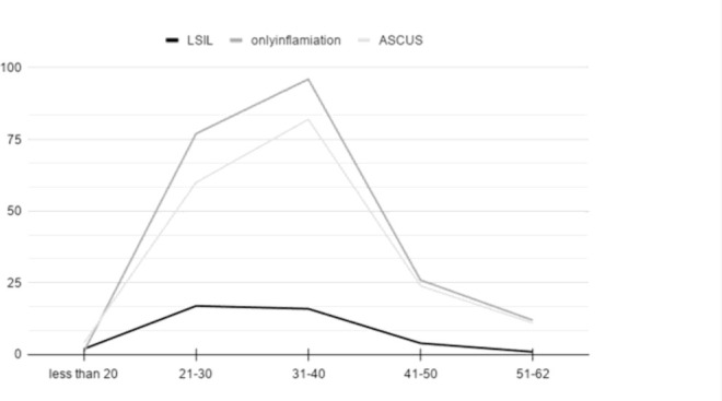
Different Pap-smear results distribution in different age groups of the patients
DISCUSSION
Conducted studies indicated that the use of Papsmear screening every 3 years in the age range from 21–65 years could reduce the incidence of cervical cancer (26). The current study aimed to evaluate different HPV genotypes in patients with different cytological results. We have found that 143 (33%) of our studied patients were infected by HPV. HPV typing showed there were HPV-6, −16 as the major high risk and low risk genotyped in our isolates, respectively. Also, multiple genotype analysis showed that 81 (57%) cases infected with one HPV type and 62 (43%) were co-infected with multiple genotypes as illustrated in Fig. 1. Interestingly, simultaneous infection with 4 and 5 genotypes was found in 12 and 9 cases, respectively.
Liu et al. (27) investigated the HPV prevalence, HPV genotypes and cytological features of 61870 patients in north China. Liu and colleges reported that, 27.9% of the patients were HPV positive. The HPV-16, 52 and 58 were the most frequent genotypes. Also, Liu et al. show a statistically significant reduction in the HPV infection frequency by age. In Liu’s study, multiple genotype co-infections were more frequent in younger patients. But also, the highest risk for HPV infection was seen in the 20–30 years age group. Single, double, triple and quadruple infections in Liu’s study were 17.9%, 6.9%, 2% and 0.5%, respectively. There was statistically different domination on high-risk genotype in most of the co-infected samples (p<0.01). In our current study, multiple genotypes were includes, 34 (24%) two HPV types, 17 (12%) cases had three HPV types, 6 (4%), four HPV types and 5 (3%) cases had co-infection with five HPV types. The age distribution in our current study was only significant for the HPV infection (p<0.01). Also, the age group analysis indicated that, most of the HPV positive cases were 21 to 40 (p<0.01). This finding of age distribution was also confirmed by Liao and colleagues (28). This difference in age distribution between the present study result and Liu and colleagues could highlight a major limitation in our present study. This limitation is due to the limited number of the study population. Furthermore, a conducted study by Brant et al. (29) concluded the HPV infection with one high-risk type had a greater risk for cervical carcinoma generation in comparison with multiple type co-infection. These findings were not supported in our current study result. These differences could be due to the study population differences.
Salehi-Vaziri and colleagues (25), reported the prevalence of the high-risk HPV types 40% in 112 cervical cancer samples in 2015. Meanwhile, another study by Salehi-Vaziri et al. (24) represents the HPV prevalence 45% in 436 cervical cancer samples. Also, the most frequent high-risk and low-risk HPV types were 16 (32%) and 6 (22%), respectively. The HPV prevalence in other countries regardless of different sample sizes and study settings are represented convincible results (30, 31). For instance, a metanalysis study represents the HPV prevalence of 25% in cervical samples (30).
The results of the current study seem to be in the confirmation for these two studies regardless of slight differences. The higher reported HPV prevalence could be due to the different sample sizes between studies. In a conducted study by Wheeler et al. (32) the HPV genotypes in the USA were assessed in 1213 patients. They reported the most common HPV genotypes are 16, 18 and 45 in these patients. Our current study showed that the HPV was present in 33% of the patients. The differences in the HPV prevalence in cervical samples from different geographical locations were reported by different studies (1, 33). Furthermore, in Denny et al. (34) study in Africa, the HPV prevalence was 86.7% in 659 patients and the most common genotypes were 16, 18, 45 and 33. Iljazović and colleagues (35) indicated that the HPV prevalence in malignant cervix tissues was 91% while more than 77% of these patients were infected with more than one genotype. These differences could be justified by the consideration of the geographical locations, methodological differences and economic development of societies. Furthermore, the reduction of cervical cancer by using HPV screening in current years was reported in different studies. This fact highlighted the importance of HPV and cytology testing in reducing cervical cancer incidence (33, 36). Conducted studies represent the presence of HPV in 4% of the cervical specimens even in the vaccinated population. The HPV type 16 was the most common high-risk HPV type (37).
Also, in study conducted by Shetty and colleagues (38) cytology assessment on 316 patients indicated that, 40% of 316 enrolled patients showed abnormal Pap-smear results and 7.6% was reported as pre-neoplastic conditions. The current study result indicates that 181 (41.8%), 212 (49%) and 40 (9.2%) of the patients show ASCUS, inflammation and LSIL, in the Pap-smear test respectively. The study results confirmed by these studies and the differences seem to be due to the assessed population and the geographical regions. The ASCUS is the most frequent diagnostic report for the Pap-smears between normal condition and LSIL or even High-grade squamous intraepithelial lesion (HSIL) (39, 40).
Furthermore, by the assessment of the HPV role in cervical, vulvar and vaginal cancer, Serrano et al. (41) suggested that the HPV vaccine against genotype 16 and 18 could be preventive 80% of these kinds of cancers. The major limitation of the current study could be referred to the limited number of the assessed patients. Also, further comprehensive studies in the HPV genotypes distribution and vaccination are suggested. Also, Kim et al. (42) show 15 new nucleotide substitutions in L1 of the HPV-18. These HPV-18 variants were not associated with clinical features but this assessment could be important in vaccine efficacy.
In conclusion, the current study indicates that the lesion pathology assessment was significantly associated with the HPV infection (p<0.01). Furthermore, the age group assessment shows that, most of the HPV positive cases were 21 to 40 (p<0.01). The HPV infection prevalence in the current study was 33% and the most frequently reported high risk and lowrisk HPV types were 16 and 6, respectively. Also, the Pap-smear test results indicated 181 (41.8%), 212 (49%) and 40 (9.2%) of the ASCUS, mild or moderate inflammation and LSIL in the patients, respectively. It could lead to the importance of the screening program. Finally, the HPV prevalence in screened patients with the Pap-smear test was 33% and 9.2% of these patients showed LSIL.
REFERENCES
- 1.Khorasanizadeh F, Hassanloo J, Khaksar N, Taheri SM, Marzaban M, Rashidi BH, et al. Epidemiology of cervical cancer and human papilloma virus infection among Iranian women—analyses of national data and systematic review of the literature. Gynecol Oncol 2013;128:277–281. [DOI] [PubMed] [Google Scholar]
- 2.McCredie MR, Sharples KJ, Paul C, Baranyai J, Medley G, Jones RW, et al. Natural history of cervical neoplasia and risk of invasive cancer in women with cervical intraepithelial neoplasia 3: a retrospective cohort study. Lancet Oncol 2008;9:425–434. [DOI] [PubMed] [Google Scholar]
- 3.Alam TM, Khan MM, Iqbal MA, Abdul W, Mushtaq M. Cervical cancer prediction through different screening methods using data mining. Int J Adv Comput Sci Appl 2019;10:388–396. [Google Scholar]
- 4.Bosch FX, Lorincz A, Muñoz N, Meijer CJ, Shah KV. The causal relation between human papillomavirus and cervical cancer. J Clin Pathol 2002;55:244–265. [DOI] [PMC free article] [PubMed] [Google Scholar]
- 5.Smith JS, Lindsay L, Hoots B, Keys J, Franceschi S, Winer R, et al. Human papillomavirus type distribution in invasive cervical cancer and high-grade cervical lesions: a meta-analysis update. Int J Cancer 2007;121:621–632. [DOI] [PubMed] [Google Scholar]
- 6.Burchell AN, Winer RL, de Sanjosé S, Franco EL. Chapter6: Epidemiology and transmission dynamics of genital HPV infection. Vaccine 2006;24 Suppl 3:S3/52-61. [DOI] [PubMed] [Google Scholar]
- 7.Roberts JN, Buck CB, Thompson CD, Kines R, Bernardo M, Choyke PL, et al. Genital transmission of HPV in a mouse model is potentiated by nonoxynol-9 and inhibited by carrageenan. Nat Med 2007;13:857–861. [DOI] [PubMed] [Google Scholar]
- 8.Baseman JG, Koutsky LA. The epidemiology of human papillomavirus infections. J Clin Virol 2005;32 Suppl 1:S16–24. [DOI] [PubMed] [Google Scholar]
- 9.Moscicki AB, Schiffman M, Burchell A, Albero G, Giuliano AR, Goodman MT, et al. Updating the natural history of human papillomavirus and anogenital cancers. Vaccine 2012;30 Suppl 5(0 5):F24–F33. [DOI] [PMC free article] [PubMed] [Google Scholar]
- 10.Bouvard V, Baan R, Straif K, Grosse Y, Secretan B, El Ghissassi F, et al. A review of human carcinogens--part B: biological agents. Lancet Oncol 2009;10:321–322. [DOI] [PubMed] [Google Scholar]
- 11.Cogliano V, Baan R, Straif K, Grosse Y, Secretan B, El Ghissassi F, et al. Carcinogenicity of human papilloma-viruses. Lancet Oncol 2005;6:204. [DOI] [PubMed] [Google Scholar]
- 12.Doorbar J, Quint W, Banks L, Bravo IG, Stoler M, Broker TR, et al. The biology and life-cycle of human papillomaviruses. Vaccine 2012;30 Suppl 5:F55–F70. [DOI] [PubMed] [Google Scholar]
- 13.Muñoz N, Bosch FX, De Sanjosé S, Herrero R, Castellsagué X, Shah KV, et al. Epidemiologic classification of human papillomavirus types associated with cervical cancer. N Engl J Med 2003;348:518–527. [DOI] [PubMed] [Google Scholar]
- 14.Bulkmans NW, Berkhof J, Bulk S, Bleeker MC, van Kemenade FJ, Rozendaal L, et al. High-risk HPV type-specific clearance rates in cervical screening. Br J Cancer 2007;96:1419–1424. [DOI] [PMC free article] [PubMed] [Google Scholar]
- 15.Bulkmans NW, Berkhof J, Rozendaal L, van Kemenade FJ, Boeke AJ, Bulk S, et al. Human papillomavirus DNA testing for the detection of cervical intraepithelial neoplasia grade 3 and cancer: 5-year follow-up of a randomised controlled implementation trial. Lancet 2007;370:1764–1772. [DOI] [PubMed] [Google Scholar]
- 16.Cutts FT, Franceschi S, Goldie S, Castellsague Xd, de Sanjose S, Garnett G, et al. Human papillomavirus and HPV vaccines: a review. Bull World Health Organ 2007;85:719–726. [DOI] [PMC free article] [PubMed] [Google Scholar]
- 17.Schiller JT, Castellsagué X, Garland SM. A review of clinical trials of human papillomavirus prophylactic vaccines. Vaccine 2012;30 Suppl 5(0 5):F123–F138. [DOI] [PMC free article] [PubMed] [Google Scholar]
- 18.Zhai L, Tumban E. Gardasil-9: A global survey of projected efficacy. Antiviral Res 2016;130:101–109. [DOI] [PubMed] [Google Scholar]
- 19.Piñeros M, Saraiya M, Baussano I, Bonjour M, Chao A, Bray F. The role and utility of population-based cancer registries in cervical cancer surveillance and control. Prev Med 2021;144:106237. [DOI] [PMC free article] [PubMed] [Google Scholar]
- 20.Arbyn M, Buntinx F, Van Ranst M, Paraskevaidis E, Martin-Hirsch P, Dillner J. Virologic versus cytologic triage of women with equivocal Pap smears: a meta-analysis of the accuracy to detect high-grade intraepithelial neoplasia. J Natl Cancer Inst 2004;96:280–293. [DOI] [PubMed] [Google Scholar]
- 21.Arbyn M, Ronco G, Anttila A, Meijer CJ, Poljak M, Ogilvie G, et al. Evidence regarding human papillomavirus testing in secondary prevention of cervical cancer. Vaccine 2012;30 Suppl 5:F88–F99. [DOI] [PubMed] [Google Scholar]
- 22.Das CR, Mahanta LB, Borah H, Hussain E, Devi A, Choudhury M, et al. A study on epidemiological factors and its association with pathological findings for precancerous symptoms of cervical cancer. Indian J Public Health Res Dev 2019;10:592–597. [Google Scholar]
- 23.Sasieni PD, Cuzick J, Lynch-Farmery E. Estimating the efficacy of screening by auditing smear histories of women with and without cervical cancer. The National Co-ordinating Network for Cervical Screening Working Group. Br J Cancer 1996;73:1001–1005. [DOI] [PMC free article] [PubMed] [Google Scholar]
- 24.Salehi-Vaziri M, Sadeghi F, Hashemi FS, Haeri H, Bokharaei-Salim F, Monavari SH, et al. Distribution of human Papillomavirus genotypes in Iranian women according to the severity of the cervical lesion. Iran Red Crescent Med J 2016;18(4):e24458. [DOI] [PMC free article] [PubMed] [Google Scholar]
- 25.Salehi-Vaziri M, Sadeghi F, Alamsi-Hashiani A, Haeri H, Monavari SH, Keyvani H. Merkel cell polyomavirus and human papillomavirus infections in cervical disease in Iranian women. Arch Virol 2015;160:1181–1187. [DOI] [PubMed] [Google Scholar]
- 26.Kavita SN, Shefali M. Visual inspection of cervix with acetic acid (VIA) in early diagnosis of cervical intraepithelial neoplasia (CIN) and early cancer cervix. J Obstet Gynaecol India 2010;60:55–60. [Google Scholar]
- 27.Liu L, Wang D, Dong H, Jin C, Jiang L, Song H, et al. Characteristics of carcinogenic HPV genotypes in North China plain and the association with cervical lesions. Medicine (Baltimore) 2019;98(37):e17087. [DOI] [PMC free article] [PubMed] [Google Scholar]
- 28.Liao G, Jiang X, She B, Tang H, Wang Z, Zhou H, et al. Multi-infection patterns and co-infection preference of 27 human Papillomavirus types among 137,943 gynecological outpatients across China. Front Oncol 2020;10:449. [DOI] [PMC free article] [PubMed] [Google Scholar]
- 29.Brant AC, Menezes AN, Felix SP, Almeida LM, Moreira MAM. Preferential expression of a HPV genotype in invasive cervical carcinomas infected by multiple genotypes. Genomics 2020;112:2942–2948. [DOI] [PubMed] [Google Scholar]
- 30.Colpani V, Soares Falcetta F, Bacelo Bidinotto A, Kops NL, Falavigna M, Serpa Hammes L, et al. Prevalence of human papillomavirus (HPV) in Brazil: A systematic review and meta-analysis. PLoS One 2020;15(2):e0229154. [DOI] [PMC free article] [PubMed] [Google Scholar]
- 31.Wang J, Tang D, Wang K, Wang J, Zhang Z, Chen Y, et al. HPV genotype prevalence and distribution during 2009–2018 in Xinjiang, China: Baseline surveys prior to mass HPV vaccination. BMC Womens Health 2019;19:90. [DOI] [PMC free article] [PubMed] [Google Scholar]
- 32.Wheeler CM, Hunt WC, Joste NE, Key CR, Quint WG, Castle PE. Human papillomavirus genotype distributions: implications for vaccination and cancer screening in the United States. J Natl Cancer Inst 2009;101:475–487. [DOI] [PMC free article] [PubMed] [Google Scholar]
- 33.Zhu B, Liu Y, Zuo T, Cui X, Li M, Zhang J, et al. The prevalence, trends, and geographical distribution of human papillomavirus infection in China: The pooled analysis of 1.7 million women. Cancer Med 2019;8:5373–5385. [DOI] [PMC free article] [PubMed] [Google Scholar]
- 34.Denny L, Adewole I, Anorlu R, Dreyer G, Moodley M, Smith T, et al. Human papillomavirus prevalence and type distribution in invasive cervical cancer in sub-Saharan Africa. Int J Cancer 2014;134:1389–1398. [DOI] [PubMed] [Google Scholar]
- 35.Iljazović E, Mena M, Tous S, Alemany L, Omeragić F, Sadiković A, et al. Human papillomavirus genotype distribution in invasive cervical cancer in Bosnia and Herzegovina. Cancer Epidemiol 2014;38:504–510. [DOI] [PubMed] [Google Scholar]
- 36.Horn J, Denecke A, Luyten A, Rothe B, Reinecke-Lüthge A, Mikolajczyk R, et al. Reduction of cervical cancer incidence within a primary HPV screening pilot project (WOLPHSCREEN) in Wolfsburg, Germany. Br J Cancer 2019;120:1015–1022. [DOI] [PMC free article] [PubMed] [Google Scholar]
- 37.Leite KRM, Pimenta R, Canavez J, Canavez F, de Souza FR, Vara L, et al. HPV genotype prevalence and success of vaccination to prevent cervical cancer. Acta Cytologica 2020;64:420–424. [DOI] [PubMed] [Google Scholar]
- 38.Shetty RS, Pi RP, Kamath VG, Manjunath AP. Screening for pre-malignant lesions of the cervix among rural women in Southern India. Indian J Res Rep Med Sci 2011;1:1–6. [Google Scholar]
- 39.Nayar R, Wilbur DC. The bethesda system for reporting cervical cytology: a historical perspective. Acta Cytol 2017;61:359–372. [DOI] [PubMed] [Google Scholar]
- 40.Ndifon CO, Al-Eyd G. Atypical squamous cells of undetermined significance (ASCUS). StatPearls [Internet]: StatPearls Publishing; 2020. [PubMed] [Google Scholar]
- 41.Serrano B, de Sanjose S, Tous S, Quiros B, Munoz N, Bosch X, et al. Human papillomavirus genotype attribution for HPVs 6, 11, 16, 18, 31, 33, 45, 52 and 58 in female anogenital lesions. Eur J Cancer 2015;51:1732–1741. [DOI] [PubMed] [Google Scholar]
- 42.Kim N, Park JS, Kim JE, Park JH, Park H, Roh EY, et al. Fifteen new nucleotide substitutions in variants of human papillomavirus 18 in Korea: Korean HPV18 variants and clinical manifestation. Virol J 2020;17:70. [DOI] [PMC free article] [PubMed] [Google Scholar]