Abstract
Purpose
Triple negative breast cancer (TNBC) is an aggressive breast cancer subtype that disproportionately affects women of African ancestry (WAA) and is often associated with poor survival. Although there is a high prevalence of TNBC across West Africa and in women of the African diaspora, there has been no comprehensive genomics study to investigate the mutational profile of ancestrally related women across the Caribbean and West Africa.
Methods
This multisite cross-sectional study used 31 formalin-fixed paraffin-embedded (FFPE) samples from Barbadian and Nigerian TNBC participants. High-resolution whole exome sequencing (WES) was performed on the Barbadian and Nigerian TNBC samples to identify their mutational profiles and comparisons were made to African American, European American and Asian American sequencing data obtained from The Cancer Genome Atlas (TCGA). Whole exome sequencing was conducted on tumors with an average of 382 × coverage and 4335 × coverage for pooled germline non-tumor samples.
Results
Variants detected at high frequency in our WAA cohorts were found in the following genes NBPF12, PLIN4, TP53 and BRCA1. In the TCGA TNBC cases, these genes had a lower mutation rate, except for TP53 (32% in our cohort; 63% in TCGA-African American; 67% in TCGA-European American; 63% in TCGA-Asian). For all altered genes, there were no differences in frequency of mutations between WAA TNBC groups including the TCGA-African American cohort. For copy number variants, high frequency alterations were observed in PIK3CA, TP53, FGFR2 and HIF1AN genes.
Conclusion
This study provides novel insights into the underlying genomic alterations in WAA TNBC samples and shines light on the importance of inclusion of under-represented populations in cancer genomics and biomarker studies.
Supplementary Information
The online version contains supplementary material available at 10.1007/s10552-022-01574-x.
Keywords: Triple negative breast cancer, Women of African ancestry, Whole exome sequencing, Genomics
Introduction
Breast cancer (BCa) is currently the second leading cause of cancer-related deaths in women worldwide [1] and is routinely categorized into different subtypes based on the amplification of human epidermal growth factor receptor 2 (HER2) and expression of estrogen receptor (ER) and progesterone receptor (PR) [2]. Tumors that lack expression for these three receptors are classified as triple negative breast cancer (TNBC). These tumors are typically more aggressive with advanced grade and stage at diagnosis and limited targeted therapies due to the absence of HER2, ER and PR [3]. Mounting evidence indicates a higher prevalence of TNBC in West African women and women of African ancestry (WAA) in the Caribbean (~ 25% in Barbados), the USA (~ 22%) and the UK (~ 22%) compared to non-Hispanic White women (11%) [4–6]. Previous studies have noted variable TNBC estimates across the African continent where West African populations have been shown to have higher TNBC estimates compared to North, East and Southern African regions [7]. The reasons for these disparities are currently unknown; however, recent studies allude to an intricate interplay of environmental and genetic risk factors [8–10].
Due to the aggressive nature of TNBC, there has been an increased interest in investigating molecular biomarkers that could be relevant for therapeutics, diagnostics, and prognostics. Recently, it was found that a subset of TNBC patients with deleterious BRCA1/2 germline mutations responded significantly better to carboplatin (platinum-based therapy) than docetaxel (taxane-based therapy) [11]. In addition to the clinical utility of germline variants, the identification of novel somatic “driver” mutations has been shown to play critical roles in the development of targeted therapies in breast and other solid tumors [12, 13]. Therefore, identification of molecular targets and subsequent development of targeted therapies will be of great importance in improving the overall survival rates of TNBC patients.
Large cancer genomics databases, such as The Cancer Genome Atlas (TCGA), have been useful in understanding the genomics landscape of a variety of tumors. However, to date, most of the TCGA breast cancer biospecimens are from women of European ancestry (~ 80%) despite higher TNBC prevalence in women of African and Hispanic ancestry [14, 15]. Due to the low percentage of African ancestry cases within the TCGA and other similar repositories, researchers have embarked on conducting independent sequencing studies on these populations to explore and understand their unique genomics landscapes [16–21]. Research consortiums such as the International Consortium for the Study of Breast Cancer Subtypes (ICSBCS), the African Caribbean Cancer Consortium (AC3), the African Organization for Research and Training in Cancer (AORTIC), and work done by the Nigerian Breast Cancer Study have begun to shed significant light on potential biomarkers among diverse populations of African ancestry [17, 18, 21–23]. Herein, we have conducted whole exome sequencing (WES) of ancestrally related WAA with TNBC in Nigeria and Barbados to further understand the genomics landscape of these groups. Previous genetic association studies have estimated a high percentage of West African (specifically Nigerian) ancestry in Barbadian cohorts with 80%-90% West African ancestry and admixture with European ancestry, thus the rationale for these two groups [24–26]. Our findings were compared with the genomic signature of TNBC cases of African American (TCGA-AA) and European American (TCGA-EA) within the TCGA database.
Methods
Patient population
Formalin-fixed paraffin-embedded (FFPE) specimens with corresponding clinical data were collected from the Pathology Department at the Queen Elizabeth Hospital (QEH) in Barbados, Lagos University Teaching Hospital (LUTH), University of Calabar Teaching Hospital (UCTH), University of Port Harcourt Teaching Hospital (UPTH), and Jos University Teaching Hospital (JUTH) in Nigeria. Fifty-nine FFPE TNBC samples were selected at random for DNA extraction and WES, however only 31 samples passed quality control. Protocols for specimen collection as outlined within the respective institutional review boards were adhered to and patients consented to give their samples. The study was approved by the institutional review boards at McMaster University, the University of the West Indies—Cave Hill, the QEH, LUTH, UCTH, UPTH and JUTH. Tissues were assessed for ER, PR and HER2 via immunohistochemistry (IHC) at their respective institutions and BCa subtype status was further confirmed at McMaster University in Canada. The Allred algorithm [27] was used to calculate the scores.
Pathologic assessment and DNA isolation
Hematoxylin and eosin (H&E) stained slides were made from FFPE samples for pathological interrogation of tumor enrichment. 10 µm FFPE tissue sections on slides were scraped and placed in NAVY RINO tubes (Next Advance, Troy, NY) with stainless steel beads and 160 µL of deparaffinization solution (Qiagen, Hilden, Germany). Samples were homogenized using the Bullet Blender (Next Advance) for 5 min at speed 12. Samples were incubated for 3 min at 56 °C and processed according to the manufacturer’s instructions. DNA was isolated from pathologically assessed tumor-enriched regions and uninvolved “normal” sections.
Whole exome sequencing
Quality and quantity of DNA was measured using the Genomic DNA Screen Tape Assay (Agilent Technologies, Santa Clara, CA) and Qubit. The concentration of genomic DNA (gDNA) larger than 200 bp was then calculated, and at least 200 ng of DNA greater than 200 bp was sheared in 50 µL of nuclease-free water with the Covaris E220 using the 96 microTUBE Plate (Covaris, Woburn, MA). The library was prepared using KAPA Hyper Prep Kit (Roche, Basel, Switzerland) and 100–500 ng of sheared DNA according to the manufacturer’s instructions. Individual tumor adapter-ligated libraries were enriched into the exome capture reaction, and for germline each adapter-ligated library was pooled before proceeding to capture using Agilent’s SureSelect Human All Exon V6 + custom probes capture library kit [28]. Samples that had successful libraries created were then sequenced on Illumina MiSeq technology for quality control to assess the ability of the libraries to be sequenced. Subsequently, each library was pooled and sequenced on Illumina’s NovaSeq 6000 (Illumina, San Diego, CA) using 300 cycle kit. Raw FASTQs were generated using the industry standard BCL2FASTQ v1.8.4
Primary informatics methods
Whole exome sequences were aligned by BWA (v0.7.17) to GRCh38. Quality score errors were detected by GATK's Base Recalibrator (v4.0.10.1). Picard Tools (v1.128) was used to merge aligned BAMs and mark duplicate reads. Germline Variant Call Format (VCF) of BAM were obtained by GATK's Haplotype Caller using GATK best practices, Samtools MPileUp together with BCFtools (v1.2), and Freebayes (v1.1.0–6-gf069ec6). Somatic variant calling was performed by MuTect2 [29] to ensure compatible comparison between with TCGA. MuTect2 somatic variant calling files (VCF) for each patient in this study were converted to MAF files using the vcf2maf v1.6.19 tool [30]. Data from TCGA were downloaded from: https://portal.gdc.cancer.gov/projects/TCGA-BRCA. The TNBC subtypes were extracted and divided into self-reported Caucasian/European American (EA) and African American (AA) race. Non-silent variants reported were validated visually using IGV (v2.7.2) [31].
Downstream bioinformatics methods
Gene frequencies in our WES data were performed by Unified Optimal Sequence Kernal Association testing in R. Visualization of somatic variants was performed using maftools (v2.6.05) [32] and R packages pheatmap (v1.0.12), ggplot2 (v3.3.3), VennDiagram (v1.6.20), and ggrepel (v0.9.1). Mutation signature from WES data were computed using Mutational Signature in Cancer (MuSiCa) [33]. Copy number analysis was performed utilizing Nexus Copy Number v10 (Biodiscovery) and focal analysis was performed by GISTIC (v2.0) [34]. CNV heatmap was plotted using the oncoprint function in the R package ComplexHeatmap (v2.6.2) and pheatmap. To deduce ancestry information from tumor DNA, 1000 Genomes Project phase 3 VCF release was used as our reference population [35]. Data were transformed to numeric genotypes using PLINK (v1.90b6.7). Principle component analysis (PCA) was performed using the R v3.6.0 function prcomp to establish ancestry distributions mapped by the anchor population.
Type I error (α) and type II error (β) were set at 0.05 and 0.1, respectively. Chi-square test and paired student t-test, as appropriate, were used to examine bivariate association of somatic differences between two cohorts. Benjamini–Hochberg was used for multiple tests. For statistical analysis and visualization, GraphPad Prism 8 was implemented (GraphPad Software, Inc.) and Rv3.6.0 packages: circlize (0.4.6), ComplexHeatmap (1.99.7), dplyr (0.8.0.1), ggplot2 (3.1.1), ggpubr (0.2), maftools (2.0.05), plyr (1.8.4), png (0.1–7), qvalue (2.16.0), reshape2 (1.4.3), stringr (1.4.0), TCGAbiolinks (2.12.6), tidyr (0.8.3), and tools (3.6.0).
Results
Participant characteristics
Mean age at diagnosis for all participants (n = 31) was 49.9 years (Table S1). Specifically, for Nigerian women (n = 12), mean age at diagnosis was 43.2 years old which was significantly younger than the mean age at diagnosis for Barbadian women (n = 19, 53.9, p < 0.05). For all participants with grade data (n = 25), 92% (n = 23) were diagnosed with intermediate and high grade (grade 2 or grade 3) carcinoma whereas only 8% (n = 2) where classified as grade 1 (Table S1). WES data were collected from The Cancer Genome Atlas (TCGA) breast cancer project, stratified by TNBC subtype and participant recorded race (African American, n = 24 [TCGA_TNBC_AA]; European American, n = 63 [TCGA_TNBC_EA]) for comparative analyses.
Mutation contributions and distributions
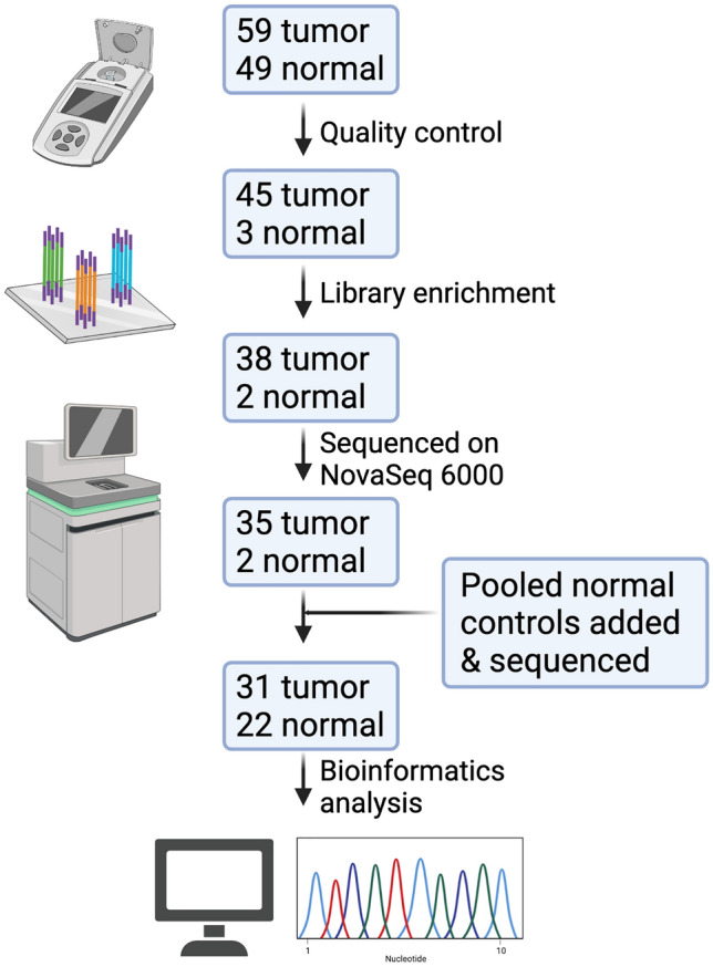
A summary of the sequencing pipeline is depicted in Fig. 1. To assess genomic alterations in TNBC in WAA we performed WES on tumors yielding a mean output of 30,625 Mbases per sample and an average of 382 × coverage (Table S2). An internal pool approach of germline DNA samples was derived from 22 internal samples to ensure better somatic estimation instead of using available Euro-centric references [36]. Germline DNA for each sample was individually indexed before being pooled into the final capture using the same probe sets as tumor samples (Table S2). The germline pool yielded 281,792 Mbase of data and an average of 4335 × coverage showing relative germline contribution to each sample (Table S2). WES identified an average of 707 non-silent somatic mutations per tumor that was higher compared to an exome-sequenced cohort of TNBC in TCGA with a mean of 87 non-silent mutations per sample. This difference is most likely due to the residual increase in private germline variants that is contributed by the diverse African genome, as well as the larger exome capture set (this study = ~ 80 Mbp compared to TCGA = ~ 34 Mbp) and the internal non-tumor pool approach used for our samples. Using ancestry informative markers [37] and principal component analysis, Barbadian samples independently clustered among themselves, the African Caribbean in Barbados (ACB) group and among the Americans of African Ancestry in Southwest USA (ASW) groups (Figure S1). Nigerian samples clustered among the Yoruba in Ibadan, Nigeria (YRI) clusters and the Esan in Nigeria (ESN) clusters (Figure S1).
Fig. 1.

Overview of sequencing pipeline. DNA was extracted from 59 tumor-enriched samples and 49 adjacent uninvolved samples. After quality control, library enrichment and sequencing on NovaSeq6000, 31 tumor samples and 22 pooled normal adjacent controls were successfully sequenced. Model Diagram created with BioRender.com
Variant analysis
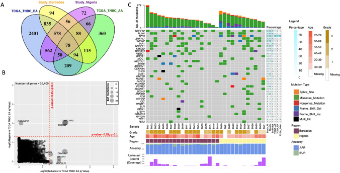
There are 94 unique non-silent somatic variants that are enriched in Barbadian TNBC tissues (n = 19), and 72 unique non-silent variants enriched in Nigerian TNBC tissues (n = 12, Fig. 2A, Table S3–S4) where Benjamini–Hochberg tests were used for multiple testing. There were also 56 commonly mutated genes (Fig. 2A) shared between the Barbadian and Nigerian study samples and 78 commonly mutated genes (Fig. 2A) shared between the four cohorts included (Barbadian, Nigerian, TCGA-TNBC-EA/AA samples). Notably, the TCGA-TNBC-EA group had 2,401 genes in isolation that were not shared with other cohorts in our study. Global comparison of somatic variants with TCGA-TNBC-EA group (Fig. 2B) identified 2 pseudogenes that exhibited an increase in variant frequency in the Nigerian and Barbados cohort compared to TCGA-EA TNBC (TNRC18P2 and DDX12P, p < 0.05; q < 0.1). However, there were no significantly mutated genes between our study samples and the TCGA-AA group (Figure S2A). Common variants in our WAA cohorts were identified in cancer-associated genes—NBPF12, PLIN4, TP53, ZNF717, TAP1, KMT2D, PIEZO1 and BRCA1 (Fig. 2C, Table S5) as well as in HMCN2 and MBD3L3 which have not been previously associated with cancer. In comparison with TCGA data for TNBC cases, these most frequently mutated genes in our WAA cohort were not as frequently mutated except for TP53 (32% in our combined cohort; 63% in TCGA-TNBC-AA; 67% in TCGA-TNBC-EA; 63% in TCGA-Asian). For TP53 specifically, there were five novel variants identified (HGVSc annotation: c.994-1_1023del, c.838_863del, c.368_374del, c.844_845insT, c.382_383del; Table S6) that were all predicted to have high impact as defined by PolyPhen2 [38].
Fig. 2.
Barbadian and Nigerian women with TNBC harbour different genetic alterations than European American (EA)and African American (AA) women with TNBC. A Global analysis of all altered genes revealed that only 78 genes are shared among the four groups (Barbadian, Nigerian, TCGA-TNBC-EA, TCGA-TNBC-AA) and 2,401 genes are unique to the EA group in comparison to the other cohorts. B Global comparison of variants with TCGA-TNBC-EA group identified 2 pseudogenes (TNRC18P2 and DDX12P) with an increase in variant frequency in the Nigerian (n = 12) and Barbadian (n = 19) cohorts. Benjamini–Hochberg tests used for multiple testing. C Genes with high frequency variants in Barbadian and Nigerian samples were not enriched in the TCGA dataset except for TP53 (third gene from the top of the gene list)
COSMIC mutational signatures
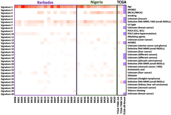
According to somatic mutation signatures as defined by COSMIC [39], most of our samples had a moderate to strong correlation with Signatures 1, 3 and 6 (Fig. 3). These signatures correspond to age, BRCA1/BRCA2 and defective DNA mismatch repair/microsatellite instability (small INDELs), respectively. Among the Barbadian samples, there was a weak correlation with Signature 10, POLE (ultra-hypermutation), that was not seen in the Nigerian samples or the TCGA groups. There were also six Nigerian samples (54%) that showed a correlation with Signature 24 (Aflatoxin) that was not observed in the Barbadian samples or the TCGA dataset.
Fig. 3.
Mutation signature contributions for Barbadian and Nigerian TNBC samples show high correlation to COSMIC Signatures 1, 3 and 6. Using COSMIC somatic mutation signatures, Age, BRCA1/BRCA2 and Defective DNA MMR/MSI (small INDELS) were enriched signatures for Barbadian and Nigerian samples. These signatures were also enriched in TCGA-AA, TCGA-EA women with TNBC and overall, all breast cancer cases within TCGA
Comparison of TNBC copy number variation
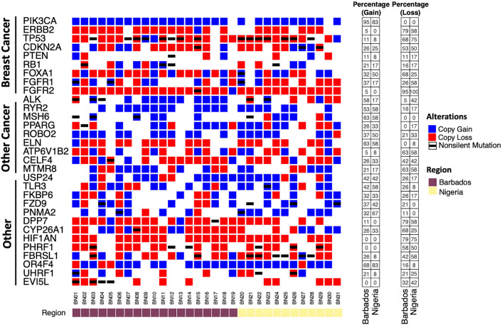
Copy number analysis identified several regions of the genome associated with common copy number gain and loss (Figure S3, Table S7-8). Twenty-eight out of the 31 samples (90%) harbored copy number gains in PIK3CA, and a copy number loss for TP53 was seen in 23 of the 31 samples (74%). Interestingly, copy number loss was seen in 30 samples (97%) for FGFR2 and in 24 samples (77%) for HIF1AN. Overall, CNVs were observed for BCa-related genes (e.g., PIK3CA, ERBB2, TP53, FOXA1), other cancer-related genes (e.g., ROBO2, ELN, CELF4) and other notable genes (e.g., DPP7, CYP26A1). To further expand upon our understanding of bi-allelic events across genes, an integrated analysis was performed across frequently altered genes. This analysis revealed a predominance of bi-allelic (copy number loss and non-silent mutation) in TP53 in our study (Fig. 4).
Fig. 4.
Copy number variation (CNV) analysis revealed no differences between Barbadian and Nigerian samples. Using NEXUS Copy Number (v10) analysis toolkits, most common CNV were investigated. The copy number gain across each sample is presented in blue and copy number loss in red. The black line indicates a non-silent somatic mutation. The bottom panel indicates region of sample origin. Left panels show percent alterations across each copy number event across two different populations. Bi-allelic mutation illustrated by black bar and either blue or red square indicating non-silent somatic mutation and either copy gain or less, respectively. No significance (p < 0.05) was detected in this cohort of samples
Discussion
Herein, we report data from WES of ancestrally related WAA (Barbadian and Nigerian) with TNBC, which revealed pathogenic and novel variants for TP53 and BRCA1 as well as in other BCa implicated genes such as MDC1 (Fig. 2C) that was observed in similar BCa studies among WAA in Nigeria [20, 40]. This is in concordance with the high mutation rate for TP53 that is typically seen in TNBC [41], and more importantly that is observed in Nigerian and African American women with TNBC [20]. Notably, we observed that 50% of our Nigerian samples (n = 12) harbored variants for TP53, which is comparable to ~ 60% of Nigerian TNBC samples (n = 54) with variants and copy number events observed in a previous study [20]. This suggests that variants in TP53 might be of importance for Nigerian women with TNBC. We also observed a high frequency of variants for NBPF12 and PLIN4. Interestingly, in silico analyses of BCa genomics data from TCGA and the International Cancer Genome Consortium have identified NBPF12 as a BCa-driver gene with an estimated 0.3% substitution rate [42]. NBPF12 belongs to the neuroblastoma breakpoint family (NBPF) of genes that are located on chromosome 1, are highly conserved across primates and are highly expressed in breast tissue [43]. PLIN4 is located on chromosome 19 and is a member of the perilipin family that is implicated in adipocyte stability and obesity [44, 45]. Notably, high PLIN4 expression has recently been implicated in TNBC chemoresistance [46]. Although beyond the scope of this study, more functional studies need to be performed to evaluate how these genes and others identified in our study are implicated in TNBC tumorigenesis and disparity in WAA. Further studies are also needed to determine the function of the 78 genes that were commonly enriched between all study groups (Barbados, Nigeria, TCGA-AA and TCGA-EA), as they might be particularly useful for TNBC drug development regardless of ancestry.
In addition to investigating variation in individual genes, we used the COSMIC database of somatic mutations and investigated individual signatures [39], which combines base substitutions with signatures such as DNA mismatch repair. Of note, there was an enrichment of signature 24 (Aflatoxin) in 6 Nigerian samples. This signature is typically observed in a subset of hepatocellular carcinoma (HCC) liver cancers with known exposures to aflatoxin, a mycotoxin that grows on grains across West Africa and is commonly consumed among these populations [47]. It was recently documented in a 10-year study that aflatoxin contamination in crops such as maize and groundnut are common across sub-Saharan African countries such as Nigeria [48], and this may be contributing to breast and liver cancers across these sub-Saharan African nations. Two independent studies reported a considerable number of liver metastases from breast cancers in two Nigerian populations [49, 50], raising the possibility that the correlation with the aflatoxin somatic signature observed in our Nigerian TNBC cases may play a role in this phenomenon. Follow-up studies on this signature in breast tumor samples should be investigated to further delineate this relationship. Nonetheless, these findings highlight the interplay of environmental risk factors with genetics, and how they could lead to tumorigenic outcomes.
When investigating copy number variation, we observed a high enrichment of PIK3CA amplifications, which was also observed in previous studies [41]. Our analysis also revealed a predominance of bi-allelic (copy number loss and non-silent) mutations in TP53 (Fig. 4) which has been previously associated with poor outcome in multiple myeloma [51]. In silico analysis of TP53 copy number loss has also highlighted its prognostic value in breast cancer [52]. Notably, almost every sample (30/31) harboured a copy number loss at 10q26.12—q26.13 that includes the FGFR2 gene. Multiple studies highlight an over-expression of FGFR2 and FGFR1 in TNBC [53–56] so this was an unexpected finding. Indeed, there is currently a clinical trial for inhibition of FGFR2 (ClinicalTrials.gov identifier: NCT04526106) in solid tumors. This copy number loss of FGFR2 highlights a novel genetic alteration in WAA not previously observed that might be protective in these populations. Further studies to investigate FGFR2 mutations and/or expression in WAA with TNBC (perhaps using different specimens—e.g., fresh frozen samples) should shed light on this phenomenon CNV data from FFPE samples can be “noisy” and CNV analyses are rapidly evolving with new tools being developed over time for better interpretation [57].
Twenty-four samples harboured a copy loss for HIF1AN which is the inhibitor of HIF-1α. In TNBC, HIF-1α is highly expressed and implicated in the renewal of cancer stem cells and epithelial-to-mesenchymal transition that is highly associated with metastasis [58]. The frequent copy loss of HIF1AN might thus be associated with the aggressive nature of TNBC observed in WAA since it prevents HIF-1α inhibition. Follow-up RNA sequencing, proteomic profiling and/or targeted sequencing experiments investigating the transcriptome and proteome of these WAA-TNBC cohorts will unveil genes and pathways of interest in WAA-TNBC with therapeutic implications.
It must be noted that social determinants of health (SDoH) should also be considered as factors in our findings since genomics testing is not routinely available among these populations. Though the costs of genomics testing are decreasing, these tests are not as accessible to resource-limited settings for routine clinical (diagnostic and prognostic) tests due to lack of infrastructure [59]. Most recent analyses from GLOBOCAN, estimated the highest BCa mortality rates in countries across West Africa and the Caribbean (with Barbados having the world’s highest mortality) [60]. This high mortality rate in Barbados might be explained by the high incidence of biologically aggressive tumours and advanced staged tumours as we previously reported [4, 61]. Indeed, a recent study investigating approaches to cancer control across the Caribbean region highlighted that there was no organised BCa national screening programme present in Barbados [62]. This might be due to lack of resources and the use of mammography services primarily for diagnostic measures rather than screening purposes. A similar scenario was observed across West African countries where financial constraints and belief in traditional medicine were contributing factors to overall BCa burden [63, 64]. Therefore, to fully understand BCa etiology and progression in these and other underrepresented populations, SDoH should be taken into consideration when highlighting genomic alterations.
Limitations
These data are specific to the samples that were included in the study, and not generalizable to all Barbadian and Nigerian women with TNBC. Further follow-up studies with more samples, appropriate germline controls and associated clinical data are needed to identify potential biomarkers and clinical utility among these populations. It should also be noted that tumor microenvironment as well as inter- and intra- tumour heterogeneity are factors that can be impacted by the sections of the tumor and non-tumor sections that were sequenced and can therefore affect variants called and any potential clinical relevance [65]. To differentiate between somatic and germline mutations, genomic DNA is routinely extracted from peripheral blood, saliva, and adjacent healthy tissue representing germline spectrum of genomics data. This however was not possible due to our retrospective study design as well as the limited resource settings in Barbados and Nigeria at the time of data collection. We acknowledge this limitation and created a pooled non-tumor sample derived from the low quantity patient’s germline adjacent normal breast tissue. This was the best approach instead of only relying on the current Euro-centric databases for germline subtraction with low representation of African ancestry genomics data [36]. We also performed a manual IGV [31] review of each highlighted variant presented in Fig. 2C to remove any false positives due to the higher potential of inflation in false positive somatic calling. We acknowledge that there may still be infiltration from naturally occurring polymorphisms in our variant calls due to not having matched tumor/normal controls. We initially used ExAC and 1000 Genomes databases for variant calls and this resulted in ~ 2 to 3-fold enrichment of potential population related germline false positives in our somatic calling when compared to our pooled reference approach (data not shown). We are confident that our pooled reference approach was appropriate given these challenges. However, further functional validation is required to assess these variant calls and associated clinical relevance. To address other issues with extracting high quality nucleic acids derived from FFPE tissues, we sequenced our tumor and non-tumor samples at high-resolution sequencing depth (382 × and 4,335 × , respectively) to increase confidence in our variant calls. Our pooled germline sample approach might be a useful application for studies of solid tumors with limited germline availability in other resource-limited populations/healthcare facilities.
Conclusions
To our knowledge this is the first study to investigate the mutational landscape of TNBC patients from populations with related African ancestry—West Africa (Nigeria) and Caribbean (Barbados). We identified pathogenic and likely pathogenic variants and novel variants in cancer-associated genes (e.g., TP53, BRCA1, MDC1) and variants in other potential genes of interest (e.g., NBPF12, PLIN4, FGFR2). These variants may be useful for development of future therapeutic options, both unique to our WAA-TNBC cohorts and universally for all women diagnosed with TNBC. Furthermore, to better reflect our global population, more collaborative studies need to be done to increase genomic data from diverse populations within genomics databases. This would allow researchers to identify genetic risks and therapeutic options for diverse populations.
Supplementary Information
Below is the link to the electronic supplementary material.
Acknowledgements
We would like to thank the staff and personnel of the Pathology and Oncology Departments at all the sites in Barbados (QEH) and Nigeria (LUTH, UCTH, UPTH, JUTH) for their assistance with collecting samples used for this project. We would also like to thank Dr. Andrew McArthur and Dr. Robert W. Cowan for Bioinformatics guidance and support.
Author contributions
SMH: conceptualization, study design, data collection, data analysis, data interpretation, co-writing original draft, review & editing. XL: data analysis, review & editing. BB: data collection, co-writing original draft, review & editing. DHAS, SSC, AD, AAFB, GE, TA, IE, JEU, CCO, ACO, MAM, DAM, ECE, JCH, AA: data collection, review & editing. IB, CM: pathological assessment. YX, YJ, SC: data analysis. JDC: data analysis, data interpretation, review. AB, GRP: data interpretation, review & editing. KRMB: data analysis, data interpretation, review & editing. ZM: study design, data analysis, data interpretation, co-writing original draft, supervision, review & editing. JMD: conceptualization, study design, data interpretation, supervision, funding acquisition, review & editing.
Funding
This study was partially funded by the Natural Sciences and Engineering Research Council of Canada (NSERC; grant number RGPIN-2020-06822), Canadian Breast Cancer Foundation/Canadian Cancer Society Research Institute (CBCF/CCSRI; grant number 316252) and Canadian Institutes of Health Research (CIHR; grant number PJT-173223) for data collection storage and trainee stipends. We have not been paid to write this article by a pharmaceutical company or any other agency.
Data availability
The manuscript has electronic data included as electronic supplementary material. Raw sequence data will be submitted to NCBI SRA upon acceptance of the manuscript.
Code availability
None.
Declarations:
Conflict of interest
All the authors declared that they have no conflict of interest.
Ethical approval
Protocols for specimen collection as outlined within the respective institutional review boards were adhered to and patients consented to give their samples. The study was approved by the institutional review boards at McMaster University, the University of the West Indies – Cave Hill, the Queen Elizabeth Hospital, Lagos University Teaching Hospital, University of Calabar Teaching Hospital, University of Port Harcourt Teaching Hospital and Jos University Teaching Hospital.
Footnotes
Publisher's Note
Springer Nature remains neutral with regard to jurisdictional claims in published maps and institutional affiliations.
Contributor Information
Shawn M. Hercules, Email: hercules@mcmaster.ca
Xiyu Liu, Email: xiyuliu@usc.edu.
Blessing B. I. Bassey-Archibong, Email: basseybi@mcmaster.ca
Desiree H. A. Skeete, Email: desiree.skeete@cavehill.uwi.edu
Suzanne Smith Connell, Email: spsmithmd@gmail.com.
Adetola Daramola, Email: aodaramola@cmul.edu.ng.
Adekunbiola A. Banjo, Email: aafbanjo@cmul.edu.ng
Godwin Ebughe, Email: gobeng02@gmail.com.
Thomas Agan, Email: evitomtu@yahoo.com.
Ima-Obong Ekanem, Email: imaekanem2013@gmail.com.
Joe Udosen, Email: josephessien@unical.edu.ng.
Christopher Obiorah, Email: christopher.obiorah@uniport.edu.ng.
Aaron C. Ojule, Email: ojuleac@gmail.com
Michael A. Misauno, Email: micoyedim@yahoo.com
Ayuba M. Dauda, Email: mdayuba10th@gmail.com
Ejike C. Egbujo, Email: anatomyejike@gmail.com
Jevon C. Hercules, Email: jevon.c.hercules@gmail.com
Amna Ansari, Email: ansara4@mcmaster.ca.
Ian Brain, Email: ian.brain@medportal.ca.
Christine MacColl, Email: christine.maccoll@medportal.ca.
Yili Xu, Email: yilixu@usc.edu.
Yuxin Jin, Email: yuxinjin@usc.edu.
Sharon Chang, Email: changsha@usc.edu.
John D. Carpten, Email: carpten@usc.edu
André Bédard, Email: abedard@mcmaster.ca.
Greg R. Pond, Email: gpond@mcmaster.ca
Kim R. M. Blenman, Email: kim.blenman@yale.edu
Zarko Manojlovic, Email: zmanojlo@usc.edu.
Juliet M. Daniel, Email: danielj@mcmaster.ca
References
- 1.Ferlay J, et al. Estimating the global cancer incidence and mortality in 2018: GLOBOCAN sources and methods. Int J Cancer. 2019;144(8):1941–1953. doi: 10.1002/ijc.31937. [DOI] [PubMed] [Google Scholar]
- 2.Prat A, et al. Clinical implications of the intrinsic molecular subtypes of breast cancer. Breast. 2015;24(Suppl 2):S26–35. doi: 10.1016/j.breast.2015.07.008. [DOI] [PubMed] [Google Scholar]
- 3.Li X, et al. Triple-negative breast cancer has worse overall survival and cause-specific survival than non-triple-negative breast cancer. Breast Cancer Res Treat. 2017;161(2):279–287. doi: 10.1007/s10549-016-4059-6. [DOI] [PubMed] [Google Scholar]
- 4.Hercules SM, et al. High triple-negative breast cancer prevalence and aggressive prognostic factors in Barbadian women with breast cancer. Cancer. 2020;126(10):2217–2224. doi: 10.1002/cncr.32771. [DOI] [PubMed] [Google Scholar]
- 5.Bowen RL, et al. Early onset of breast cancer in a group of British black women. Br J Cancer. 2008;98(2):277–281. doi: 10.1038/sj.bjc.6604174. [DOI] [PMC free article] [PubMed] [Google Scholar]
- 6.DeSantis CE, et al. Breast cancer statistics, 2015: convergence of incidence rates between black and white women. CA Cancer J Clin. 2016;66(1):31–42. doi: 10.3322/caac.21320. [DOI] [PubMed] [Google Scholar]
- 7.Newman LA, Kaljee LM. Health disparities and triple-negative breast cancer in African American women: a review. JAMA Surg. 2017;152(5):485–493. doi: 10.1001/jamasurg.2017.0005. [DOI] [PubMed] [Google Scholar]
- 8.Linnenbringer E, et al. Associations between breast cancer subtype and neighborhood socioeconomic and racial composition among Black and White women. Breast Cancer Res Treat. 2020;180(2):437–447. doi: 10.1007/s10549-020-05545-1. [DOI] [PMC free article] [PubMed] [Google Scholar]
- 9.Qin B, et al. Neighborhood social environmental factors and breast cancer subtypes among Black women. Cancer Epidemiol Biomark Prev. 2020;30:344. doi: 10.1158/1055-9965.EPI-20-1055. [DOI] [PMC free article] [PubMed] [Google Scholar]
- 10.Williams DR, Mohammed SA, Shields AE. Understanding and effectively addressing breast cancer in African American women: unpacking the social context. Cancer. 2016;122(14):2138–2149. doi: 10.1002/cncr.29935. [DOI] [PMC free article] [PubMed] [Google Scholar]
- 11.Tutt A, et al. Carboplatin in BRCA1/2-mutated and triple-negative breast cancer BRCAness subgroups: the TNT Trial. Nat Med. 2018;24(5):628–637. doi: 10.1038/s41591-018-0009-7. [DOI] [PMC free article] [PubMed] [Google Scholar]
- 12.Pereira B, et al. The somatic mutation profiles of 2,433 breast cancers refines their genomic and transcriptomic landscapes. Nat Commun. 2016;7:11479. doi: 10.1038/ncomms11479. [DOI] [PMC free article] [PubMed] [Google Scholar]
- 13.Yu B, O'Toole SA, Trent RJ. Somatic DNA mutation analysis in targeted therapy of solid tumours. Transl Pediatr. 2015;4(2):125–138. doi: 10.3978/j.issn.2224-4336.2015.04.04. [DOI] [PMC free article] [PubMed] [Google Scholar]
- 14.Yin L, et al. Triple-negative breast cancer molecular subtyping and treatment progress. Breast Cancer Res. 2020;22(1):61. doi: 10.1186/s13058-020-01296-5. [DOI] [PMC free article] [PubMed] [Google Scholar]
- 15.Yuan J, et al. Integrated analysis of genetic ancestry and genomic alterations across cancers. Cancer Cell. 2018;34(4):549–560.e9. doi: 10.1016/j.ccell.2018.08.019. [DOI] [PMC free article] [PubMed] [Google Scholar]
- 16.Brewster AM, Chavez-MacGregor M, Brown P. Epidemiology, biology, and treatment of triple-negative breast cancer in women of African ancestry. Lancet Oncol. 2014;15(13):e625–e634. doi: 10.1016/S1470-2045(14)70364-X. [DOI] [PMC free article] [PubMed] [Google Scholar]
- 17.Davis M, et al. Identification of distinct heterogenic subtypes and molecular signatures associated with African ancestry in triple negative breast cancer using quantified genetic ancestry models in admixed race populations. Cancers. 2020;12(5):1220. doi: 10.3390/cancers12051220. [DOI] [PMC free article] [PubMed] [Google Scholar]
- 18.Martini R, et al. Investigation of triple-negative breast cancer risk alleles in an International African-enriched cohort. Sci Rep. 2021;11(1):9247. doi: 10.1038/s41598-021-88613-w. [DOI] [PMC free article] [PubMed] [Google Scholar]
- 19.Newman LA, et al. Hereditary susceptibility for triple negative breast cancer associated with western sub-Saharan African ancestry results from an international surgical breast cancer collaborative. Ann Surg. 2019;270(3):484–492. doi: 10.1097/SLA.0000000000003459. [DOI] [PubMed] [Google Scholar]
- 20.Pitt JJ, et al. Characterization of Nigerian breast cancer reveals prevalent homologous recombination deficiency and aggressive molecular features. Nat Commun. 2018;9(1):4181. doi: 10.1038/s41467-018-06616-0. [DOI] [PMC free article] [PubMed] [Google Scholar]
- 21.George SHL, et al. Gene sequencing for pathogenic variants among adults with breast and ovarian cancer in the caribbean. JAMA Netw Open. 2021;4(3):e210307. doi: 10.1001/jamanetworkopen.2021.0307. [DOI] [PMC free article] [PubMed] [Google Scholar]
- 22.Adedokun B, et al. Prevalence of inherited mutations in breast cancer predisposition genes among women in Uganda and Cameroon. Cancer Epidemiol Biomark Prev. 2020;29(2):359–367. doi: 10.1158/1055-9965.EPI-19-0506. [DOI] [PMC free article] [PubMed] [Google Scholar]
- 23.Adedokun B, et al. Cross-ancestry GWAS meta-analysis identifies six breast cancer loci in African and European ancestry women. Nat Commun. 2021;12(1):4198. doi: 10.1038/s41467-021-24327-x. [DOI] [PMC free article] [PubMed] [Google Scholar]
- 24.Murray T, et al. African and non-African admixture components in African Americans and an African Caribbean population. Genet Epidemiol. 2010;34(6):561–568. doi: 10.1002/gepi.20512. [DOI] [PMC free article] [PubMed] [Google Scholar]
- 25.Benn-Torres J, et al. Admixture and population stratification in African Caribbean populations. Ann Hum Genet. 2008;72(Pt 1):90–98. doi: 10.1111/j.1469-1809.2007.00398.x. [DOI] [PubMed] [Google Scholar]
- 26.Cropp CD, et al. 8q24 risk alleles and prostate cancer in African-Barbadian men. Prostate. 2014;74(16):1579–1588. doi: 10.1002/pros.22871. [DOI] [PMC free article] [PubMed] [Google Scholar]
- 27.Allred DC, et al. Prognostic and predictive factors in breast cancer by immunohistochemical analysis. Mod Pathol. 1998;11(2):155–168. [PubMed] [Google Scholar]
- 28.Zhrebker L, et al. Case report: whole exome sequencing of primary cardiac angiosarcoma highlights potential for targeted therapies. BMC Cancer. 2017;17(1):17. doi: 10.1186/s12885-016-3000-z. [DOI] [PMC free article] [PubMed] [Google Scholar]
- 29.Cibulskis K, et al. Sensitive detection of somatic point mutations in impure and heterogeneous cancer samples. Nat Biotechnol. 2013;31(3):213–219. doi: 10.1038/nbt.2514. [DOI] [PMC free article] [PubMed] [Google Scholar]
- 30.Kandoth C et al (2018) mskcc/vcf2maf: vcf2maf v1.6.16
- 31.Robinson JT, et al. Integrative genomics viewer. Nat Biotechnol. 2011;29(1):24–26. doi: 10.1038/nbt.1754. [DOI] [PMC free article] [PubMed] [Google Scholar]
- 32.Mayakonda A, et al. Maftools: efficient and comprehensive analysis of somatic variants in cancer. Genome Res. 2018;28(11):1747–1756. doi: 10.1101/gr.239244.118. [DOI] [PMC free article] [PubMed] [Google Scholar]
- 33.Diaz-Gay M, et al. Mutational signatures in cancer (MuSiCa): a web application to implement mutational signatures analysis in cancer samples. BMC Bioinform. 2018;19(1):224. doi: 10.1186/s12859-018-2234-y. [DOI] [PMC free article] [PubMed] [Google Scholar]
- 34.Mermel CH, et al. GISTIC2.0 facilitates sensitive and confident localization of the targets of focal somatic copy-number alteration in human cancers. Genome Biol. 2011;12(4):41. doi: 10.1186/gb-2011-12-4-r41. [DOI] [PMC free article] [PubMed] [Google Scholar]
- 35.Genomes Project C et al. A global reference for human genetic variation. Nature. 2015;526(7571):68–74. doi: 10.1038/nature15393. [DOI] [PMC free article] [PubMed] [Google Scholar]
- 36.Halperin RF, et al. A method to reduce ancestry related germline false positives in tumor only somatic variant calling. BMC Med Genomics. 2017;10(1):61. doi: 10.1186/s12920-017-0296-8. [DOI] [PMC free article] [PubMed] [Google Scholar]
- 37.Manojlovic Z, et al. Comprehensive molecular profiling of 718 Multiple Myelomas reveals significant differences in mutation frequencies between African and European descent cases. PLoS Genet. 2017;13(11):e1007087. doi: 10.1371/journal.pgen.1007087. [DOI] [PMC free article] [PubMed] [Google Scholar]
- 38.Adzhubei I, Jordan DM, Sunyaev SR. Predicting functional effect of human missense mutations using PolyPhen-2. Curr Protoc Hum Genet. 2013 doi: 10.1002/0471142905.hg0720s76. [DOI] [PMC free article] [PubMed] [Google Scholar]
- 39.Forbes SA, et al. COSMIC: high-resolution cancer genetics using the catalogue of somatic mutations in cancer. Curr Protoc Hum Genet. 2016;91:10.11.1–10.11.37. doi: 10.1002/cphg.21. [DOI] [PubMed] [Google Scholar]
- 40.Friebel TM, et al. BRCA1 and BRCA2 pathogenic sequence variants in women of African origin or ancestry. Hum Mutat. 2019;40:1781. doi: 10.1002/humu.23804. [DOI] [PMC free article] [PubMed] [Google Scholar]
- 41.Shi Y, et al. Therapeutic landscape in mutational triple negative breast cancer. Mol Cancer. 2018;17(1):99. doi: 10.1186/s12943-018-0850-9. [DOI] [PMC free article] [PubMed] [Google Scholar]
- 42.Rajendran BK, Deng CX. Characterization of potential driver mutations involved in human breast cancer by computational approaches. Oncotarget. 2017;8(30):50252–50272. doi: 10.18632/oncotarget.17225. [DOI] [PMC free article] [PubMed] [Google Scholar]
- 43.Vandepoele K, et al. A novel gene family NBPF: intricate structure generated by gene duplications during primate evolution. Mol Biol Evol. 2005;22(11):2265–2274. doi: 10.1093/molbev/msi222. [DOI] [PubMed] [Google Scholar]
- 44.Wolins NE, et al. Adipocyte protein S3–12 coats nascent lipid droplets. J Biol Chem. 2003;278(39):37713–37721. doi: 10.1074/jbc.M304025200. [DOI] [PubMed] [Google Scholar]
- 45.Richardson K, et al. The PLIN4 variant rs8887 modulates obesity related phenotypes in humans through creation of a novel miR-522 seed site. PLoS ONE. 2011;6(4):e17944. doi: 10.1371/journal.pone.0017944. [DOI] [PMC free article] [PubMed] [Google Scholar]
- 46.Sirois I, et al. A unique morphological phenotype in chemoresistant triple-negative breast cancer reveals metabolic reprogramming and PLIN4 expression as a molecular vulnerability. Mol Cancer Res. 2019;17(12):2492–2507. doi: 10.1158/1541-7786.MCR-19-0264. [DOI] [PubMed] [Google Scholar]
- 47.Ladep NG, et al. Problem of hepatocellular carcinoma in West Africa. World J Hepatol. 2014;6(11):783–792. doi: 10.4254/wjh.v6.i11.783. [DOI] [PMC free article] [PubMed] [Google Scholar]
- 48.Bandyopadhyay R, et al. "Ground-truthing" efficacy of biological control for aflatoxin mitigation in farmers' FIELDS in Nigeria: from field trials to commercial usage, a 10-year study. Front Microbiol. 2019;10:2528. doi: 10.3389/fmicb.2019.02528. [DOI] [PMC free article] [PubMed] [Google Scholar]
- 49.Adisa AO, et al. Metastatic breast cancer in a Nigerian tertiary hospital. Afr Health Sci. 2011;11(2):279–284. [PMC free article] [PubMed] [Google Scholar]
- 50.Abur PP, Yusufu LM, Odigie VI. Pattern of visceral metastasis from breast cancer patients in Ahmadu Bello University Teaching Hospital, Zaria, North Western Nigeria. Arch Med Surg. 2019;4(2):31. doi: 10.4103/archms.archms_15_19. [DOI] [Google Scholar]
- 51.Thanendrarajan S, et al. The level of deletion 17p and bi-allelic inactivation of. Haematologica. 2017;102(9):e364–e367. doi: 10.3324/haematol.2017.168872. [DOI] [PMC free article] [PubMed] [Google Scholar]
- 52.Smith JC, Sheltzer JM. Systematic identification of mutations and copy number alterations associated with cancer patient prognosis. Elife. 2018;7:e39217. doi: 10.7554/eLife.39217. [DOI] [PMC free article] [PubMed] [Google Scholar]
- 53.Jafarian AH, et al. The relationship between fibroblastic growth factor receptor-1 (FGFR1) gene amplification in triple negative breast carcinomas and clinicopathological prognostic factors. Iran J Pathol. 2019;14(4):299–304. doi: 10.30699/IJP.2019.96713.1952. [DOI] [PMC free article] [PubMed] [Google Scholar]
- 54.Kim S, et al. FGFR2 promotes breast tumorigenicity through maintenance of breast tumor-initiating cells. PLoS ONE. 2013;8(1):e51671. doi: 10.1371/journal.pone.0051671. [DOI] [PMC free article] [PubMed] [Google Scholar]
- 55.Cunningham DL, et al. Differential responses to kinase inhibition in FGFR2-addicted triple negative breast cancer cells: a quantitative phosphoproteomics study. Sci Rep. 2020;10(1):7950. doi: 10.1038/s41598-020-64534-y. [DOI] [PMC free article] [PubMed] [Google Scholar]
- 56.Fletcher MN, et al. Master regulators of FGFR2 signalling and breast cancer risk. Nat Commun. 2013;4:2464. doi: 10.1038/ncomms3464. [DOI] [PMC free article] [PubMed] [Google Scholar]
- 57.Filia A, et al. High-resolution copy number patterns from clinically relevant FFPE material. Sci Rep. 2019;9(1):8908. doi: 10.1038/s41598-019-45210-2. [DOI] [PMC free article] [PubMed] [Google Scholar]
- 58.Cecil DL, et al. Immunization against HIF-1α inhibits the growth of basal mammary tumors and targets mammary stem cells. Clin Cancer Res. 2017;23(13):3396–3404. doi: 10.1158/1078-0432.CCR-16-1678. [DOI] [PMC free article] [PubMed] [Google Scholar]
- 59.Helmy M, Awad M, Mosa KA. Limited resources of genome sequencing in developing countries: challenges and solutions. Appl Transl Genom. 2016;9:15–19. doi: 10.1016/j.atg.2016.03.003. [DOI] [PMC free article] [PubMed] [Google Scholar]
- 60.Sung H, et al. Global Cancer Statistics 2020: GLOBOCAN estimates of incidence and mortality worldwide for 36 cancers in 185 countries. CA Cancer J Clin. 2021;71(3):209–249. doi: 10.3322/caac.21660. [DOI] [PubMed] [Google Scholar]
- 61.Bassey-Archibong BI, et al. Kaiso is highly expressed in TNBC tissues of women of African ancestry compared to Caucasian women. Cancer Causes Control. 2017;28(11):1295–1304. doi: 10.1007/s10552-017-0955-2. [DOI] [PMC free article] [PubMed] [Google Scholar]
- 62.Spence D, et al. Cancer control in the Caribbean island countries and territories: some progress but the journey continues. Lancet Oncol. 2019;20(9):e503–e521. doi: 10.1016/S1470-2045(19)30512-1. [DOI] [PubMed] [Google Scholar]
- 63.Foerster M, et al. Inequities in breast cancer treatment in sub-Saharan Africa: findings from a prospective multi-country observational study. Breast Cancer Res. 2019;21(1):93. doi: 10.1186/s13058-019-1174-4. [DOI] [PMC free article] [PubMed] [Google Scholar]
- 64.Gyan K, et al. African clinicians' prioritization of needs in international breast cancer partnerships. JAMA Surg. 2019;154(2):182–184. doi: 10.1001/jamasurg.2018.4208. [DOI] [PMC free article] [PubMed] [Google Scholar]
- 65.Lüönd F, Tiede S, Christofori G. Breast cancer as an example of tumour heterogeneity and tumour cell plasticity during malignant progression. Br J Cancer. 2021;125(2):164–175. doi: 10.1038/s41416-021-01328-7. [DOI] [PMC free article] [PubMed] [Google Scholar]
Associated Data
This section collects any data citations, data availability statements, or supplementary materials included in this article.
Supplementary Materials
Data Availability Statement
The manuscript has electronic data included as electronic supplementary material. Raw sequence data will be submitted to NCBI SRA upon acceptance of the manuscript.
None.


