Abstract
Aim:
This study was aimed to analyze the stress generation and distribution for “polyether ether ketone (PEEK)” and metal cobalt-chromium (Co-Cr) at different locations of the tooth using finite element analysis (FEA), when they are casted-off as “Richmond crowns.”
Materials and Methods:
The model of the tooth was designed using “computer-aided design/computer-aided manufacturing” followed by generating the “Mesh” of the tooth to analyze the stress caused by applying vertical and oblique loads of 100N and 40N, respectively, in cubical nodes for both PEEK and metal endodontic post-based Richmond crown. The “3-dimensional von Mises criteria” was used to compare stresses of both elements using FEA. The material properties for each component were designated by respective modulus of elasticity and Poisson's ratio. The statistical test of the stress generation in various locations of PEEK and Metal (Co-Cr) Richmond crown was done by independent t-test.
Results:
From the FEA analysis of Richmond crown, it is evident that maximum stress was generated by “Metal” of about 66.418 MPa when compared to “PEEK” (15.826 MPa). “PEEK Richmond crown” produced minimal stress on the tooth and the other surrounding regions than “Metal Richmond crown” with a statistically significant difference (P < 0.05).
Conclusion:
The results proved that the “Metal Richmond crown” postsystem had a tendency to produce more stress on the tooth and the other surrounding regions than the PEEK. The FEA proved the pros of using “PEEK post Richmond crown,” which is a big boon for the modern era dentistry.
Keywords: Endodontically treated teeth, finite element analysis, polyether ether ketone, Richmond crown, stress
INTRODUCTION
The main objective of restorative dentistry is to restore the characteristic and esthetic features of the teeth that have severely damaged or compromised their tooth structure due to trauma, caries, and fracture.[1] Wherever the existing weakened or impaired crown structure is inadequate to retain a full-coverage crown, then the “post and core” system is needed to improve retention and resistance.[2] However, “post and core” protocols can lead to problems such as dislocation of assembly, fracture of post or root, deprivation of the restorative seal, periodontal ligament, and bone injury.[3]
The Richmond crown is a good alternative treatment to restore the injured teeth. The main purpose of a “post” is to retain a core in a tooth that has lost its crown structure extensively.[4] The main application of Richmond crown is that it can be used in the badly broken single tooth where the remaining height of the crown is very low. The advantages of using this design include custom fitting to the root structure, low or no stress generation at the cervical margin, and high strength when the “post and core” are two separate entities. The “post” reflection under functional forces produces stress on the postcore interface which leads to the separation due to the “post” deformation. “Core” breakdown leads to caries or crown dislodge. Therefore, uniting the post, core, and crown as a single component is good for retaining long-term stability.[5] This system is not popularly used because the high-stress generation surrounding the post leads to failure which can be eliminated by changing the material.[1] Polyether ether ketone (PEEK) and semi-crystalline thermoplastic material with good mechanical and chemical resistance properties with a low-elasticity modulus (8 − 10 GPa) and is of low plaque affinity can be a material of choice for endodontic post and Richmond crown system.[6]
The finite element analysis (FEA) is an approved method for inducing biophysical phenomena in computerized teeth models and their periodontium in the finite-element method is considered an extremely useful tool for simulating the mechanical effects of chewing forces acting on the periodontal ligament and the hard-dental tissue.[7] Deformations and stresses can be assessed at any point within the model and regions with higher stresses can be analyzed. Each dimension in the elastic modulus and the Poisson's ratio are defined for the model materials. A system of simultaneous equations is developed and solved throughout a structure to yield predictable stress distribution.[8] Even though many previous literatures are present on “FEA,” this will be the first study to report on “Richmond crown.” The main aim of this study was to compare the stress generation and distribution between PEEK and metal cobalt-chromium (Co-Cr) as a Richmond crown using the FEA.
MATERIALS AND METHODS
The study protocol was approved by the Institutional Review Board with the ethical committee number IHEC/SDC/UG-1842/21/118. This study was carried out in a tertiary care Dental College and Hospital in Tamilnadu, India.
Computer-aided design/computer-aided manufacturing modeling
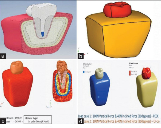
This study was done by initially designing an axisymmetric model representing the various parts of the structure, i.e., gutta-percha, post, core, root, and crown of the posterior tooth (mandibular second premolar) with a proper geometry of each component using computer-aided design/computer-aided manufacturing modeling process [Figure 1a and b]. The dimensions of various parts in the finite element model were taken as given by various researchers and obtained from standard dental literature.[1,7,9] Three dimensional FEA model was simulated, and elastic moduli and Poisson's ratio of all the materials were fed to the software and the “finite element model geometry” was obtained on the computer screen by the provision of various entities such as grids, lines, and patches.
Figure 1.

Model preparation and load application. (a) Axisymmetric model representing the various parts in cross section. (b) The final element model geometry obtained on the computer screen by the provision of various entities such as grids, lines, and patches. (c) The mesh of the tooth model was created by segmenting the entire tooth structure into small cubical forms to analyze the stress caused in each cube. (d) A static load of a 100N and 40N were simultaneously applied on the second premolar tooth in a vertical and oblique direction (30° angulations), respectively for both the materials (i.e., polyether ether ketone and Metal)
Mesh generation
The element form was defined as the solid brick, the four-node element, and the material properties were the options for the meshing purpose. The material properties specified were the modulus of elasticity and Poisson's ratio [Table 1]. Using the material properties for each material and the element specified, the model was meshed with the help of the meshing tool. Meshing generated 159,627 elements interconnected by 36,108 nodes. Then, the mesh of the tooth model was created by segmenting the entire tooth structure into small cubical forms to analyze the stress caused in each cube, and then we finally summarized the stress caused on the cubes connecting different elements of the teeth. These corner points of cubes on which the stress is measured are called nodes which were done using the MeshLab software [Figure 1c].
Table 1.
Modulus of elasticity and Poisson’s ratio for each component of the tooth
| Material | Youngs modulus (Gpa) | Poisson’s ratio |
|---|---|---|
| PEEK | 5.1 | 0.4 |
| Cr-Co | 210 | 0.33 |
| Gingiva | 0.003 | 0.45 |
| Alveolar bone | 1.37 | 0.3 |
| Cortical bone | 1.37 | 0.3 |
| Tooth (dentine) | 18.6 | 0.31 |
| PDL | 0.069 | 0.45 |
| Spongy bone | 1.37 | 0.3 |
| Gutta-percha | 0.69 | 0.45 |
PEEK: Polyether ether ketone, PDL: Periodontal ligament
Application of loads
A static load of 100N and 40N were simultaneously applied on the tooth in a vertical and oblique direction (30° angulations), respectively, for both the materials (i.e., PEEK and Metal). The Richmond crown was previously designed by varying the modulus of elasticity and Poisson's ratio for each material and component [Figure 1d].
Finite element analysis
The data files created in the preprocessor were used to provide solutions and displacement. FEA was performed for both PEEK and Richmond crown. The results and data obtained from the study were analyzed using the 3-dimensional (3D) von Mises criteria.[10] The output data for each material and type of post was obtained as the color-coded diagrams where similar colors depict the same range of stresses generated and warmer colors represent higher stresses (i.e., the blue color which indicates the least stress to red color which indicates the maximum stress) caused on that particular element using finite element modeling and postprocessing software. This was done to determine the employment of durable and comfortable material in patients. The stress pattern and maximum value of the generated von Mises stress were used to give the output of the study as it indicates the site where yielding of ductile material is likely to occur. The statistical test to find association of stress generation in various locations for PEEK and Metal (Co-Cr) Richmond crown was done by “independent t-test” using the IBM-SPSS (Statistical Package for the Social Sciences) Version 24.0 (IBM Corporation, Chicago, USA).
RESULTS
Polyether ether ketone
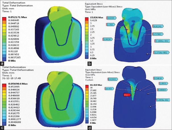
“Deformation” is the displacement caused due to increased stress as a result of increased load in which the PEEK Richmond crown exposed 0.0521 mm of total deformity. From the FEA analysis, it is evident that PEEK Richmond crown showed the maximum of 15.826 Mpa stress where mean stress 2.81 Mpa around the post, 0.57 Mpa around the trabecular bone, and 3.526 Mpa stress generation around the cortical bone [Table 2, Figure 2a and b].
Table 2.
Comparative evaluation of stress generation around the post between polyether ether ketone and Metal Richmond crown in which P value derived from independent t-test
| Group | Mean±SD | t | P |
|---|---|---|---|
| PEEK (around post) | 2.813±0.166 | 20.28 | 0.030* |
| Metal (around post) | 16.113±1.123 | ||
| PEEK (around trabecular bone) | 0.570±0.010 | 68.068 | 0.001* |
| Metal (around trabecular bone) | 1.004±0.004 | ||
| PEEK (around cortical bone) | 3.526±0.661 | 3.408 | 0.027* |
| Metal (around cortical bone) | 5.887±1.001 |
*Statistically significant. PEEK: Polyether ether ketone, SD: Standard deviation
Figure 2.
Finite element analysis. (a) Total deformation caused in polyether ether ketone Richmond crown. (b) Maximum stress caused in PEEK Richmond crown. (c) Total deformation caused in the Metal Richmond crown. (d) Maximum stress caused in Metal Richmond crown
Metal cobalt-chromium
Deformation in Metal (Co-Cr) Richmond crown exposed 0.0569 mm of total deformity. From the FEA analysis, it is evident that maximum stress is caused around the Metal Richmond crown which is 66.418 Mpa, where mean stress is 16.113 Mpa around the post, 1.004 Mpa around the trabecular bone, and 5.887 Mpa stress generation around the cortical bone [Table 2, Figure 2c and d].
On statistical analysis (independent t-test), the difference of stress generation between Metal and PEEK was statistically significant (P < 0.05).
DISCUSSION
A root fracture is an unwanted incident for a postcore restored endodontically treated tooth. When a metal “post and core” is used as an intraradicular “post and core,” vertical root fractures often occur, and leading to tooth extraction. A prefabricated fiberglass post and resin core is commonly used as a “post and core” device to avoid several vertical root fractures. Because fiberglass has a lower elastic modulus than metal, fiberglass postsystems of similar strength induce desirable stress distributions within the root and typically exhibit a repairable horizontal fracture mode when root fractures occur.[3] Although fiberglass has a lower elastic modulus than metal, its elastic modulus is higher than dentin.
The Richmond crown is a single-unit “post and core.” This design has advantages such as being fitted to the root configuration, having little or no stress on the cervical margin. “Post and core” restorations are multicomponent complex systems wherein the stress distribution within the structure is multiaxial, nonuniform, and depends on the magnitude and direction of the applied external loads.[11] A well-known computational tool for measuring stress distribution within such a complex structure is the FEM, which enables researchers to determine the effect of model parameter variance once the basic model has been properly designed.[9] Hence the present study was conducted using 3D FEA to test the biomechanical behavior and long-term protection of the PEEK “postcore” system as a single unit “Richmond crown” in the radicular tooth structure and also the stress distribution analysis was carried out using the von Mises criterion as a quantitative association of maximum stress.[12]
It was found that these materials, when subject to specific loading conditions, have an effect on the pattern of stress distribution within the restore-tooth complex. When a restoration-tooth complex is subjected to loading, a combination of shear and maximum principal stress develops within the system (known as the complex stress state).[13] Since the von Mises criteria method depends on the entire stress field, it has been commonly used as an indicator for the possibility of damage occurrence.[10]
Furthermore, previous literature showed that stress on the fiber post is distributed more evenly in the posttip area, whereas in the metal posthigh stress is concentrated around the teeth.[14,15,16] Another FEA study proved the stress accumulation within the cast “post and core” system is more than fiber post.[17] According to the previous literature, “fiber post” showed more homogenous stress distribution and better biomechanical performance than “metallic post” because the stiffness of fiber post is similar to dentin.[18,19] This proves that both metal and fiber posts shave high-stress distribution.
In the present study, “PEEK” had given the best result in regard to stress distribution which was least when compared to a “metal post.” The experimental results of stress analysis showed a high correlation with FEA predictions, which is in accordance with the previous study conducted by Değer et al., which concluded that “glass and carbon posts” display high-stress distribution, tensile strength, and have Young's modulus when compared to dentin.[20] Due to the reason that the vertical static loads with “fiber posts” restored teeth substantially stronger than those with “metal posts.” This suggested that metallic posts could cause root fractures in pulpless teeth under the influence of vertical static loading but fiber posts did not trigger or accelerate vertical root fracture. This study has shown to be an effective outcome at testing the “PEEK Richmond crown” showing the benefit of using it over metal one.
A study conducted by Pegoretti et al. observed the influence of different postdesigns and composition on stress distribution on maxillary central incisor: finite analysis of the elements showed that the limitations related to the assumption that stress distributions in all vertical sections are identically parallel to the selected two-dimensional model (plane strain assumption), hence we used 3D analysis in this present study which is more accurate to obtain standardized results when compared to two-dimensional models. Many authors also considered that 3D models are definitely more accurate in describing the actual stress conditions.[21]
Therefore, it is evident that “PEEK Richmond crown” can resist tooth fracture due to high modulus of elasticity and it also increases the lifespan of the prosthesis which will act as a viable option for upcoming digital dentistry, as a new horizon in dental technology and material science. Possible limitations of this study include that “PEEK Richmond crown” is very costly, time-consuming, and they are mostly applicable for highly demanding applications. To sum up, further extensive clinical research is required to know about the efficacy and successful outcome of this treatment.
Clinical implications
Endodontically treated tooth with “Metal Richmond crown” can cause tooth fracture due to high-stress generation, whereas “PEEK Richmond crown” aids in distributing less stress around the tooth to prevent fracture and also increases the lifespan of the tooth.
CONCLUSION
From the FEA analysis, it is evident that maximum stress was generated by “Metal Richmond crown” when compared to “PEEK Richmond crown.” Furthermore, surrounding the cortical and trabecular bones, the stress generated was least in “PEEK Richmond crown.” Hence, this study proves that “PEEK Richmond crown” can be used to restore endodontically treated teeth, generating least stress in the clinical approach for great success outcome.
Financial support and sponsorship
Nil.
Conflicts of interest
There are no conflicts of interest.
REFERENCES
- 1.Shetty PP, Meshramkar R, Patil KN, Nadiger RK. A finite element analysis for a comparative evaluation of stress with two commonly used esthetic posts. Eur J Dent. 2013;7:419–22. doi: 10.4103/1305-7456.120668. [DOI] [PMC free article] [PubMed] [Google Scholar]
- 2.Asmussen E, Peutzfeldt A, Sahafi A. Finite element analysis of stresses in endodontically treated, dowel-restored teeth. J Prosthet Dent. 2005;94:321–9. doi: 10.1016/j.prosdent.2005.07.003. [DOI] [PubMed] [Google Scholar]
- 3.Eid R, Juloski J, Ounsi H, Silwaidi M, Ferrari M, Salameh Z. Fracture resistance and failure pattern of endodontically treated teeth restored with computer-aided design/computer-aided manufacturing post and cores: A pilot study. J Contemp Dent Pract. 2019;20:56–63. [PubMed] [Google Scholar]
- 4.Gogna R, Jagadish S, Shashikala K, Keshava Prasad B. Restoration of badly broken, endodontically treated posterior teeth. J Conserv Dent. 2009;12:123–8. doi: 10.4103/0972-0707.57637. [DOI] [PMC free article] [PubMed] [Google Scholar]
- 5.Vinothkumar TS, Kandaswamy D, Chanana P. CAD/CAM fabricated single-unit all-ceramic post-core-crown restoration. J Conserv Dent. 2011;14:86–9. doi: 10.4103/0972-0707.80730. [DOI] [PMC free article] [PubMed] [Google Scholar]
- 6.Panayotov IV, Orti V, Cuisinier F, Yachouh J. Polyetheretherketone (PEEK) for medical applications. J Mater Sci Mater Med. 2016;27:118. doi: 10.1007/s10856-016-5731-4. [DOI] [PubMed] [Google Scholar]
- 7.Patil DB, Reddy ER, Rani ST, Kadge SS, Patil SD, Madki P. Evaluation of stress in three different fiber posts with two-dimensional finite element analysis. J Indian Soc Pedod Prev Dent. 2021;39:178–82. doi: 10.4103/JISPPD.JISPPD_240_20. [DOI] [PubMed] [Google Scholar]
- 8.Mahmoudi M, Saidi A, Gandjalikhan Nassab SA, Hashemipour MA. A three-dimensional finite element analysis of the effects of restorative materials and post geometry on stress distribution in mandibular molar tooth restored with post-core crown. Dent Mater J. 2012;31:171–9. doi: 10.4012/dmj.2011-138. [DOI] [PubMed] [Google Scholar]
- 9.Srirekha A, Bashetty K. A comparative analysis of restorative materials used in abfraction lesions in tooth with and without occlusal restoration: Three-dimensional finite element analysis. J Conserv Dent. 2013;16:157–61. doi: 10.4103/0972-0707.108200. [DOI] [PMC free article] [PubMed] [Google Scholar]
- 10.Aslan T, Üstün Y, Esim E. Stress distributions in internal resorption cavities restored with different materials at different root levels: A finite element analysis study. Aust Endod J. 2019;45:64–71. doi: 10.1111/aej.12275. [DOI] [PubMed] [Google Scholar]
- 11.Upadhyaya V, Bhargava A, Parkash H, Chittaranjan B, Kumar V. A finite element study of teeth restored with post and core: Effect of design, material, and ferrule. Dent Res J (Isfahan) 2016;13:233–8. doi: 10.4103/1735-3327.182182. [DOI] [PMC free article] [PubMed] [Google Scholar]
- 12.Kang N, Wu YY, Gong P, Yue L, Ou GM. A study of force distribution of loading stresses on implant-bone interface on short implant length using 3-dimensional finite element analysis. Oral Surg Oral Med Oral Pathol Oral Radiol. 2014;118:519–23. doi: 10.1016/j.oooo.2014.05.021. [DOI] [PubMed] [Google Scholar]
- 13.Narayanaswamy S, Meena N, Shetty A, Kumari A, Dn N. Finite element analysis of stress concentration in Class V restorations of four groups of restorative materials in mandibular premolar. J Conserv Dent. 2008;11:121–6. doi: 10.4103/0972-0707.45251. [DOI] [PMC free article] [PubMed] [Google Scholar]
- 14.Reinhardt RA, Krejci RF, Pao YC, Stannard JG. Dentin stresses in post-reconstructed teeth with diminishing bone support. J Dent Res. 1983;62:1002–8. doi: 10.1177/00220345830620090101. [DOI] [PubMed] [Google Scholar]
- 15.Hsu ML, Chen CS, Chen BJ, Huang HH, Chang CL. Effects of post materials and length on the stress distribution of endodontically treated maxillary central incisors: A 3D finite element analysis. J Oral Rehabil. 2009;36:821–30. doi: 10.1111/j.1365-2842.2009.02000.x. [DOI] [PubMed] [Google Scholar]
- 16.Cailleteau JG, Rieger MR, Akin JE. A comparison of intracanal stresses in a post-restored tooth utilizing the finite element method. J Endod. 1992;18:540–4. doi: 10.1016/S0099-2399(06)81210-0. [DOI] [PubMed] [Google Scholar]
- 17.Eskitaşcioğlu G, Belli S, Kalkan M. Evaluation of two post core systems using two different methods (fracture strength test and a finite elemental stress analysis) J Endod. 2002;28:629–33. doi: 10.1097/00004770-200209000-00001. [DOI] [PubMed] [Google Scholar]
- 18.Barjau-Escribano A, Sancho-Bru JL, Forner-Navarro L, Rodríguez-Cervantes PJ, Pérez-Gónzález A, Sánchez-Marín FT. Influence of prefabricated post material on restored teeth: Fracture strength and stress distribution. Oper Dent. 2006;31:47–54. doi: 10.2341/04-169. [DOI] [PubMed] [Google Scholar]
- 19.Coelho CS, Biffi JC, Silva GR, Abrahão A, Campos RE, Soares CJ. Finite element analysis of weakened roots restored with composite resin and posts. Dent Mater J. 2009;28:671–8. doi: 10.4012/dmj.28.671. [DOI] [PubMed] [Google Scholar]
- 20.Değer Y, Adigüzel Ö, Yiğit Özer S, Kaya S, Seyfioğlu Polat Z, Bozyel B. Evaluation of temperature and stress distribution on 2 different post systems using 3-dimensional finite element analysis. Med Sci Monit. 2015;21:3716–172. doi: 10.12659/MSM.896132. [DOI] [PMC free article] [PubMed] [Google Scholar]
- 21.Pegoretti A, Fambri L, Zappini G, Bianchetti M. Finite element analysis of a glass fibre reinforced composite endodontic post. Biomaterials. 2002;23:2667–82. doi: 10.1016/s0142-9612(01)00407-0. [DOI] [PubMed] [Google Scholar]


