Key Points
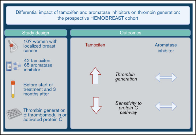
Initiation of tamoxifen is associated with greater thrombin generation and reduced sensitivity to the protein C pathway.
Initiation of aromatase inhibitors does not impact these biomarkers, suggesting a lack of influence on VTE risk.
Visual Abstract
Abstract
Tamoxifen and aromatase inhibitors (AIs) are potent antitumoral agents against breast cancer. Tamoxifen increases the risk of venous thromboembolism (VTE), but the influence of AIs on the risk of VTE remains unclear. To inform clinical decisions, we evaluated associations of tamoxifen or AIs with changes of surrogate hemostatic biomarkers. This prospective cohort included 107 women with localized breast cancer starting tamoxifen (n = 42) or an AI (n = 65). Thrombin generation (CAT) its sensitivity to thrombomodulin (TM) or activated protein C (APC), and specific coagulation parameters, were measured before and 10-16 weeks after initiation of treatmen Compared with baseline, endogenous thrombin potential and thrombin peak increased in tamoxifen users (+86 nM × min; 95% confidence interval [CI], 30-142; and +33 nM; 95% CI, 21-45) but not in AI users (n = 65; +44 nM × min; 95% CI, −4 to 93; and +7 nM; 95% CI, −3 to 17). Normalized TM sensitivity ratios increased with tamoxifen (+0.26; 95% CI, 0.19-0.33y) but not with AI (+0.02; 95% CI, −0.03 to 0.07). Plasma levels of fibrinogen, antithrombin, protein C, and Tissue Factor Pathway Inhibitor decreased, and free protein S increased with tamoxifen but not with AIs. The observed shift toward increased coagulability associated with tamoxifen is in line with its known increased risk of VTE. In contrast, AIs do not appear to impact hemostasis, suggesting a lack of associated VTE risk. The trial was registered at www.clinicaltrials.gov as #NCT03381963.
Introduction
Breast cancer (BC) immensely impacts women’s health,1 as about 1 in 8 women and 1 in 39 women will suffer and die of breast cancer, over the course of their lifetime, respectively.2 Endocrine treatment has become standard for women with estrogen-receptor positive (ER+) BC, which comprises 80% of all BCs, with reductions of cancer recurrence and cancer-related mortality.3 Contemporary estrogen-suppressing treatments are selective estrogen receptor modulators (tamoxifen) and aromatase inhibitors (AIs), comprising anastrozole, exemestane, and letrozole, which are prescribed for 5 to 10 years after adjuvant treatment of ER+ BC. Generally, AIs are preferred for postmenopausal women and some premenopausal women with a high risk of cancer recurrence, in combination with a gonadotropin-releasing hormone agonist. Tamoxifen is prescribed to premenopausal women, and some postmenopausal women with tolerance or contraindication to AIs.
Cancer-associated venous thromboembolism (VTE) creates substantial morbidity and potential mortality and adds complexity to the medical care in 0.5% to 5% of women with localized BC, early after diagnosis.4 Tamoxifen causes some of the VTE burden, with an estimated attributable excess VTE risk of ∼0.3% to 1.9% per year.5,6 The well-demonstrated thrombogenic impact of tamoxifen contrasts with the scarce evidence available for AIs and whether AIs influence the occurrence of VTE remain unclear.7-9
This gap of knowledge creates a challenge to clinicians, leading to uninformed decisions for the prescription of AIs in women at high risk of VTE and the management of AIs after acute VTE.
Therefore, we designed a prospective cohort to primarily estimate the influence of AIs and tamoxifen on global and estrogen-specific hemostatic laboratory outcomes, acting as intermediate phenotype markers for VTE risk. We hypothesized that the use of AIs would not lead to a procoagulant laboratory profile, whereas the use of tamoxifen would.
Methods
The HEMOBREAST study is a prospective cohort set in the Geneva University Hospitals, a large tertiary hospital. The ethics committee approved the study, and all women provided written informed consent.
Population
We approached adult women with a new diagnosis of non-metastatic BC and a planned adjuvant endocrine treatment. Exclusion criteria were an ongoing anticoagulation, a personal history of VTE, recent (<3 months) use of exogenous estrogens, an initiation of endocrine treatment <4 weeks after BC surgery, and a planned chemotherapy, to avoid other factors influencing coagulation parameters.4 Women with a new diagnosis of localized BC and no exclusion criteria (n = 120) were referred for study inclusion, before the start of endocrine treatment, from 2 breast cancer centers (Geneva University Hospitals and Clinique des Grangettes, Geneva), from March 2018 until August 2019.
Endocrine treatment
Following standard care, women were prescribed tamoxifen 20 mg once daily, anastrozole 1 mg once daily, letrozole 2.5 mg once daily, or exemestane 25 mg once daily. They were instructed to start the endocrine treatment after study inclusion, compliance was reported by the participants at 3 months. The selection of the most appropriate drug was left at the discretion of the oncologist. Premenopausal women could also receive a pharmacologic suppression of ovarian function with goserelin, a gonadotropin-releasing hormone agonist.
Data collection and variables
Participants were evaluated at baseline, before initiation of the endocrine treatment (T1), and after 3 months (10-16 weeks) of treatment (T2). At the 2 study evaluations, demographic, medical, and treatment variables were collected through direct interview and medical chart review, and blood was collected. Participants were specifically questioned about their compliance with the endocrine treatment at the 3-month visit.
Endocrine treatment was classified as tamoxifen or AIs (anastrozole, letrozole, exemestane).
Outcomes
The primary outcomes were the endogenous thrombin potential (ETP) and thrombin peak measured with a thrombin generation assay (CAT) and sensitivity of ETP to the activated protein C (APC) pathway, assessed with addition of APC or thrombomodulin (TM) through normalized APC or TM sensitivity ratios (nAPCsr or nTMsr). ETP reflects the overall generation of thrombin activity. Thrombin peak reflects the dynamics of thrombin generation. Both are valid intermediate phenotypes of VTE risk.10,11 The APC pathway is influenced by exogenous estrogens, such as combined oral contraceptives and oral menopausal hormone therapy, and its activity parallels VTE risk in these situations.12,13
Secondary outcomes included other measures of thrombin generation, individual coagulation factors selected a priori (antithrombin, protein C, free protein S, Tissue Factor Pathway Inhibitor [TFPI], factor VII, factor VIII), D-dimer, and clot lysis time, based on prior literature on tamoxifen.14-16 For mechanistic insights of the findings, we also measured factor II (prothrombin) because the basic determinants of thrombin potential are factor II and antithrombin.17 As clinical outcomes, we recorded all VTE and cardiovascular events during the study period.
Laboratory analyses
Blood samples were collected through a peripheral phlebotomy, in nonfasting state, into 0.109-M citrate tubes (BD Vacutainer, Pymouth, UK), after having discarded the first few milliliters. Blood was processed with double-centrifugation (2 × 2000g for 10 minutes), at <90 minutes after collection, and then aliquots were frozen at −80°C.
Clotting times and measurements of D-dimer plasma levels (Vidas D-Dimer exclusion kit; bioMérieux, Marcy l'Etoile, France) were performed on the day of blood draw. All other analyses were performed in batches by laboratory technicians who were blinded to the type of endocrine treatment, with plasma samples frozen for a median of 290 days (interquartile ratio, 162-452) for thrombin generation study.
Coagulation assays were performed on a Atellica Coag 360 (Siemens, Munich, Germany) automate, using the following reagents: Pathromtin SL (Siemens) for the activated partial thromboplastin time (aPTT), Innovin (Siemens) for the prothrombin time (PT), FII-deficient plasma (Siemens) for factor II, FVII-deficient plasma (Siemens) for factor VII, FVIII-deficient plasma (Siemens) for factor VIII, Staclot Protein C (Stago, Asnières, France) for anticoagulant activity of protein C, Berichrom Protein C (Siemens) for chromogenic activity of protein C, VIDAS PC for protein C antigen, Asserachrom Free Protein S for free protein S antigen, and Berichrom antithrombin (Siemens) for anthrombin activity. Plasma level of fibrinogen was measured by the Clauss method using Multifibern U reagent (Siemens). That of TFPI-1 was measured with an enzyme-linked immunosorbent assay (ThermoFisher Scientific, Whaltam, MA). Factor V Leiden and PTG20210A were identified with GeneXpert instrument and cartridges (Cepheid, Thalwil, Switzerland).
Thrombin generation was studied with the CAT method (Stago) using a fluorimeter (Fluroscan Ascent; ThermoLab Systems) equipped with an automatic dispenser.18 Coagulation was initiated with 5 pM tissue factor and 4 μM phospholipids (PPP reagent; Stago), with Thrombin Calibrator (Thrombinoscope BV) for calibration for each patient’s sample. A 5-pM final concentration of tissue factor (TF) was chosen because it is the most studied, in particular, when the APC-operated negative feedback is taken into account, and because it obviates the contribution of the contact phase. In addition, those experimental conditions (TF 5 pM + TM) were shown to be sensitive to elevated plasma levels of factor VIII.19 All plates (Immulon 2HB; Stago) were incubated at 37°C for 10 minutes before addition of the fluorogenic substrate and calcium chloride (CaCl2; FluCa-Kit; Stago), and tests were performed in triplicate. Preliminary experiments allowed us to determine the final concentration of APC (3 nM; Stago) and rabbit lung TM (2 nM; BioMedica Diagnostic), each added to the PPP reagent, needed to reduce by 50% the ETP of healthy control plasma. For each assay, lag time, thrombin peak height, ETP, and velocity index were calculated. Raw data were analyzed with Thrombinoscope V5 software (Stago). The intra-assay and interassay coefficients of variation for ETP were 3.5% and 6.8%, respectively. Normalized APC/TM sensitivity ratios were measured as the ratio of ETP without APC/TM over ETP with APC/TM in participants’ plasma, divided by the same ratio in a control plasma.
Clot lysis time was assessed with turbidity assays in plasma as previously described.20 Clotting and fibrinolysis were monitored at 405 nm every 5 seconds for 120 minutes using a microplate reader (VersaMax; Molecular Devices), after incubation with 0.1 U/mL human thrombin (Merck KGAa), 5 mM CaCl2, and 85 ng/mL tissue plasminogen activator (Technoclone GmbH) in a final volume of 150 μL. Clot lysis time corresponds to the time from the threshold of 50% of the maximum fibrin polymerization to the threshold of 50% of fibrinolysis. Data were analyzed with the Shiny App tool.21
Sample size and statistical analysis
We estimated the sample size (50 participants in each group) with a power of 90% to detect an increase of 30% of nAPCsr between T1 and T2, assuming a correlation of 0.4 between the 2 study times within participants, an α of 0.05. The conservative assumption of 0.4 for the correlation was lower than the correlation observed in our data (0.6-0.8). Retrospectively, using these correlations, the obtained sample size of 42 and 65 yielded a greater power than anticipated (>99% in each group).
Baseline characteristics were described using simple proportions or means and standard deviations. Pure, APC-based, and TM-based ETP and thrombin peak values were normalized, using the values measured with the same commercial plasma on all plates, to minimize interplate variability.
All primary and secondary outcomes were compared between T1 and T2 using a paired t test, within groups of tamoxifen and AIs. Absolute changes of thrombin generation outcomes were directly compared between groups of those using tamoxifen and AIs, using a linear regression model with robust estimator of variance and a priori selected adjustment variables: age (continuous), body mass index (BMI; continuous), current smoking (binary), and the baseline value (T1). This analysis evaluated whether the observed changes in users of tamoxifen were different from the changes in users of AIs while accounting for possible differences in the baseline value between groups. In a post hoc analysis, we explored the associations of letrozole and of exemestane, the 2 most commonly used AIs, with the primary outcome.
We did not formally test the normality of the data but based the use of parametric tests on the central limit theorem in our sample size of moderate size.22 We corrected the α threshold for multiple testing. Based on a marginal α of 0.05, for the primary and secondary outcomes, we used Bonferroni-corrected P < .0041 (12 analyses) and < .0016 (32 analyses), respectively, for significant threshold.
Results
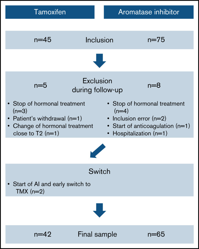
Among the 120 included participants, we excluded 13 from the study (Figure 1). Two participants started with AI but were switched to tamoxifen shortly after inclusion, at least 2 months before the end of the study, and were analyzed as tamoxifen users. The specific AI drugs were letrozole (n = 47), exemestane (n = 17), and anastrazole (n = 1). Four women receiving tamoxifen were also treated with goserelin.
Figure 1.
Flowchart of the study.
At baseline, compared with users of tamoxifen (n = 42) and as expected, users of AI (n = 65) were older (65.5 vs 49.5 years), more likely to be postmenopausal, and had a greater prevalence of cardiovascular risk factors (Table 1). The prevalence of previous vascular disease was low in both groups. The modal cancer stage was IA in tamoxifen users (52.4%) and AI users (50.8%). All women but 1 had undergone breast surgery, at least 4 weeks before study inclusion, mostly skin-sparing tumorectomies combined with radiotherapy. The average time from the end of radiotherapy to study inclusion was 30 (tamoxifen) and 40.2 days (AIs) and from baseline (T1) to end of study (T2) was 14.3 weeks (tamoxifen) and 13.0 weeks (AIs). Heterozygous factor V Leiden and PTG20210A variants were found in 2.8% and 1.9% of patients, respectively.
Table 1.
Baseline characteristics of the 107 participants
| Users of tamoxifen (n=42) | Users of aromatase inhibitors (n=65) | |
|---|---|---|
| Age, y | 49.5 (8.9) | 65.5 (9.4) |
| Weight, kg | 65.7 (11.0) | 69.8 (13.9) |
| BMI, kg/m2 | 24.7 (4.5) | 26.4 (4.9) |
| Parity | 1.7 (0.9) | 1.7 (1.2) |
| Nonmenopausal status | 31 (73.8%) | 0 (0%) |
| Previous hysterectomy | 1 (2.4%) | 11 (16.9%) |
| Diabetes | 1 (2.4%) | 7 (10.9%) |
| Hypertension | 2 (4.8%) | 26 (40%) |
| Dyslipidemia | 4 (9.5%) | 19 (29.2%) |
| Current smoking | 8 (19.1%) | 10 (15.4%) |
| Previous coronary heart disease | 0 (0%) | 3 (4.6%) |
| Previous cerebrovascular disease | 0 (0%) | 2 (3.1%) |
| Breast cancer stage | ||
| 0 | 7 (16.7%) | 2 (3.1%) |
| IA | 22 (52.4%) | 33 (50.8%) |
| IB | 2 (4.8%) | 4 (6.2%) |
| IIA | 5 (11.9%) | 16 (24.6%) |
| IIB | 3 (7.1%) | 8 (12.3%) |
| IIIA | 2 (4.5%) | 1 (1.5%) |
| IIIB | 0 (0%) | 1 (1.5%) |
| IIIC | 1 (2.4%) | 0 (0%) |
| Previous cancer treatment | ||
| Breast surgery | 42 (100%) | 64 (98.5%) |
| Time from surgery to inclusion, days | 107 (64) | 104 (73) |
| Breast radiotherapy before inclusion | 33 (78.6%) | 57 (87.7%) |
Numbers are mean (standard deviation) or n (%).
Thrombin generation
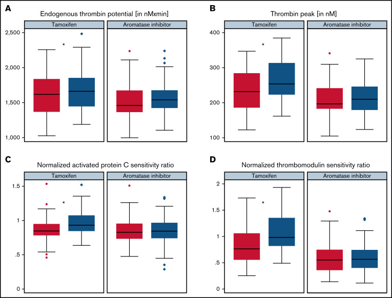
In within-group comparisons of before and after treatment initiation, measures of ETP and thrombin peak increased significantly in tamoxifen users (Table 2; Figure 2A-B). In AI users, ETP and thrombin peak increased somewhat, but without statistical evidence of a difference during the study (Table 2; Figure 2A-B). Differences in changes in lag time and time to peak were found between those using tamoxifen and AIs, as both temporal parameters shortened only in tamoxifen users (supplemental Table 1).
Table 2.
Change (T2 − T1) in thrombin generation and its sensitivity to APC or TM, stratified by use of tamoxifen and AIs
| Tamoxifen (n=42) | Aromatase inhibitors (n=65) | |||||||
|---|---|---|---|---|---|---|---|---|
| T1 mean (SD) | T2 mean (SD) | Absolute difference (95% CI) | P | T1 mean (SD) | T2 mean (SD) | Absolute difference (95% CI) | P | |
| ETP, nM × min | 1596.8 (298.5) | 1683.0 (297.8) | +86.2 (+30.3 to +142.2) | .003 | 1531.3 (252.7) | 1575.7 (233.2) | +44.4 (−4.2 to +93.1) | .07 |
| Thrombin peak, nM | 235.4 (65.8) | 268.3 (62.5) | +32.9 (+21.1 to +44.7) | <.001 | 207.2 (48.4) | 214.4 (47.2) | +7.2 (−2.5 to +16.9) | .13 |
| nAPCsr | 0.86 (0.19) | 0.97 (0.19) | +0.11 (+0.06 to +0.16) | <.001 | 0.84 (0.19) | 0.84 (0.19) | 0.0 (−0.05 to + 0.05) | .91 |
| nTMsr | 0.81 (0.35) | 1.07 (0.36) | +0.26 (+0.19 to +0.33) | <.001 | 0.58 (0.28) | 0.60 (0.27) | +0.02 (−0.03 to + 0.07) | .37 |
Figure 2.
Change of thrombin generation with use of tamoxifen and aromatase inhibitors. Box plots of the measures of ETP (A), thrombin peak (B), nAPCsr (C), and nTMsr at baseline and after 3 months in users of tamoxifen and in users of AIs (D). *P < .0041 (threshold of statistical significance).
In between-group comparisons with comparative adjusted regression models, the increase in ETP and thrombin peak was numerically greater in those using tamoxifen than AIs, with strong statistical evidence for greater thrombin peak (Table 3; +34.4 nM; 95% confidence interval [CI], 17.1-51.7 nM; P = .003) but not for ETP (P = .13).
Table 3.
Unadjusted/adjusted differences in change (T2 − T1) of these measures between the groups
| Unadjusted difference (95% CI) | Adjusted difference* (95% CI) | P | |
|---|---|---|---|
| ETP, nM × min | +41.8 (−31.5 to +115.1) | +62.6 (−19.5 to +144.7) | .13 |
| Thrombin peak, nM | +25.7 (+10.6 to +40.8) | +34.4 (+17.1 to +51.7) | <.001 |
| nAPCsr | +14% (+4 to +23) | +17.6% (+5.5 to +29.7) | .005 |
| nTMsr | +34% (+17 to +50) | +48.3% (+30.5 to +66.6) | <.001 |
Interpreted with a Bonferroni-corrected significant P value threshold of <.0041.
Adjusted for age, BMI, current smoking, and baseline value.
No meaningful difference was seen between users of letrozole and of exemestane (data not shown).
Sensitivity to APC and TM
The normalized sensitivity ratios of APC and TM increased after treatment initiation in those using tamoxifen and remained stable in those using AIs (Table 2; Figure 2C-D).
Compared with AIs, tamoxifen was associated with a marginal increase of 17.6% (95% CI, 5.5-29.7) and statistically significant increase of 35.4% (95% CI, 16.1-54.8) in nAPCsr and nTMsr, respectively. ETP measured with APC or TM followed the same pattern, with a significant increase in those using tamoxifen but not in those using AIs (supplemental Table 1). No meaningful difference was seen between letrozole and exemestane (data not shown).
Individual coagulation factors
With Bonferroni-adjusted α thresholds of <0.0016, we observed a significant decrease in plasma levels of fibrinogen (−0.7 g/L; 95% CI, −0.9 to −0.5), antithrombin (−8.1%; 95% CI, −10.8 to −5.5), and TFPI (−49.2 ng/mL; 95% CI, −69.0 to −29.5) and a significant increase in free protein S (+8.1%; 95% CI, 5.1-11.1) associated with tamoxifen use (Table 4). AI treatment did not significantly affect these parameters. Clot lysis time and D-dimer plasma levels decreased in both those using tamoxifen and those using AIs; however, there was no statistical significance.
Table 4.
Change in individual hemostasis parameters and in clot lysis time, stratified by use of tamoxifen and AIs
| Tamoxifen (n=42) | Aromatase inhibitors (n=65) | |||||||
|---|---|---|---|---|---|---|---|---|
| T1 mean (SD) | T2 mean (SD) | Absolute difference (95% CI) | P | T1 mean (SD) | T2 mean (SD) | Absolute difference (95% CI) | P | |
| PT, % | 95.8 (6.2) | 95.9 (7.2) | 0 (−1.5 to 1.6) | .98 | 96.8 (6.0) | 96.7 (6.1) | 0 (−1.2 to 1.0) | .89 |
| aPTT, s | 28.4 (2.6) | 26.0 (2.4) | −2.5 (−3.2 to−1.8) | <.001* | 28.8 (3.7) | 28.2 (4.0) | −0.6 (−1.2 to −0.1) | .033 |
| Fibrinogen, g/L | 3.2 (0.6) | 2.5 (0.4) | −0.7 (−0.9 to−0.5) | <.001* | 3.6 (0.7) | 3.4 (0.7) | −0.1 (−0.3 to 0.0) | .096 |
| D-dimer, ng/mL | 446.2 (423.8) | 292.4 (137.7) | −153.7 (−285.2 to−22.3) | .023 | 710.6 (642.8) | 560.6 (282.2) | −150.0 (−309.2 to 9.2) | .064 |
| FII, % | 103.5 (17.8) | 97.3 (17.9) | −6.2 (−10.8 to−1.7) | .008 | 109.0 (20.4) | 109.9 (17.4) | −0.8 (−5.0 to 3.3) | .7 |
| FVII, % | 95.4 (21.4) | 93.1 (21.7) | −2.2 (−5.98 to 1.4) | .22 | 105.1 (27.0) | 103.8 (24.7) | −1.3 (−5.3 to 2.7) | .52 |
| FVIII, % | 135.2 (42.0) | 138.3 (44.7) | +3.0 (−3.5 to 9.6) | .36 | 152.3 (38.3) | 152.8 (42.5) | +0.5 (−5.7 to 6.6) | .88 |
| AT, % | 96.0 (9.5) | 88.0 (10.8) | −8.1 (−10.8 to−5.5) | <.001* | 98.4 (12.9) | 100.2 (12.4) | +1.8 (−0.5 to 4.1) | .12 |
| Free protein S, % | 83.0 (10.2) | 91.1 (12.4) | +8.1 (5.1−11.1) | <.001* | 91.2 (13.8) | 93.6 (11.9) | +2.4 (0.5 to 4.3) | .017 |
| Protein C, % | 105.0 (23.8) | 98.8 (21.2) | −6.2 (−10.6 to−1.9) | .006 | 113.8 (23.0) | 113.5 (22.1) | −0.3 (−3.6 to 3.0) | .85 |
| TFPI, pg/mL | 365.0 (96.0) | 315.7 (85.8) | −49.2 (−69.0 to−29.5) | <.001* | 507.0 (137.5) | 510.8 (135.2) | +3.7 (−21.8 to 29.3) | .77 |
| Clot lysis time, s | 909.3 (194.4) | 839.0 (172.4) | −70.3 (−121.3 to−19.3) | .008 | 975 (201.9) | 1024.8 (385.4) | +48.9 (−49.7 to 147.5) | .33 |
AT, antithrombin; CTL, clot lysis time.
Statistically significant with a Bonferroni-corrected α threshold of <.0016.
Clinical events
There was no arterial cardiovascular event, but a single VTE event occurred during the study. A 76-year-old woman had a lobar pulmonary embolism and popliteal deep vein thrombosis, about 7 weeks after the start of letrozole. Her thrombotic risk factors were a heterozygous factor V Leiden, obesity (BMI, 33 kg/m2), and a non-O blood group. At baseline, her ETP, thrombin peak, and D-dimer levels were not greater than average levels at 1366 nM × min, 194 nM, and 690 ng/mL, respectively. She was excluded from the study before T2 because of initiation of therapeutic anticoagulation.
Discussion
In this prospective comparative cohort of women with localized BC, tamoxifen treatment appeared to influence in vitro coagulation toward hypercoagulability, as evidenced by increased thrombin generation and its decreased sensitivity to APC and TM. In contrast, AI treatment was not associated with any significant changes in these parameters. There was strong evidence for a different impact for thrombin peak and sensitivity to TM between tamoxifen and AI treatment.
The changes of thrombin generation and sensitivity to TM or APC associated with tamoxifen appear somewhat less marked than that associated with exogenous estrogens, although between-studies comparisons remain hazardous because of laboratory differences in the way to measure thrombin generation. The increase in ETP was ∼30 to 140 nM × min in our study and around 200 nM × min in studies of combined oral contraceptives23 and of oral hormone therapy.24 Nevertheless, thrombin generation and its sensitivity to the protein C pathway appear to be valid surrogate markers of VTE risk, even in the general population.25
Tamoxifen
Our findings confirm and expand our knowledge on the influence of tamoxifen on hemostasis. A decrease in thrombin generation–based sensitivity to APC with tamoxifen had already been shown in a prospective observational cohort of 25 women with BC, with a 41% relative decrease.26 Several randomized trials have previously documented an impact of tamoxifen on specific coagulation factors or inhibitors. In 2 substudies of trials randomizing tamoxifen to placebo in women after hysterectomy15 and in women without cancer,14 at 6 months, the use of tamoxifen significantly decreased antithrombin plasma levels (−6% to −10%). Another trial randomized women with ER+ BC to 1, 5, or 20 mg tamoxifen for 4 weeks and found decreased fibrinogen and antithrombin plasma levels in a dose–response relationship.27 Finally, a fourth randomized trial of oral estrogen, tamoxifen, and raloxifene in postmenopausal women without cancer found that tamoxifen decreased plasma levels of antithrombin (−10%) and protein C (−7%), and increased factor VIII (+34%) and had a differential effect on total protein S (−15%) and free protein S (+6%), with no effect on prothrombin or D-dimer plasma levels.16
The large decrease in TFPI levels in those using tamoxifen in our study is in line with a previous cohort.28 Low levels of TFPI have been associated with the use exogenous estrogens29 and with VTE risk30 and may be an important explanation of the procoagulant risk of tamoxifen.
Overall, our novel findings demonstrate an association of tamoxifen use with increased thrombin generation and its decreased sensitivity to APC and TM. With regard to possible mechanisms, we found decreases in plasma levels of antithrombin and TFPI, which are important determinants of thrombin generation under the experimental conditions we chose.17 Contrary to our anticipation, free protein S levels increased in the tamoxifen group, and we hypothesize that the magnitude of change of protein S is not sufficient to counteract the decrease in antithrombin, TFPI, and APC plasma levels responsible, at least in part, for the prothrombotic profile induced with tamoxifen use.
AIs
Our findings suggest that AIs do not induce a prothrombotic state, as assessed by the abovementioned parameters. This issue has not been studied in detail in the past, with little available comparative data. Fadrozole, a second-generation AI no longer used, was not associated with changes in antithrombin, APC, and free protein S plasma levels in a prospective cohort of 21 women with advanced BC.31
Biological plausibility
Tamoxifen is a selective estrogen receptor modulator, which competitively binds estrogen receptors with mixed agonist and antagonist activities on different targets. Intranuclear estrogen receptors, which are ubiquitous, activate or repress expression of genes related to the physiologic actions of estrogens. Similar to exogenous estrogens, the main effect of tamoxifen on hemostasis lies in the hepatocyte, where most coagulation factors and inhibitors are produced. Interestingly, the observed decrease in TFPI with tamoxifen suggests an effect of the drug on endothelial cells, the most important source of TFPI, in a similar fashion as oral contraceptives.32 With regard to cardiovascular disease, tamoxifen lowers low-density lipoprotein cholesterol,27 but this has likely little impact on the risk of VTE and thrombin generation.
In contrast, third-generation AIs lower estrogen plasma levels in postmenopausal women through a very effective inhibition of the enzyme aromatase, which converts androgens to estrogens.33 After menopause, residual estrogen production comes from subcutaneous fat with peripheral aromatase activity, without the glandular production. The natural fall in estrone and estradiol levels during menopause is further accentuated by use of AIs.34 The lack of a postulated mechanism between AIs and the hemostatic productive system parallels the lack of association between AI use and hemostatic global or specific biomarkers in our study.
Clinical implications
We believe that our findings on surrogate markers of VTE risk have clinical implications. The lack of effect of AI on hemostasis parameters used in this study, relevant to hypercoagulability, and the contrast with the effect of tamoxifen brings reassurance to women with BC at risk of VTE using AIs. Moreover, it is likely safe to continue AIs in women with VTE during the treatment, and AIs may possibly be considered an alternative treatment in women with a VTE event on tamoxifen. However, an epidemiologic study with VTE as primary outcome would be necessary to definitively support this approach.
Our results are in line with the findings of 2 randomized trials demonstrating that women with BC who had completed 5 years of endocrine therapy and were allocated to anastrozole/letrozole treatment did not show any increase in VTE incidence compared with placebo.35,36 Two large cohort studies further support these latter findings. Using the English primary care database, linked to hospital and cancer registries, Walker et al6 studied 13 2020 women with BC for a median of 5.3 years, of whom 82% used endocrine therapy and with an incidence of VTE of 8.4 of 1000 women-years. Among several identified risk factors for VTE, the use of tamoxifen was associated with an adjusted hazard ratio of 1.9 to 5.5, but no increased risk was observed in users of AIs (hazard ratio, 0.3-0.8). Similarly, Xu et al37 prospectively studied 12 904 postmenopausal women with ER+ BC in a large health maintenance organization in California for a median of 5.4 years (incidence of VTE of 7.4/1000 person-years). AI use was associated with a lower risk of VTE than tamoxifen use (adjusted hazard ratio, 0.59; 95% CI, 0.43-0.81), and analyses stratified by duration of drug use showed an increased early risk of VTE for those using tamoxifen but not those using AIs.
Altogether, epidemiologic data and our laboratory findings concur toward the safety of AIs in terms of VTE risk.
Strengths and limitations
Strengths of this study include the use of both integrative functional assays of coagulation and fibrinolysis and specific hemostatic parameters, which have been associated with clinical risks of VTE; the prospective pre-post design reducing the potential for confounding, with high preanalytic and analytic quality processes; and the homogenous group of women with localized BC, at least 4 weeks after breast surgery, and without chemotherapy. Between-group analyses were adjusted for baseline values to account for differences in thrombin generation and APC/TM sensitivity at baseline between users of tamoxifen and AIs.
The study has, however, some limitations. First, this observational study did not randomize women to tamoxifen or AIs, and users of AIs were logically older than tamoxifen users. Randomized trials have already shown the superiority of AIs over tamoxifen in postmenopausal patients with BC and are unlikely to be conducted in the future. Second, participants were representative of women with localized BC, because metastatic BC was excluded. Third, our prospective study explored in vitro clinically relevant laboratory end points but was not designed to assess clinical end points, for which a much larger sample size would be needed.
In conclusion, this study demonstrates that in women with BC, procoagulant changes are found following the use of tamoxifen but not the use of AIs.
Supplementary Material
The full-text version of this article contains a data supplement.
Acknowledgments
The authors thank the Centre du Sein de Genève (Hirslanden, Clinique des Grangettes) and in particular Anne Hugli, and the SONGe (Réseau de sénologie et Onco-gynécologie Genevois) for help with study recruitement. The authors also thank Oana Bulla and Céline Gonthier for expertise and work with the hemostatic measurements, Saadia Bouzhir and Raquel Novoa for their help during the study, the residents and chief residents of the Division of Gyneco-oncology of the Geneva University Hospitals, and the women who kindly participated in this study.
This work was partly supported by the research fund of the Department of Internal Medicine of the University Hospital and the Faculty of Medicine of Geneva; this fund received an unrestricted grant from AstraZeneca, Switzerland.
Authorship
Contribution: M.B., A.B., T.L., M.R., P.F., and A.C. designed the study; M.B., L.T., and A.C. collected data; M.B. and A.C. analyzed the data; M.B. wrote the paper; and all authors performed research, critically interpreted data, and approved the last version of this manuscript.
Conflict-of-interest disclosure: The authors declare no competing financial interests.
Correspondence: Marc Blondon, Geneva University Hospitals, 14 rue Gabrielle-Perret-Gentil, 1205 Geneva, Switzerland; e-mail: marc.blondon@hcuge.ch.
References
- 1.Centers for Disease Control and Prevention. United States Cancer Statistics: data visualizations: leading cancer cases and death, all races and ethnicities, male and female, 2018. Available at: https://gis.cdc.gov/Cancer/USCS/#/AtAGlance/. Accessed 10 November 2021.
- 2.American Cancer Society. Key statistics for breast cancer. Available at: https://www.cancer.org/cancer/breast-cancer/about/how-common-is-breast-cancer.html. Accessed 10 November 2021.
- 3.Davies C, Godwin J, Gray R, et al. ; Early Breast Cancer Trialists’ Collaborative Group (EBCTCG) . Relevance of breast cancer hormone receptors and other factors to the efficacy of adjuvant tamoxifen: patient-level meta-analysis of randomised trials. Lancet. 2011;378(9793):771-784. [DOI] [PMC free article] [PubMed] [Google Scholar]
- 4.Mandalà M, Tondini C. Adjuvant therapy in breast cancer and venous thromboembolism. Thromb Res. 2012;130(suppl 1):S66-S70. [DOI] [PubMed] [Google Scholar]
- 5.Hernandez RK, Sørensen HT, Pedersen L, Jacobsen J, Lash TL. Tamoxifen treatment and risk of deep venous thrombosis and pulmonary embolism: a Danish population-based cohort study. Cancer. 2009;115(19):4442-4449. [DOI] [PubMed] [Google Scholar]
- 6.Walker AJ, West J, Card TR, et al. When are breast cancer patients at highest risk of venous thromboembolism? A cohort study using English health care data. Blood. 2016;127(7):849-857. [DOI] [PMC free article] [PubMed] [Google Scholar]
- 7.Amir E, Seruga B, Niraula S, Carlsson L, Ocaña A. Toxicity of adjuvant endocrine therapy in postmenopausal breast cancer patients: a systematic review and meta-analysis. J Natl Cancer Inst. 2011;103(17):1299-1309. [DOI] [PubMed] [Google Scholar]
- 8.Zhao X, Liu L, Li K, Li W, Zhao L, Zou H. Comparative study on individual aromatase inhibitors on cardiovascular safety profile: a network meta-analysis. OncoTargets Ther. 2015;8:2721-2730. [DOI] [PMC free article] [PubMed] [Google Scholar]
- 9.Matthews A, Stanway S, Farmer RE, et al. Long term adjuvant endocrine therapy and risk of cardiovascular disease in female breast cancer survivors: systematic review. BMJ. 2018;363:k3845-k3911. [DOI] [PMC free article] [PubMed] [Google Scholar]
- 10.Lutsey PL, Folsom AR, Heckbert SR, Cushman M. Peak thrombin generation and subsequent venous thromboembolism: the Longitudinal Investigation of Thromboembolism Etiology (LITE) study. J Thromb Haemost. 2009;7(10):1639-1648. [DOI] [PMC free article] [PubMed] [Google Scholar]
- 11.van Hylckama Vlieg A, Christiansen SC, Luddington R, Cannegieter SC, Rosendaal FR, Baglin TP. Elevated endogenous thrombin potential is associated with an increased risk of a first deep venous thrombosis but not with the risk of recurrence. Br J Haematol. 2007;138(6):769-774. [DOI] [PubMed] [Google Scholar]
- 12.Blondon M, van Hylckama Vlieg A, Wiggins KL, et al. Differential associations of oral estradiol and conjugated equine estrogen with hemostatic biomarkers. J Thromb Haemost. 2014;12(6):879-886. [DOI] [PMC free article] [PubMed] [Google Scholar]
- 13.Smith NL, Blondon M, Wiggins KL, et al. Lower risk of cardiovascular events in postmenopausal women taking oral estradiol compared with oral conjugated equine estrogens. JAMA Intern Med. 2014;174(1):25-31. [DOI] [PMC free article] [PubMed] [Google Scholar]
- 14.Cushman M, Costantino JP, Bovill EG, et al. Effect of tamoxifen on venous thrombosis risk factors in women without cancer: the Breast Cancer Prevention Trial. Br J Haematol. 2003;120(1):109-116. [DOI] [PubMed] [Google Scholar]
- 15.Mannucci PM, Bettega D, Chantarangkul V, Tripodi A, Sacchini V, Veronesi U. Effect of tamoxifen on measurements of hemostasis in healthy women. Arch Intern Med. 1996;156(16):1806-1810. [PubMed] [Google Scholar]
- 16.Cosman F, Baz-Hecht M, Cushman M, et al. Short-term effects of estrogen, tamoxifen and raloxifene on hemostasis: a randomized-controlled study and review of the literature. Thromb Res. 2005;116(1):1-13. [DOI] [PubMed] [Google Scholar]
- 17.Dielis AWJH, Castoldi E, Spronk HMH, et al. Coagulation factors and the protein C system as determinants of thrombin generation in a normal population. J Thromb Haemost. 2008;6(1):125-131. [DOI] [PubMed] [Google Scholar]
- 18.Hemker HC, Giesen P, AlDieri R, et al. The calibrated automated thrombogram (CAT): a universal routine test for hyper- and hypocoagulability. Pathophysiol Haemost Thromb. 2002;32(5-6):249-253. [DOI] [PubMed] [Google Scholar]
- 19.Sinegre T, Duron C, Lecompte T, et al. Increased factor VIII plays a significant role in plasma hypercoagulability phenotype of patients with cirrhosis. J Thromb Haemost. 2018;16(6):1132-1140. [DOI] [PubMed] [Google Scholar]
- 20.Pieters M, Philippou H, Undas A, de Lange Z, Rijken DC, Mutch NJ; Subcommittee on Factor XIII and Fibrinogen, and the Subcommittee on Fibrinolysis . An international study on the feasibility of a standardized combined plasma clot turbidity and lysis assay: communication from the SSC of the ISTH. J Thromb Haemost. 2018;16(5):1007-1012. [DOI] [PubMed] [Google Scholar]
- 21.Longstaff C; Subcommittee on Fibrinolysis . Development of Shiny app tools to simplify and standardize the analysis of hemostasis assay data: communication from the SSC of the ISTH. J Thromb Haemost. 2017;15(5):1044-1046. [DOI] [PubMed] [Google Scholar]
- 22.Kwak SG, Kim JH. Central limit theorem: the cornerstone of modern statistics. Korean J Anesthesiol. 2017;70(2):144-156. [DOI] [PMC free article] [PubMed] [Google Scholar]
- 23.Westhoff CL, Pike MC, Cremers S, Eisenberger A, Thomassen S, Rosing J. Endogenous thrombin potential changes during the first cycle of oral contraceptive use. Contraception. 2017;95(5):456-463. [DOI] [PMC free article] [PubMed] [Google Scholar]
- 24.Scarabin P-Y, Hemker HC, Clément C, Soisson V, Alhenc-Gelas M. Increased thrombin generation among postmenopausal women using hormone therapy: importance of the route of estrogen administration and progestogens. Menopause. 2011;18(8):873-879. [DOI] [PubMed] [Google Scholar]
- 25.Tans G, van Hylckama Vlieg A, Thomassen MCLGD, et al. Activated protein C resistance determined with a thrombin generation-based test predicts for venous thrombosis in men and women. Br J Haematol. 2003;122(3):465-470. [DOI] [PubMed] [Google Scholar]
- 26.Rühl H, Schröder L, Müller J, et al. Tamoxifen induces resistance to activated protein C. Thromb Res. 2014;133(5):886-891. [DOI] [PubMed] [Google Scholar]
- 27.Decensi A, Robertson C, Viale G, et al. A randomized trial of low-dose tamoxifen on breast cancer proliferation and blood estrogenic biomarkers. J Natl Cancer Inst. 2003;95(11):779-790. [DOI] [PubMed] [Google Scholar]
- 28.Erman M, Abali H, Oran B, et al. Tamoxifen-induced tissue factor pathway inhibitor reduction: a clue for an acquired thrombophilic state? Ann Oncol. 2004;15(11):1622-1626. [DOI] [PubMed] [Google Scholar]
- 29.Høibraaten E, Mowinckel MC, de Ronde H, Bertina RM, Sandset PM. Hormone replacement therapy and acquired resistance to activated protein C: results of a randomized, double-blind, placebo-controlled trial. Br J Haematol. 2001;115(2):415-420. [DOI] [PubMed] [Google Scholar]
- 30.Dahm A, Van Hylckama Vlieg A, Bendz B, Rosendaal F, Bertina RM, Sandset PM. Low levels of tissue factor pathway inhibitor (TFPI) increase the risk of venous thrombosis. Blood. 2003;101(11):4387-4392. [DOI] [PubMed] [Google Scholar]
- 31.Costa LAM, Kopreski MS, Demers LM, et al. Effect of the potent aromatase inhibitor fadrozole hydrochloride (CGS 16949A) in postmenopausal women with breast carcinoma. Cancer. 1999;85(1):100-103. [DOI] [PubMed] [Google Scholar]
- 32.Harris GM, Stendt CL, Vollenhoven BJ, Gan TE, Tipping PG. Decreased plasma tissue factor pathway inhibitor in women taking combined oral contraceptives. Am J Hematol. 1999;60(3):175-180. [DOI] [PubMed] [Google Scholar]
- 33.Forrest ARW. Aromatase inhibitors in breast cancer. N Engl J Med. 2003;349(11):1090-1090. [DOI] [PubMed] [Google Scholar]
- 34.Bonanni B, Serrano D, Gandini S, et al. Randomized biomarker trial of anastrozole or low-dose tamoxifen or their combination in subjects with breast intraepithelial neoplasia. Clin Cancer Res. 2009;15(22):7053-7060. [DOI] [PubMed] [Google Scholar]
- 35.Jakesz R, Greil R, Gnant M, et al. ; Austrian Breast and Colorectal Cancer Study Group . Extended adjuvant therapy with anastrozole among postmenopausal breast cancer patients: results from the randomized Austrian Breast and Colorectal Cancer Study Group Trial 6a. J Natl Cancer Inst. 2007;99(24):1845-1853. [DOI] [PubMed] [Google Scholar]
- 36.Goss PE, Ingle JN, Martino S, et al. Randomized trial of letrozole following tamoxifen as extended adjuvant therapy in receptor-positive breast cancer: updated findings from NCIC CTG MA.17. J Natl Cancer Inst. 2005;97(17):1262-1271. [DOI] [PubMed] [Google Scholar]
- 37.Xu X, Chlebowski RT, Shi J, Barac A, Haque R. Aromatase inhibitor and tamoxifen use and the risk of venous thromboembolism in breast cancer survivors. Breast Cancer Res Treat. 2019;174(3):785-794. [DOI] [PubMed] [Google Scholar]
Associated Data
This section collects any data citations, data availability statements, or supplementary materials included in this article.


