Abstract
PURPOSE
This study investigated the accuracy of full-arch intraoral scans obtained by various scan strategies with the segmental scan and merge methods.
MATERIALS AND METHODS
Seventy intraoral scans (seven scans per group) were performed using 10 scan strategies that differed in the segmental scan (1, 2, or 3 segments) and the scanning motion (straight, zigzag, or combined). The three-dimensional (3D) geometric accuracy of scan images was evaluated by comparison with a reference image in an image analysis software program, in terms of the arch shape discrepancies. Measurement parameters were the intermolar distance, interpremolar distance, anteroposterior distance, and global surface deviation. One-way analysis of variance and Tukey honestly significance difference post hoc tests were carried out to compare differences among the scan strategy groups (α = .05).
RESULTS
The linear discrepancy values of intraoral scans were not different among scan strategies performed with the single scan and segmental scan methods. In general, differences in the scan motion did not show different accuracies, except for the intermolar distance measured under the scan conditions of a 3-segmental scan and zigzag motion. The global surface deviations were not different among all scan strategies.
CONCLUSION
The segmental scan and merge methods using two scan parts appear to be reliable as an alternative to the single scan method for full-arch intraoral scans. When three segmental scans are involved, the accuracy of complete arch scan can be negatively affected.
Keywords: Intraoral scan, Scan strategy, Accuracy, Image stitching, Segmental scan, Scan motion
INTRODUCTION
The use of intraoral optical scans has increased in daily clinical practice.1 The intraoral scan operation begins with the emission of a light beam (laser or structured light) onto the surface of the target object, and receptors in the tip of the scanner detect the pattern of the light deformed according to the surface geometry of the object.2 Then, using a processing software, the recognized shape of light is calculated into 3D coordinates (x, y, z) of point clouds, resulting in a mesh image.3,4 To obtain a whole scan image of the object, a series of each scan shot generated with each scan frame are combined by stitching the overlapping image areas together in each scan frame.3,4 The point cloud registration and subsequent image stitching process enable the whole 3D reconstruction of the scanned object.
The intraoral scan directly digitizes the anatomic structures in the oral cavity, which reduces clinical and laboratory steps that were previously required for analog impressions, such as selecting the impression tray, preparing the impression material, and pouring a stone model.5 Moreover, the digital impression can increase patient convenience by eliminating allergic reactions to the impression material and possible contact between the impression tray and intraoral tissues.6
The accuracy of intraoral scans is essential when applying a complete digital workflow for dental prosthetic treatment. Previous studies reported that scan strategy and movement of the scanner tip while scanning affected the quality and reliability of the scanned image.1,7 The effect of scan strategy on the accuracy of scanning is more significant in comprehensive full dental arch scans because the possible error in image stitching is proportionately increased as the scan size is increased. Scan strategies during intraoral scanning have been of an increasing interest, but optimal scan motions or scan paths are still controversial in the literature. To reduce the cumulated error in image stitching and the difficulty in single scan performance, segmental scan and merge methods were developed.8 These methods partially scan the full dental arch in several segments and combine the separate segments by image stitching. The purpose of this study was to investigate the accuracy of full-arch intraoral scans obtained by various scan strategies with the segmental scan and merge methods. The first null hypothesis was that the geometric accuracy of full-arch scans would not be different between single scan and segmental scan methods. The second null hypothesis was that scanner movement strategy while intraoral scanning would not affect the accuracy of full-arch scans.
MATERIALS AND METHODS
A maxillary dentiform model with a dentate arch (500A-M; Nissin, Kyoto, Japan) was prepared as a study model for this study. The model was attached to a phantom head (PH-1-DK; Shinhung, Seoul, Korea) to simulate clinical circumstances.
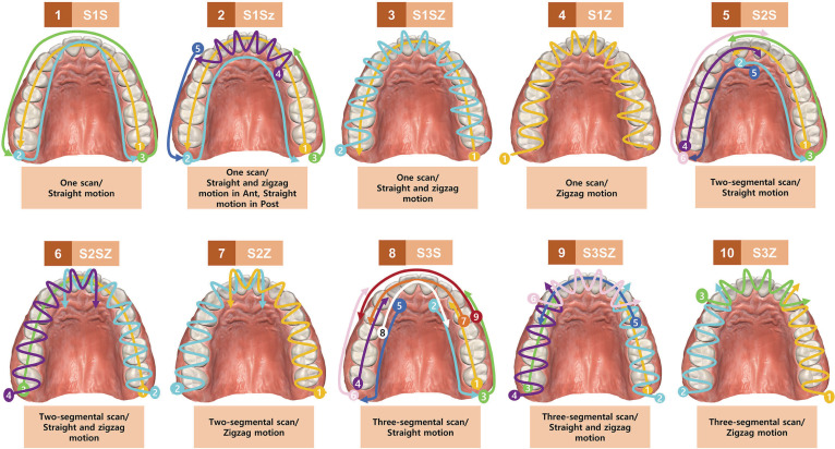
The scan strategy in each group was characterized by the number of scan segments (1, 2, or 3 segments) and the motion of scanning (straight, zigzag, or combined). By combining the experimental scanning factors, 10 scan strategies were established (Table 1, Fig. 1). The scan segments were whole, half, or one-third arch. Regarding the scan motion, straight motion indicates the scanner moves over the surface of each tooth (occlusal, buccal, or lingual) straight and following the arch. The zigzag motion indicates that the scanner scans a tooth using cross-sectional movement and then moves on to the next tooth.
Table 1. Intraoral scan strategy.
| Scan strategy | Scan segment in arch portion (N) | Scan motion | Image merging area |
|---|---|---|---|
| S1S | Whole (1) | Straight | NA |
| S1Sz | Whole (1) | Straight and zigzag in Ant, Straight in Post | NA |
| S1SZ | Whole (1) | Straight and zigzag | NA |
| S1Z | Whole (1) | Zigzag | NA |
| S2S | Rt and Lt half (2) | Straight | Incisor |
| S2SZ | Rt and Lt half (2) | Straight and zigzag | Incisor |
| S2Z | Rt and Lt half (2) | Zigzag | Incisor |
| S3S | Ant, Rt Post, Lt Post (3) | Straight | Canine, 1st premolar |
| S3SZ | Ant, Rt Post, Lt Post (3) | Straight and zigzag | Canine, 1st premolar |
| S3Z | Ant, Rt Post, Lt Post (3) | Zigzag | Canine, 1st premolar |
NA: Not applicable
Fig. 1. Schematic illustrations of the scan strategies in this study.
Digitization of the model was performed by an intraoral scanner (i700; Medit, Seoul, Korea) using different scan strategies, and the scan images were recorded in a dedicated software program (Meditlink v2.3; Medit). According to the manufacturer's data, this intraoral scanner is a structured light scanner, and data is acquired based on the principle of 3D-in-motion video technology/3D full color streaming capture. Scanning was performed once using the scan strategy of each group in order, and then again by the scan strategy of each group. A total of seventy scans were obtained in the software program (seven scans per group) and saved in the polygon file format (PLY) on the computer. The interval between each scan performance was 20 minutes. The crossover measurement and the washout period were performed to minimize the carryover effect caused by the repeated performance. The scanning procedures in all groups were carried out by an experienced clinician (D.-H.L.)
The 3D geometric accuracy of scan images was evaluated with regards to the arch shape (Fig. 2). All the intraoral scans were delivered to an image analysis software program (Geomagic DesignX; 3D Systems, Rock Hill, SC, USA) in which they were aligned to a reference image with reference to the whole tooth area using an area-designated best-fit algorithm. The reference image of the study model was created by digitizing the surface of the model using a high-performance lab-based model scanner with an accuracy of 7 µm and 200,000 pixel of the camera sensor’s point according to the manufacturer’s instructions (DOF Freedom HD; DOF, Seoul, Korea). The outcome parameters for evaluating the geometric discrepancies of intraoral scans were intermolar distance, premolar distance, anteroposterior distance, and global surface deviation. The intermolar distance was assessed by measuring the most external points of the buccal surfaces of the left and right first molars in a virtual horizontal cross-sectional plane made at the middle level of the whole tooth structure. The premolar distance was evaluated by measuring the distance between the buccal surfaces of the left and right first premolars in the virtual horizontal plane. The anteroposterior distance was recorded by measuring the perpendicular distance from the posterior line of the arch to the buccal surface of central incisors in the horizontal plane. The global surface deviation was evaluated by calculating the average root-mean-square error (RMSE) between the test scan images and the reference image. The distribution of surface deviation was visualized using color-coded mapping. All accuracy measurements were conducted by a single investigator who was blinded to the objective of this study.
Fig. 2. Evaluation of the geometric discrepancies of intraoral scans. (A) 3D linear measurement of the arch, (B) Global surface deviation.
Using the recorded values, the absolute discrepancies of the intraoral scans from the reference image (baseline) were determined for the outcome parameters. The means ± standard deviations of the absolute discrepancies in each scan strategy were reported. One-way analysis of variance (ANOVA) and the Tukey honestly significance difference post hoc test were carried out to compare the differences among the different scan strategy groups. All calculations were performed using a statistical software program (IBM SPSS Statistics v25.0; IBM Corp., Armonk, NY, USA) (α = .05).
RESULTS
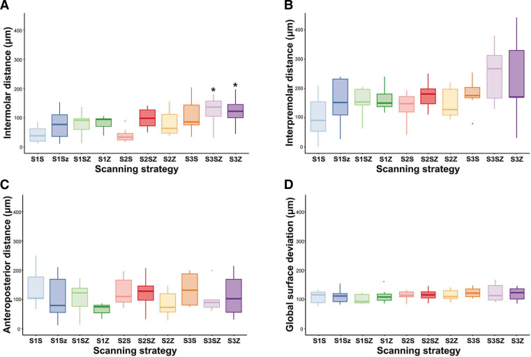
Table 2 and Figure 3 show the mean and standard deviation for geometric discrepancies and global surface deviations of the full-arch scan images. The mean geometric discrepancies in the intermolar distance ranged from 39 µm (S2S scan strategy) to 130 µm (S3SZ scan strategy), whereas the global surface deviations ranged from 109 µm (S1SZ scanning strategy) to 125 µm (S3S and S3SZ scan strategies).
Table 2. Geometric discrepancies of full-arch scan images using different scan strategies.
| Scan strategy | Geometric discrepancies (μm) | |||||||
|---|---|---|---|---|---|---|---|---|
| Intermolar distance | Interpremolar distance | Anteoposterior distance | Global surface deviation | |||||
| M ± SD | %RSD | M ± SD | %RSD | M ± SD | %RSD | M ± SD | %RSD | |
| S1S | 46 ± 34a,b | 74 | 151 ± 77 | 51 | 122 ± 61 | 50 | 116 ± 17 | 15 |
| S1Sz | 73 ± 60a,b | 82 | 151± 77 | 51 | 90 ± 70 | 78 | 116 ± 18 | 16 |
| S1SZ | 76 ± 45a,b | 59 | 160 ± 63 | 39 | 89 ± 51 | 57 | 109 ± 12 | 11 |
| S1Z | 90 ± 26a,b | 29 | 154 ± 40 | 26 | 51 ± 19 | 37 | 117 ± 19 | 16 |
| S2S | 39 ± 30a | 77 | 132 ± 49 | 37 | 104 ± 46 | 44 | 118 ± 12 | 10 |
| S2SZ | 109 ± 39a,b | 36 | 167 ± 46 | 28 | 99 ± 53 | 54 | 119 ± 14 | 12 |
| S2Z | 91 ± 49a,b | 54 | 143 ± 50 | 35 | 68 ± 42 | 62 | 119 ± 13 | 11 |
| S3S | 112 ± 72a,b | 64 | 168 ± 51 | 30 | 110 ± 53 | 48 | 125 ± 12 | 10 |
| S3SZ | 130 ± 58b | 45 | 233 ± 90 | 39 | 74 ± 46 | 62 | 125 ± 23 | 18 |
| S3Z | 127 ± 56b | 44 | 211 ± 134 | 64 | 91 ± 66 | 73 | 122 ± 17 | 14 |
| P-value | .008 | .260 | .371 | .787 | ||||
Different superscript alphabetical letters within columns indicate a statistical difference between groups.
M: Mean; SD: Standard; RSD: Relative standard deviation.
Fig. 3. Box plots for geometric discrepancies of the full arch scan images. (A) Intermolar distance, (B) Interpremolar distance, (C) Anteroposterior distance, (D) Global surface deviation.
*Statistical difference
Linear discrepancy values for the measurement of intermolar distance were not different among scan strategies performed with a single scan or two-segmental scan. The scan strategy using a three-segmental scan and straight motion showed similar mean linear discrepancy to scan strategies with a single scan and two-segmental scan. However, two groups with the three-segmental scan and zigzag scan motion (S3SZ and S3Z) had significantly lower accuracy than the other scan strategies (P = .008). Regarding the evaluation of the interpremolar distance and anteroposterior distance, no statistical difference was found among discrepancy values from all scan strategies (P = .260 and .371, respectively).
Regarding global surface deviation, RMSE values were similar for all scan strategies. Post hoc analysis revealed no statistical difference among groups (P = .787).
DISCUSSION
This study was designed to assess the accuracy of a full-arch intraoral scan generated using the segmental scan and merge methods. The results of this study showed that the dimensional discrepancy values of scans were not different in single and two-segmental scan methods in terms of all linear measurements and global surface deviation outcome parameters regardless of scan motions. The method of scanning the arch segmentally in two parts and merging the scan images of each part using the image stitching function was comparable with the one-time scan method in accuracy. Meanwhile, when scanning was performed in three parts with the zigzag motion, the accuracy of full-arch image was decreased in the intermolar distance evaluation. Thus, the first null hypothesis, the geometric accuracy of full-arch scans would not be different between single scan and segmental scan methods, was partially rejected. The results of the present study generally correspond well with those reported by an earlier experimental study.8 In the study, intraoral scans for six implants placed to the edentulous model were taken using two methods, a single scan and segmental scan-and-merge. The scan strategy with image stitching had better accuracy and exhibited a smaller dimensional deviation compared to the single scan method. Thus, the segmental scan and merge methods could be an alternative to the single-scan method when continuous scanning is clinically difficult for a comprehensive area.
To increase the accuracy of the resulting scan image, specific scanner movements are needed to superimpose the individual image of each snap with congruent features with sufficient reliability.9,10 Several studies showed that a straight or linear scan movement along the dental arch had higher accuracy than the zigzag scan movement.11,12,13 During the progressive and consecutive straight scan movements, congruent areas between each captured image might be easily recognized and properly stitched to each other. A simple motion seems to be optimal to obtain accurate scanning images in general areas, although straight motions can be imprecise in interproximal areas.14 However, zigzag scan movements consist of crossing the buccal, occlusal, and lingual faces of each tooth successively with the rotation of the scanner tip. The rotation of the scanning tip facilitates the scan procedure in the interproximal zones, prepared teeth, and high curvatures of anterior teeth.15,16,17 Medina-Sotomayor et al.15 reported the zigzag scan motion was better than the straight scan motion in accuracy. However, a study by Oh et al.12 reported that rotation of the scan tip caused a sudden change in direction and interrupted image-stitching processes in consecutive images, which lowered the scan accuracy. Difficulty in keeping some distance between the scanner tip and target tooth during a rotation scan is another possible reason for inaccuracy because the inconsistent distance hinders the focus when capturing clear images, which could generate errors in the process of overlapping images.17 In our study, the difference in scan strategy, straight or zigzag, did not influence the geometric accuracy of scans in all measurement outcome parameters, except when multiple segmental scans and the zigzag scan motion were involved. Accordingly, the second null hypothesis, scanner movement strategy during intraoral scanning would not affect the accuracy of full-arch scans, was partially rejected. This finding implies that the scan motion may not affect the accuracy of digitization of the oral cavity in the scanning system used in this study. The hardware and software of the intraoral scan system were highlighted as a conditioning factor for scanning performance.9,18 It is thought that developments in imaging engineering have decreased the effect of scan motion on the accuracy of the scan.
Linear measurements and 3D surface comparisons have been described as major methods for investigating the accuracy of 3D images obtained by the intraoral scanners.7 Typically, the linear measurement method measures the linear distance from the selected points on the test model to the corresponding points on the reference.19 Since point selection is conducted only based on visual observations, correctly selecting identical points on the test and reference models may be difficult, particularly in the case of a completely edentulous arch due to the lack of distinguishable landmarks. In contrast, the 3D surface comparison method calculates the distances of all corresponding points between the two models by using the root mean square formula and displays the deviations in a color code according to a range of deviation values.20 In the present study, the 3D geometric accuracy of the intraoral scan images was evaluated regarding their ability to represent the trueness of the captured dental arch shape of the scan images. For this purpose, linear and 3D measurements were used to evaluate the dental arch forms and dimensions. Unlike the traditional linear measurement method, in this study, the linear distance was assessed by measuring the distance between corresponding points of the test and reference models in a virtual horizontal cross-sectional plane made at the middle level of whole teeth structures. The advantage of this approach is that the cross-sectional images were drawn as a schematic diagram with clear borderlines and points that prevented errors in detecting and selecting measurement points.21 Thus, the use of this modified linear measurement method may enhance the trueness and precision of the measurements while reducing the time and effort required for decision-making during the measuring process.
In this study, intraoral scan experiments were performed by using a dentiform model in a phantom head. The in vitro study design facilitates setting experimental variables and controlling confounding factors, providing valid scientific insights.20,22 At the same time, limitations of the in vitro design, such as the lack of an oral environment and patient factors, should also be considered.23,24 Although all the scans were conducted by a single clinician to eliminate operator-related factors, deviations in measurement data at each scan strategy condition were observed. It is thought that these results were involved because the scan process could not be duplicated exactly in each trial in terms of speed, scan distance, and time, even though the intraoral scan was performed in the same sequence. These scan parameters can also influence the accuracy of final image of intraoral scan while stitching numerous individual images, especially in large-sized scans. In addition, various conditions of the dental arch should be included in future studies. Clinical situations in the intact dentate arch and partially edentulous arches are different, which could require more diversified scan strategies for specific cases. To validate the results of the present study and expand the scope of the study subject, comprehensive patient-based clinical studies will be needed with multiple clinicians. Meanwhile, a lab-based model scanner was used to create a reference image of study model. Lab scanners have been used for obtaining accurate images in the literature; however, discrepancies in measurements between lab scanners and coordinate measuring machines were also reported.25,26 Verification process on the scan accuracy is recommended when the scanner is used for obtaining reference images.
CONCLUSION
Within the limitations of this experiment, the accuracy of full-arch intraoral scan generated by the segmental scan and merge method using two scan parts was comparable with that of the single scan method regardless of scan strategies and motions. When three segmental scans were involved, the accuracy of complete arch scan can be negatively affected.
Footnotes
This work was supported by the Korea Medical Device Development Fund grant funded by the Korea government (the Ministry of Science and ICT, the Ministry of Trade, Industry and Energy, the Ministry of Health & Welfare, Republic of Korea, the Ministry of Food and Drug Safety) (202011A02).
References
- 1.Wulfman C, Naveau A, Rignon-Bret C. Digital scanning for complete-arch implant-supported restorations: A systematic review. J Prosthet Dent. 2020;124:161–167. doi: 10.1016/j.prosdent.2019.06.014. [DOI] [PubMed] [Google Scholar]
- 2.Gjelvold B, Chrcanovic BR, Korduner EK, Collin-Bagewitz I, Kisch J. Intraoral digital impression technique compared to conventional impression technique. A randomized clinical trial. J Prosthodont. 2016;25:282–287. doi: 10.1111/jopr.12410. [DOI] [PubMed] [Google Scholar]
- 3.Diker B, Tak Ö. Comparing the accuracy of six intraoral scanners on prepared teeth and effect of scanning sequence. J Adv Prosthodont. 2020;12:299–306. doi: 10.4047/jap.2020.12.5.299. [DOI] [PMC free article] [PubMed] [Google Scholar]
- 4.Nulty AB. A comparison of full arch trueness and precision of nine intra-oral digital scanners and four lab digital scanners. Dent J (Basel) 2021;9:75. doi: 10.3390/dj9070075. [DOI] [PMC free article] [PubMed] [Google Scholar]
- 5.Ahlholm P, Sipilä K, Vallittu P, Jakonen M, Kotiranta U. Digital versus conventional impressions in fixed prosthodontics: a review. J Prosthodont. 2018;27:35–41. doi: 10.1111/jopr.12527. [DOI] [PubMed] [Google Scholar]
- 6.Burzynski JA, Firestone AR, Beck FM, Fields HW, Jr, Deguchi T. Comparison of digital intraoral scanners and alginate impressions: Time and patient satisfaction. Am J Orthod Dentofacial Orthop. 2018;153:534–541. doi: 10.1016/j.ajodo.2017.08.017. [DOI] [PubMed] [Google Scholar]
- 7.Abduo J, Elseyoufi M. Accuracy of Intraoral Scanners: A Systematic Review of Influencing Factors. Eur J Prosthodont Restor Dent. 2018;26:101–121. doi: 10.1922/EJPRD_01752Abduo21. [DOI] [PubMed] [Google Scholar]
- 8.Mandelli F, Gherlone EF, Keeling A, Gastaldi G, Ferrari M. Full-arch intraoral scanning: comparison of two different strategies and their accuracy outcomes. J Osseointegration. 2018;10:65–74. [Google Scholar]
- 9.Paratelli A, Vania S, Gómez-Polo C, Ortega R, Revilla-León M, Gómez-Polo M. Techniques to improve the accuracy of complete-arch implant intraoral digital scans: a systematic review. J Prosthet Dent. 2021;21:00486-8. doi: 10.1016/j.prosdent.2021.08.018. [DOI] [PubMed] [Google Scholar]
- 10.Zimmermann M, Mehl A, Mörmann WH, Reich S. Intraoral scanning systems - a current overview. Int J Comput Dent. 2015;18:101–129. [PubMed] [Google Scholar]
- 11.Latham J, Ludlow M, Mennito A, Kelly A, Evans Z, Renne W. Effect of scan pattern on complete-arch scans with 4 digital scanners. J Prosthet Dent. 2020;123:85–95. doi: 10.1016/j.prosdent.2019.02.008. [DOI] [PubMed] [Google Scholar]
- 12.Oh KC, Park JM, Moon HS. Effects of scanning strategy and scanner type on the accuracy of intraoral scans: a new approach for assessing the accuracy of scanned data. J Prosthodont. 2020;29:518–523. doi: 10.1111/jopr.13158. [DOI] [PubMed] [Google Scholar]
- 13.Passos L, Meiga S, Brigagão V, Street A. Impact of different scanning strategies on the accuracy of two current intraoral scanning systems in complete-arch impressions: an in vitro study. Int J Comput Dent. 2019;22:307–319. [PubMed] [Google Scholar]
- 14.Richert R, Goujat A, Venet L, Viguie G, Viennot S, Robinson P, Farges JC, Fages M, Ducret M. Intraoral scanner technologies: a review to make a successful impression. J Healthc Eng. 2017;2017:8427595. doi: 10.1155/2017/8427595. [DOI] [PMC free article] [PubMed] [Google Scholar]
- 15.Medina-Sotomayor P, Pascual-Moscardó A, Camps I. Accuracy of four digital scanners according to scanning strategy in complete-arch impressions. PLoS One. 2018;13:e0202916. doi: 10.1371/journal.pone.0202916. [DOI] [PMC free article] [PubMed] [Google Scholar]
- 16.Müller P, Ender A, Joda T, Katsoulis J. Impact of digital intraoral scan strategies on the impression accuracy using the TRIOS Pod scanner. Quintessence Int. 2016;47:343–349. doi: 10.3290/j.qi.a35524. [DOI] [PubMed] [Google Scholar]
- 17.van der Meer WJ, Andriessen FS, Wismeijer D, Ren Y. Application of intra-oral dental scanners in the digital workflow of implantology. PLoS One. 2012;7:e43312. doi: 10.1371/journal.pone.0043312. [DOI] [PMC free article] [PubMed] [Google Scholar]
- 18.Zhang YJ, Shi JY, Qian SJ, Qiao SC, Lai HC. Accuracy of full-arch digital implant impressions taken using intraoral scanners and related variables: A systematic review. Int J Oral Implantol (Berl) 2021;14:157–179. [PubMed] [Google Scholar]
- 19.Oliva B, Sferra S, Greco AL, Valente F, Grippaudo C. Three-dimensional analysis of dental arch forms in Italian population. Prog Orthod. 2018;19:34. doi: 10.1186/s40510-018-0233-1. [DOI] [PMC free article] [PubMed] [Google Scholar]
- 20.Kuhr F, Schmidt A, Rehmann P, Wöstmann B. A new method for assessing the accuracy of full arch impressions in patients. J Dent. 2016;55:68–74. doi: 10.1016/j.jdent.2016.10.002. [DOI] [PubMed] [Google Scholar]
- 21.Mai HN, Lee KE, Ha JH, Lee DH. Effects of image and education on the precision of the measurement method for evaluating prosthesis misfit. J Prosthet Dent. 2018;119:600–605. doi: 10.1016/j.prosdent.2017.05.022. [DOI] [PubMed] [Google Scholar]
- 22.Park HN, Lim YJ, Yi WJ, Han JS, Lee SP. A comparison of the accuracy of intraoral scanners using an intraoral environment simulator. J Adv Prosthodont. 2018;10:58–64. doi: 10.4047/jap.2018.10.1.58. [DOI] [PMC free article] [PubMed] [Google Scholar]
- 23.Kurz M, Attin T, Mehl A. Influence of material surface on the scanning error of a powder-free 3D measuring system. Clin Oral Investig. 2015;19:2035–2043. doi: 10.1007/s00784-015-1440-5. [DOI] [PubMed] [Google Scholar]
- 24.Nedelcu RG, Persson AS. Scanning accuracy and precision in 4 intraoral scanners: an in vitro comparison based on 3-dimensional analysis. J Prosthet Dent. 2014;112:1461–1471. doi: 10.1016/j.prosdent.2014.05.027. [DOI] [PubMed] [Google Scholar]
- 25.Pan Y, Tam JM, Tsoi JK, Lam WY, Huang R, Chen Z, Pow EH. Evaluation of laboratory scanner accuracy by a novel calibration block for complete-arch implant rehabilitation. J Dent. 2020;102:103476. doi: 10.1016/j.jdent.2020.103476. [DOI] [PubMed] [Google Scholar]
- 26.Mandelli F, Gherlone E, Gastaldi G, Ferrari M. Evaluation of the accuracy of extraoral laboratory scanners with a single-tooth abutment model: A 3D analysis. J Prosthodont Res. 2017;61:363–370. doi: 10.1016/j.jpor.2016.09.002. [DOI] [PubMed] [Google Scholar]


