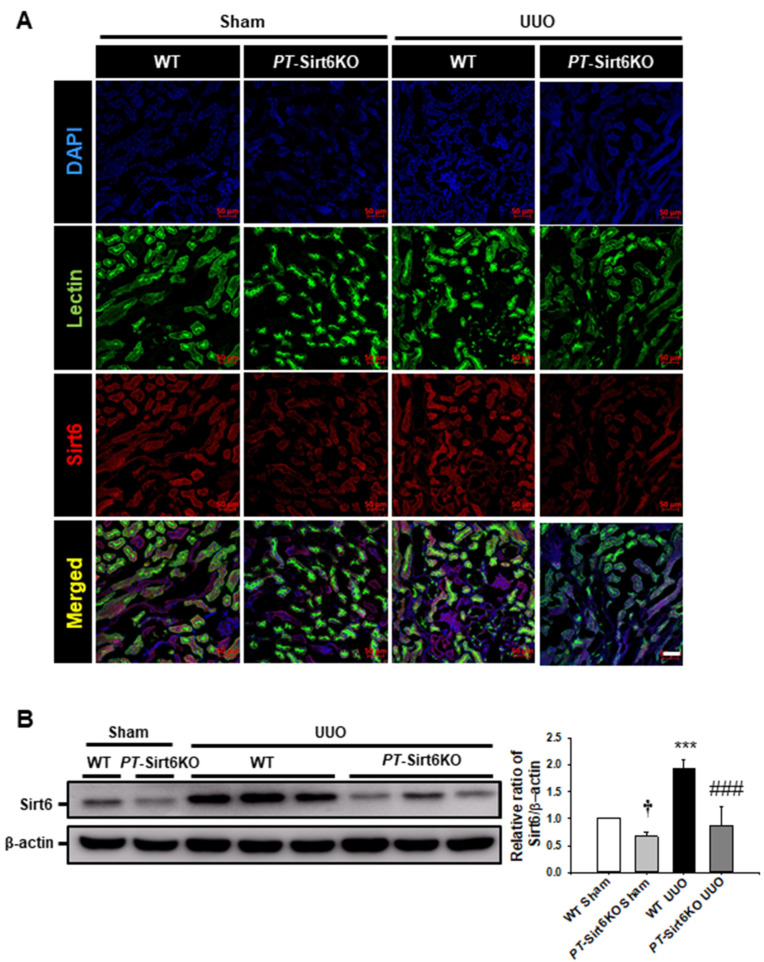
Figure 1.
Establishment of proximal tubule-specific Sirt6 knockout (Sirt6flox/flox; Ggt1-Cre+) mice (A) Proximal tubule-specific loss of Sirt6 was confirmed by immunofluorescence staining of Sirt6 in proximal tubules. Lotus tetragonolobus lectin was used as a proximal tubule marker. Scale bar = 50 μm. (B) Representative Western blot analysis of Sirt6 in the kidneys from sham- and UUO-operated Sirt6flox/flox; Ggt1-Cre- (WT) and Sirt6flox/flox; Ggt1-Cre+ (PT-Sirt6KO) mice. The bar graph shows the densitometric quantification presented as the relative ratio of each protein to β-actin. The relative ratio measured in the kidneys from sham-operated WT mice is arbitrarily presented as 1. Data are expressed as mean ± SD. †, p < 0.05 versus WT sham; ***, p < 0.001 versus WT sham or PT-Sirt6KO sham; ###, p < 0.001 versus WT UUO. Sham, sham-operated mice; UUO, unilateral ureteral obstruction; WT, wild-type; PT-Sirt6KO, proximal tubule-specific Sirt6 knockout; lectin, Lotus tetragonolobus lectin.

