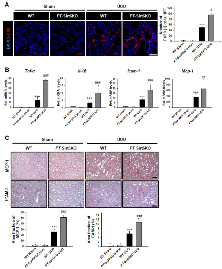
Figure 5.
Proximal tubule-specific loss of Sirt6 aggravates UUO-induced inflammation. (A) Representative sections of kidneys from sham- and UUO-operated Sirt6flox/flox; Ggt1-Cre- (WT) and Sirt6flox/flox; Ggt1-Cre+ (PT-Sirt6KO) mice were immunofluorescence stained with F4/80 (red). The nucleus was stained by DAPI (blue). Scale bar = 50 μm. The bar graph shows the number of F4/80 (+) cells in the sham- and UUO-operated kidneys from ten randomly chosen, non-overlapping fields at a magnification of 400× (n = 15 per group). (B) Quantitative RT-PCR analyses for Tnf-α, Il-1β, Icam-1, and Mcp-1 were performed using mRNA from the sham- and UUO-operated WT and PT-Sirt6KO kidneys (n = 15). Data are expressed as mean ± SD. (C) Representative sections of kidneys from sham- and UUO-operated Sirt6flox/flox; Ggt1-Cre- (WT) and Sirt6flox/flox; Ggt1-Cre+ (PT-Sirt6KO) mice were stained with MCP-1 and ICAM-1. Scale bar = 100 μm. The bar graph shows area fractions (%) of MCP-1 and ICAM-1 in the sham and UUO kidneys from ten randomly chosen, non-overlapping fields at a magnification of 200× (n = 15 per group). ***, p < 0.001 versus WT sham or PT-Sirt6KO sham; #, p < 0.05, ##, p < 0.01 and ###, p < 0.001 versus WT UUO. Sham, sham-operated mice; UUO, unilateral ureteral obstruction; WT, wild-type; PT-Sirt6KO, proximal tubule-specific Sirt6 knockout; Tnf-α, tumor necrosis factor-α; Il-1β, interleukin-1β; Icam-1, intercellular adhesion molecule-1; Mcp-1, monocyte chemoattractant protein-1.

