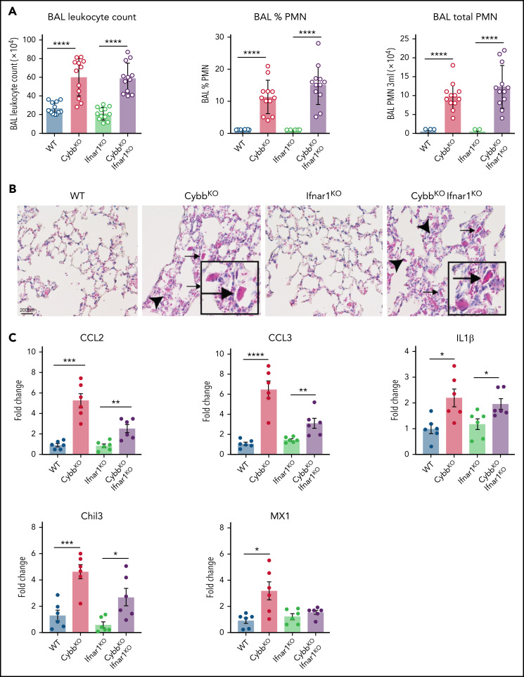
Figure 5.
Type I IFN signaling is not necessary for NOX2-deleted mice to develop spontaneous lung inflammation and activated AMs. (A) BAL leukocyte count, percentage of polymorphonuclear leukocytes (PMNs), and total PMNs in 3 mL of BAL from WT, CybbKO, Ifnar1KO, and CybbKO Ifnar1KO mice (n = 12 in each group). (B) Hematoxylin-eosin–stained lung tissues showing the presence of eosinophilic macrophages (arrowhead) and extracellular crystals (arrow) in CybbKO and CybbKOIfnar1KO mice. Scale bar, 200 μm and the scale is same for all panels. Representative image from more than 4 samples in each group. (C) qRT-PCR analysis of gene expression in BAL cells isolated from WT, CybbKO, Ifnar1KO, and CybbKOIfnar1KO mice (n = 6 in each group). Bar graph data are expressed as the mean ± standard error of the mean. Experiments were repeated at least twice and the Student t test was performed for samples distributed between 2 groups. *P < .05; **P < .01; ***P < .001; ****P < .0001.

