Abstract
Polymers, due to their high molecular weight, tunable architecture, functionality, and buffering effect for endosomal escape, possess unique properties as a carrier or prophylactic agent in preventing pandemic outbreak of new viruses. Polymers are used as a carrier to reduce the minimum required dose, bioavailability, and therapeutic effectiveness of antiviral agents. Polymers are also used as multifunctional nanomaterials to, directly or indirectly, inhibit viral infections. Multifunctional polymers can interact directly with envelope glycoproteins on the viral surface to block fusion and entry of the virus in the host cell. Polymers can indirectly mobilize the immune system by activating macrophages and natural killer cells against the invading virus. This review covers natural and synthetic polymers that possess antiviral activity, their mechanism of action, and the effect of material properties like chemical composition, molecular weight, functional groups, and charge density on antiviral activity. Natural polymers like carrageenan, chitosan, fucoidan, and phosphorothioate oligonucleotides, and synthetic polymers like dendrimers and sialylated polymers are reviewed. This review discusses the steps in the viral replication cycle from binding to cell surface receptors to viral-cell fusion, replication, assembly, and release of the virus from the host cell that antiviral polymers interfere with to block viral infections.
Keywords: antiviral polymers, natural, synthetic, polysaccharides, nucleic acid polymers, dendrimers, sialylated polymers, viral infection
1. Introduction
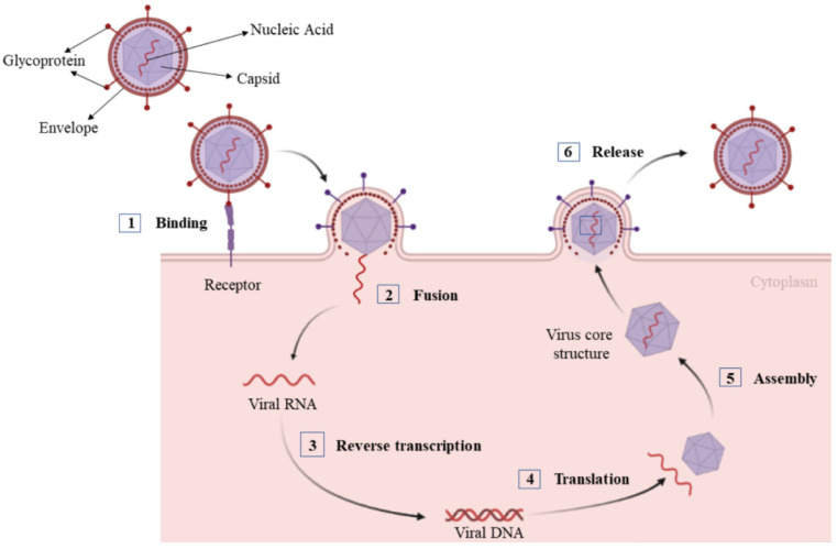
Due to their rapid mutation and evolution, viral infections have always been a global health challenge threatening both human and animal health. A virus is a nonliving microscopic structure that contains the genomic material for replication encapsulated and protected by a proteinaceous membrane. Unlike living organisms, viruses must invade a living cell, like a human or animal cell, and hijack its metabolic system for energy and nutrients to replicate and grow [1]. Some viruses even recruit the metabolic system of cancer cells to support their reproduction [2]. Some viruses are surprisingly simple like the Ebola virus, which is made up of only seven different proteins, even though this virus has had devastating consequences on human health [3,4]. Conversely, giant viruses like the Mimivirus possess genes for metabolizing proteins and serve as a bridge between nonliving viruses and living organisms [5]. Although there are considerable differences among virus types, viral replication consists of six basic steps as shown in Figure 1 [6]. These include (1) interaction between the proteins on the viral surface with surface receptors on the host cell for viral attachment; (2) fusion of the virus and host cell membranes for viral penetration in the host cell; (3) release of the viral genomic material inside the host cell by uncoating the virus; (4) synthesis of viral components like proteins, RNAs and DNAs by the host cell for viral replication; (5) assembly of the synthesized components into viral particles; and (6) exocytosis of the particles from the host cell to the interstitial space for viral spreading to other cells.
Figure 1.
Steps in the viral replication cycle.
The function of an antiviral agent is to block one or more of the basic steps in the viral replication cycle without affecting the normal metabolic activities of the cell [7]. As viruses use the host cell for replication, it is challenging to develop safe and effective antiviral agents that do not affect the function of the host cell. As a result, many commercially available antiviral agents are limited by undesired side effects [8]. Further, the effectiveness of antiviral agents and vaccines is severely constrained by viral mutation [9,10].
Polymers, due to their tunable chemical structure and composition, high molecular weight, and their buffering effect possess unique capabilities as antiviral agents. Many polymers have been recently developed to meet the global demand for antiviral agents and to treat viral infections [11,12,13,14,15,16,17,18,19,20,21,22,23,24,25]. Although the mechanism of action of antiviral polymers is not completely understood, it is known that their potency and effectiveness can be tailored against a specific virus by varying the polymer molecular weight, chain architecture, composition, or functional groups [26,27]. Two approaches are used to utilize polymers as part of an antiviral system to fight against infections. In the first approach, the polymer is used as a matrix to protect, stabilize, and deliver the antiviral agent to the site of infection. In the second approach, the functionalized polymer is used as an antiviral agent to bind to the surface of viral particles to inhibit infections. The polymer functional groups that take part in binding to viral particles include among others phenolic, sulfate, amine, and carboxylic acid groups. The focus of this review is on the latter in which the polymer acts directly as an antiviral agent to fight against viral infections. Keeping an eye on current and future pandemics, this review covers repeating functional units in the structure of macromolecules that impart antiviral activity to natural and synthetic polymers.
2. Natural Polymers
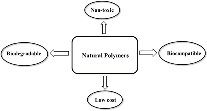
Natural polymers or biopolymers are classified into polysaccharides, polypeptides (proteins), and nucleic acid polymers (polynucleotides) [28]. Natural polymers as components of living systems are derived from plants, animals, and microorganisms [28]. Advantages of natural polymers over synthetic polymers include biocompatibility, non-toxicity, biodegradability, and intrinsic antiviral properties as shown in Figure 2 [29]. Among different classes of biopolymers, polysaccharides and nucleic acid polymers are capable of interfering with the virus-host cell interaction to block fusion, entry, and replication of the virus in the host cell [30].
Figure 2.
Key benefits of natural polymers as antiviral materials.
2.1. Polysaccharides
Polysaccharides are an abundant source of renewable and biodegradable polymers. Polysaccharides are made up of more than 10 monosaccharide repeat units connected by glycosilic linkages in branched or linear chains with molecular weights ranging from tens of thousands to a few millions [31]. Like other natural polymers, polysaccharides play vital functions in living systems from cell adhesion to intracellular signaling and maintenance of biological and mechanical properties of tissues [32]. Natural plant polysaccharides are composed of long sequences of monosaccharides with different biological activities including anti-inflammatory, immunomodulatory, antioxidant, and antiviral activities [33,34]. Table 1 presents the general properties of carrageenan, chitin, chitosan, and fucoidan as widely studied polysaccharides with antiviral activities.
Table 1.
Polysaccharides, their origin, and molecular characteristics.
| Polysaccharide | Origin | Occurrence/Function | Molecular Characteristics | Ref. |
|---|---|---|---|---|
| Carrageenan | Algae | Structural polysaccharides of marine red algae | Heteropolysaccharide, linear, anionic | [35] |
| Chitin/chitosan | Animal | Chitin: structural polysaccharide from exoskeleton of insects and shells of crustaceans Chitosan: derivative of chitin prepared by deacetylation |
Chitin: homopolysaccharide, linear, neutral Chitosan: heteropolysaccharide, linear, cationic |
[36] |
| Fucoidan | Algae | Structure and composition dependent on species with diverse structures | Heteropolysaccharide, anionic, linear. | [37] |
Polysaccharides possess different types of functional groups including hydroxyl, amine, and carboxylic acid groups which can be used for polymer modification [33,34]. These functional groups are used to tailor the properties of polysaccharides to specific biological applications [38,39]. The inhibitory effect of negatively charged polysaccharides on viral infections is well documented [40,41,42]. Polysaccharides interact with Trans-Activator of Transcription (Tat) regulatory protein of viruses to inhibit their interaction with cell surface receptors like heparan sulfates [43]. As an example, carrageenan which is a naturally sulfated polysaccharide of red algae, inhibits many viral infections including human papillomavirus (HPV), herpes simplex virus type-1 (HSV-1), and influenza A virus (IAV) by interacting with the viral Tat protein [44,45]. Numerous studies have reported the inhibitory effect of plant polysaccharides on replication of human and animal viruses including SARS associated coronaviruses (SARS-CoV) [46,47,48,49]. These include brown algae, Auricularia auricula, Pinus massoniana, and Acanthopanax polysaccharides [50]. Recently, the antiviral activity of highly sulfated synthetic oligomers and polymers has been investigated for inhibition of viral infections [23]. These studies show that both long and short chain, highly sulfated polysaccharides possess inhibitory effect on viral infections. According to these results, highly sulfated long-chain glycomimetic polymers prevented human papillomavirus (HPV16) infection in vitro and in vivo by interaction with the virus prior to cell attachment whereas highly sulfated short-chain glycomimetic oligomers inhibited viral infection post cell attachment [23]. The polyanionic nature of sulfated polysaccharides facilitates interaction with the positively charged domains of the virus envelope glycoproteins to form a non-reversible complex, thus making the viral glycoprotein sites inaccessible to interact with surface receptors of the host cell [51]. This non-reversible polysaccharide-viral interaction blocks binding and fusion of the virus with the host cell which inhibits viral entry in the cell [51]. Seaweed polysaccharides with high anionic sulfate content are attractive as antiviral agents to inhibit infections [52,53,54]. The structural diversity and complexity of marine polysaccharides and their derivatives contribute to their antiviral activity at different stages of viral infections [55]. The antiviral activity of polysaccharides is affected by their molecular weight. As the antiviral activity arises from the interaction of polysaccharides with viral envelope proteins through reversible secondary interactions like electrostatic, nonpolar, and hydrogen bonding, long chains that can form irreversible complexes substantially increase antiviral activity. In one study, polymeric sulfated carrageenan was more effective in blocking HPV16 viral infection in mice as compared to oligomeric sulfated carrageenan [23]. In another study, high molecular weight fucoidan polysaccharide showed antiviral activity against influenza A virus at low doses whereas low molecular weight fractions showed antiviral activity only at medium to high doses [56,57]. As a result, high molecular weight polysaccharides in general show higher antiviral activity [58].
2.1.1. Carrageenan
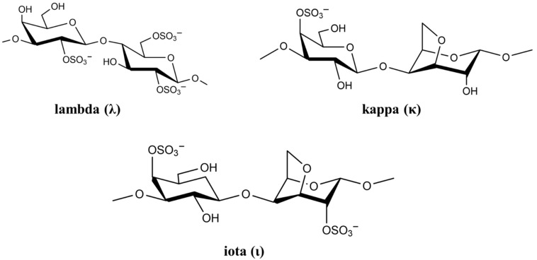
The cell wall of marine red seaweed contains the polysaccharides agaran and carrageenan that form approximately 50% of the dry mass of seaweed. As a high molecular weight sulfated polysaccharide, carrageenan has long been used as a gelling agent and stabilizer in food [59]. However, carrageenan has many biological activities including anticoagulation, antioxidation, antiviral, and immunostimulatory [60]. Based on sulfate content, there are more than 10 types of carrageenan, but only the types lambda (λ), kappa (κ), and iota (ι) are of particular interest in viral and medical applications [61]. The chemical structure of the three carrageenan types is shown in Figure 3.
Figure 3.
Chemical structure of lambda (λ), kappa (κ), and iota (ι) carrageenan.
The negatively-charged sulfate groups of carrageenan neutralize the positive charge of the cell surface, leading to inhibition of viral attachment, penetration, and viral uncoating [62]. The biological activity of carrageenan strongly depends on its degree of sulfation, which depends on carrageenan (λ, κ and ι) as well as virus type [63]. List of studies on antiviral activity of λ, κ, and ι carrageenan are presented in Table 2.
Table 2.
Antiviral activity of λ, κ, and ι carrageenan against different viruses.
| Type | Virus | Genome | Cell Line | IC50/EC50 (µg/mL) | Ref. |
|---|---|---|---|---|---|
| κ | DENV-1 | RNA | Vero | >50 (EC50) | [64] |
| κ | DENV-2 | RNA | Vero | 1.8 (EC50) | [64] |
| κ | HSV-1 | DNA | Vero | 1.9 (EC50) | [65] |
| κ | HSV-2 | DNA | Vero | 1.6 (EC50) | [65] |
| κ | HIV-1 | RNA | MT-4 | 12 (IC50) | [66] |
| ι | DENV-1 | RNA | Vero | 41 (EC50) | [64] |
| ι | DENV-2 | RNA | Vero | 0.40 (EC50) | [64] |
| ι | H1N1 | RNA | MDCK | 0.39 (IC50) | [67] |
| ι | SARS-CoV-2 | RNA | Vero | 0.05 (IC50) | [68] |
| λ | HIV-1 | RNA | MT-4 | 1.9 (IC50) | [66] |
| λ | H1N1 | RNA | MDCK | 0.04 (IC50) | [44] |
| λ | VSV | RNA | HeLa | 4.0 (IC50) | [66] |
In general, a higher degree of sulfation leads to a higher antiviral activity [55]. It is reported that λ-carrageenan having 35% sulfation exhibits approximately ten times higher inhibition against HIV infection as compared to κ-carrageenan with 25% sulfation [69]. Aside from sulfation, the source of polysaccharide affects its antiviral activity [70]. Carrageenan is capable of affecting virus replication directly through the sulfate groups leading to a decrease in viral activity [71]. For example, carrageenan binding changes the structure of HSV virus, namely the structure of glycoproteins B and C, which results in virus deactivation. Vissani et al. demonstrated that the antiviral activity of ι-carrageenan against herpesvirus-3 is linked to envelope glycoprotein binding [72]. Functional characterization showed that ι-carrageenan and N-sulfonated derivatives of poly (allylamine hydrochloride) blocked the release of human metapneumovirus (hMPV) from infected cells, which reduced viral spreading [73]. A nasal spray for the delivery of carrageenan has been developed for treating patients infected with rhinovirus (HRV), coronavirus (HCoV), and influenza A (IAV) virus. The nasal spray decreased viral symptoms in infected patients and it was most effective against the human coronavirus [74]. It should be pointed out that ι-carrageenan is an approved antiviral drug for use against not only HRV, IAV-H1N1 and HCoV-OC43 but also against viral throat infection [75]. A study on the use of ι-carrageenan against viral throat infection showed 85% and 91% decrease in symptoms in IAV and HCoV-OC43 infected patients, respectively [75].
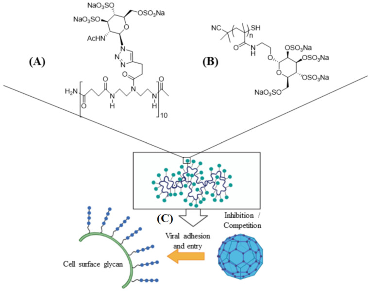
Carrageenan has also been used against human papillomavirus (HPV) which is a non-enveloped small DNA virus capable of infecting mucosal membrane or skin [76]. In one study, carrageenan showed antiviral activity against HPV pseudovirus in a luciferase mouse model without exhibiting a cytotoxic effect [77]. In another study, the antiviral activity of carrageenan was compared with heparin sulfate and highly sulfated synthetic glycomimetic polymers in vitro and in vivo [23]. Both short chain oligomeric sulfated saccharides with 2–10 sugar units and long polymeric saccharides with 40–80 sugar units were synthesized to mimic different properties of glycopolymers, as shown in Figure 4A,B. The in vitro evaluation of the antiviral agents were done with HPV16-eGFP pseudovirus incubated with HeLa cervical cancer cells whereas the in vivo evaluations were done with HPV16-luciferase inoculated in the mouse vagina [23]. The addition of carrageenan, heparin sulfate, and polymeric sulfated saccharides inhibited the attachment of HPV16 pseudovirus to the surface of HeLa cells to block viral infection (Figure 4C) [23]. The effectiveness of polymeric sulfated saccharides with 40 units in inhibiting viral attachment was slightly less than that with 80 units. The in vivo imaging results showed that both carrageenan and heparin sulfate were effective in inhibiting HPV16 infection after 2 days of inoculation [23]. According to in vivo imaging results, the polymeric sulfated glycomimetic saccharides were effective in blocking the activity of HPV16 virus whereas the oligomeric saccharides were ineffective [23]. Carrageenan was effective in reducing viral infection to below 50% at concentrations as low as 100 ng/mL [23]. The results demonstrate the importance of molecular weight of sulfated polysaccharides in blocking viral infections.
Figure 4.
Schematic representation of short ((A), 10 sugar units) and long ((B), n units) chain oligomeric sulfated saccharides to mimic different properties of glycopolymers; (C) the sulfated glycopolymers block viral infection by interfering with virus attachment to the cell surface. Reprinted with permission from Ref. [23]. Copyright 2020, American Chemical Society.
Recently, the antiviral activity of different carrageenan types against SARS-CoV-2 has been reported [68,78,79]. In one study, it was shown that λ-carrageenan purified from red seaweed was effective against influenza A and B and SARS-CoV-2 [78]. In another study, a carrageenan nasal spray inhibited SARS-CoV-2 infection in Vero E6 monkey kidney epithelial cells expressing TMPRSS2 receptor, which was attributed to the polyanionic nature of ι- and κ-carrageenan [79]. In another study, ι-carrageenan delivered using a nasal spray inhibited the entry of SARS-CoV-2 spike protein pseudo-typed lentiviral particles to Vero B4 embryonic monkey kidney cells [68]. In that study, ι-carrageenan also showed effectiveness against rhinoviruses and endemic coronaviruses with an IC50 value of 2.6 µg/mL [68]. These studies show that ι-carrageenan can potentially serve as a prophylactic agent in the treatment of pandemic outbreaks of new viruses in the absence of vaccines.
2.1.2. Chitosan and Its Derivatives
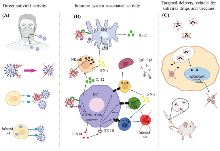
Chitin and chitosan due to their biocompatibility, degradability, and non-immunogenicity are used in the development of antimicrobial wound dressing as well as other medical applications [80,81]. Chitin, comprised of N-acetyl-D-glucosamine and D-glucosamine units, is isolated from the shell of arthropods like shrimp, lobster, crab, and insects or produced by fermentation [82]. Chitosan is produced from chitin by partial deacetylation or by enzymatic hydrolysis [83]. Although the antimicrobial properties of chitosan is well-documented, less is known about the antiviral activity of this biopolymer [84]. The degree of acetylation and amination, chitosan concentration, molecular weight, overall charge as well as charge distribution, and functional group modification affects antiviral activity of chitosan [85,86]. The positively charged derivatives of chitosan possess antiviral activity whereas the negatively charged derivatives are not antiviral [81]. Chitosan can inhibit viral infection directly and indirectly or it can be used as a carrier in the delivery of antiviral agents, as shown in Figure 5 [87]. In the direct approach, chitosan interferes with the receptors that maintain viral activity and growth [87]. For example, the anionic sulfate groups on glucosamine residues of chitosan interact electrostatically with positively-charged envelope glycoprotein GP120 on the virus surface to prevent its fusion with the host cell, which effectively inhibits virus entry and replication [88]. In the indirect approach, chitosan mobilizes the immune response by stimulating proliferation and activation of macrophages and natural killer cells against the invading virus. In this regard, the N-acetyl-glucosamine residues in chitosan increase the level of reactive oxygen species in macrophages after uptake, which subsequently increase the synthesis of IFN-γ by lymphocytes in the spleen, which in turn prevent translation of viral genomic RNA [89].
Figure 5.
The direct (A) and indirect (B) approaches to using chitosan to inhibit viral infection; (C) chitosan is also used as a carrier for delivery of antiviral agents. Reprinted with permission from Ref. [83]. Copyright 2021, Elsevier.
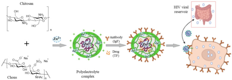
A sulfated derivative of chitin, namely N-carboxymethyl chitosan-N-O-sulfate (NCMCS), was shown to inhibit uptake and replication of human immunodeficiency virus-1 (HIV-1) in CD4+ human T-lymphoma cells, which was attributed to the interference of NCMCS with the interaction of viral GP120 envelope glycoproteins with CD4+ cell surface receptors [90]. In another study, a sulfated derivative of carboxymethyl chitin (SCM-chitin) was shown to inhibit penetration and subsequent pathogenesis of herpes simplex type-1 virus (HSV-1) in vitro in monkey kidney epithelial cells (Vero) as compared to carboxymethyl chitin which showed no activity [91]. The sulfated derivative of chitosan was also found to protect Vero cells from cytopathic infection by Coxsackie and Rift Valley fever RNA viruses [92]. Chitosan and its derivatives are used as a carrier in delivery of antiviral drugs to reduce the minimum required dose and increase bioavailability, thus improving the drugs’ therapeutic effectiveness (Figure 5C) [93]. In one study, curcumin encapsulated in chitosan nanoparticles (NPs) was shown to have higher antiviral activity against hepatitis C virus genotype 4a (HCV-4a) in human hepatoma cells as compared to unencapsulated curcumin [94]. A delivery system based on zinc-stabilized nanocomplex of chitosan and chondroitin sulfate for delivery of tenofovir was developed for inhibition of HIV-1 viral infection, as shown in Figure 6 [95]. The tenofovir-loaded nanocomplex showed strong antiviral activity against HIV-1 in human peripheral blood mononuclear cells (PBMCs) without a cytotoxic effect. The delivery system showed considerable antiviral activity in the absence of tenofovir [95].
Figure 6.
A nanocomplex for delivery of tenofovir based on zinc-stabilized chitosan and chondroitin sulfate. Abbreviations are TF for tenofovir, CS for chitosan, Chons for chondroitin sulfate, IgA for antibody, PECs for polyelectrolyte complexes, and PBMCs for human peripheral blood mononuclear cells. Reprinted with permission from Ref. [95]. Copyright 2016, American Chemical Society.
Recently, the antiviral activity of quaternary ammonium modified chitosan such as N-(2-hydroxypropyl)-3-trimethylammonium chitosan (HTCC) and N-palmitoyl-N-monomethyl-N,N-dimethyl-N,N,N-trimethyl-6-O-glycolchitosan (GCPQ) is reported against SARS-CoV-2, as shown in Figure 7 [96,97]. Due to the quaternary ammonium functional groups in the structure of HTCC and GCPQ, these positively charged molecules bind electrostatically to coronavirus S proteins to block not only viral entry to host cells but also to prevent viral replication. List of studies on antiviral activity of chitosan and its derivatives are presented in Table 3.
Figure 7.
Chemical structures of N-(2-hydroxypropyl)-3-trimethylammonium chitosan (HTCC) and N-palmitoyl-N-monomethyl-N,N-dimethyl-N,N,N-trimethyl-6-O-glycolchitosan (GCPQ) with quaternary ammonium functional groups.
Table 3.
Antiviral activity of chitosan and its derivatives.
| Chitosan | Virus | Cell Line | Concentration | Ref. |
|---|---|---|---|---|
| Zinc stabilized chitosan | HIV-1 | Human peripheral blood mononuclear | 0.1–0.2% w/v chitosan in acetic acid | [95] |
| Curcumin-loaded chitosan nanocomposite | Hepatitis C | human hepatoma | 20 µg/mL curcumin in 4% chitosan composite | [94] |
| Chitosan with medium molecular weight | AD169 strain of HCMV | Human embryonic lung fibroblasts | 9–14 mg Fostcanet antiviral drug in 4.5 mg chitosan |
[98] |
| Silver-loaded chitosan | H1N1 Influenza A | Madin-Darby canine kidney | Ag NPs in 10 mg/mL chitosan | [99] |
| Nanosilver-loaded polyquaternary phosphonium oligochitosan | Hepatitis A | Vero | 100 µL/mL silver NPs | [100] |
| 3,6-O-sulfated chitosan | HPV | 293FT, Hela, HaCaT | 2.4, 4.7, 4.2 µg/mL chitosan | [101] |
| HTCC | SARS-CoV2 | human airway epithelium (HAE) | 12.5 µg/mL HTCC | [96] |
| GCPQ | SARS-CoV2 | Vero E6 and A549 | 10 µg/mL GCPQ | [97] |
2.1.3. Fucoidan
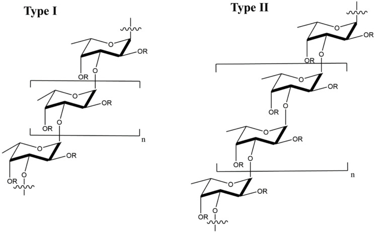
Fucoidan is a sulfated, acidic polysaccharide composed of L-fucose and galactose monosaccharides with smaller proportions of xylose and mannose [102]. It is isolated from the cell wall of marine brown seaweeds like Ascopbyllum nodosum, Fucus vesiculosus, Saccharina japonica, and Sargassum thunbergia. Based on molecular structure, there are two types of fucoidan, as shown in Figure 8, namely type I having (1→3)-l-fucopyranose repeats and type II having (1→3)- and (1→4)-l-fucopyranose repeat units [103]. Depending on degree and pattern of sulfation, molecular weight and composition, fucoidans possess different biological properties including antiviral [56,57,104,105,106,107], anti-inflammatory [108], immunologic [109] and antitumor [110,111] properties.
Figure 8.
Molecular structure of type I and type II fucoidans from brown seaweed; the R groups represent sulfate, glucuronic acid, or fucopyranose groups.
The mechanism of action for antiviral activity of fucoidans from different sources has been recently studied [56,112]. Fucoidan inactivates the virus before or after cell internalization [113]. After internalization, fucoidan prevents viral transcription as well as other intracellular targets, thus blocking viral replication [113]. List of studies on antiviral activity of fucoidans from different sources are presented in Table 4. In one recent study, Kjellmaniella crassifolia fucoidan with a molecular weight of 540 kDa and 30% sulfate content was targeted to inhibition influenza A virus (IAV) [56]. According to in vitro results, the fucoidan outperformed the anti-IAV drug amantadine in inhibiting viral infection [56]. In another study, low molecular weight (LMW) fucoidans with 30–42% fucose, 19–24% galactose, 3–5% uronic acid, and 30–32% sulfate were synthesized and their antiviral activity was evaluated [57]. The LMW fucoidans showed antiviral activity in HeLa, Hep-2, and MDCK cells infected with influenza A virus at medium and high fucoidan doses in the range of 0.15–2.4 mg/mL whereas low doses were ineffective against influenza A [57]. Further, the survival time of the virus-infected mice increased and their lung index improved with intravenous administration of fucoidan [57]. In another study, the influence of Fucus evanescens fucoidan on inhibition of hepatitis B virus (HBV) was investigated in an HBV-infected mouse model [114]. In this study, fucoidan acted as an adjuvant to suppress HBV DNA, HBV surface antigen (HBsAG) and active HBV proteins implicated in viral replication, namely HBeAg and HBcAg [114]. The treatment of HBV-infected mice with 100 mg of fucoidan upregulated the expression of interferon-α (INF-α) via activation of mitogen-activated protein kinase (MAPK-ERK1/2) pathway, which reduced DNA production and viral replication [114]. Despite numerous studies demonstrating the therapeutic potential of fucoidan for treating HBV infections, further research is needed to validate the results in large animal models prior to translation to clinical practice.
Table 4.
Antiviral activity of fucoidans from different species of seaweed.
| Fucoidan | Virus | Cell Line | IC50 (μg/mL) | Ref. |
|---|---|---|---|---|
| Cladosiphon okamuranus | Newcastle Disease (NDV) | Vero | 0.75 | [115] |
| Scytosiphon lomentaria | HSV-1, HSV-2 | Vero | 1.12–1.22 | [112] |
| Sargassum henslowianum | HSV-1, HSV-2 | Vero | 0.48–0.89 | [113] |
| Sporophyll of Undaria pinnatifida (Mekabu) | Influenza A | MDCK | 15 | [116] |
| Brown Algae: Fucus evanescens | HSV-1, HSV-2, HIV-1 | MT-4 | 25–80 | [117] |
2.2. Nucleic Acid Polymers
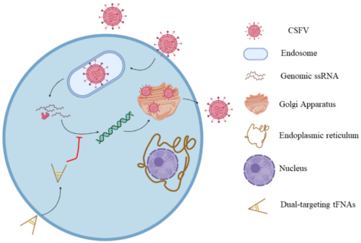
Nucleic acid polymers (NAPs), as phosphorothioate oligonucleotides, possess an-tiviral activity [118]. Oligonucleotides and NAPs can be stabilized against degradation by nucleases made amphipathic by phosphorothioation of non-bridging oxygen atoms through phosphodiester linkages [118]. The antiviral activity of phosphorothioated NAPs was first reported in a study on mechanism of action and the effect of NAPs’ structure on inhibition of HIV-1 infection [114]. It was found that NAPs inhibited viral entry to the host cell and the extent of inhibition was controlled by the size of NAPs, not their sequence distribution. According to this study, NAPs with >20 nucleotides showed antiviral activity [114]. The antiviral activity of NAPs against HIV-1 was at-tributed to the interaction of NAPs with the amphipathic alpha helix triplets found in the core of metastable envelope glycoprotein gp41 of HIV to inhibit viral entry to the host cell. The first step in the viral inhibition is decomplexation of the amphipathic al-pha helices of gp41 by NAPs followed by interaction with hydrophobic moieties of the alpha helices, which explains the dependence of NAP inhibition on size and phos-phorothioation [119]. The interaction of NAPs with amphipathic alpha helix triplets of gp41 was used for targeting and inactivating other viruses including cytomegalovirus, herpesvirus, and lymphocytic choriomeningitis virus [120,121,122,123]. A similar mechanism is proposed for the role of NAPs in blocking the entry of hepatitis C virus (HCV) into host cells [122]. However, one study reported that NAPs did not prevent HCV attach-ment to the host cells but blocked the post-binding step required for viral entry [123]. The post-binding step is posited to involve the interaction of NAPs with other viral proteins including the hypervariable region of glycoprotein E2 or apolipoprotein E [124,125]. The antiviral activity of NAPs assessed against ductal hepatitis B virus (DHBV) with primary duck hepatocytes (PDH) as the host was shown to be dependent on NAP size and amphipathicity [126]. Specifically, NAPs with >40 nucleotides showed higher antiviral activity against DHBV. The therapeutic potential of two NAPs, namely REP 2055 and REP 2139-Ca, were evaluated in patients with HBeAg positive chronic HBV infection in two clinical studies [127,128]. The patients treated with NAP monotherapy showed 2–7 log reduction in serum HBsAg, 3–9 log reduction in serum HBV DNA, and the presence of serum anti-HBsAg antibodies in the patients’ blood [127,128]. These results indicated that NAPs could potentially be used as a component in combination therapies in treating patients with HBV. In another study, the safety and therapeutic efficacy of REP 2139 and REP 2165 NAPs combined with commonly used medication to treat patients with chronic hepatitis B, namely tenofovir disoproxil fumarate (TDF) and pegylated interferon α-2a (PegIFN), were assessed in a randomized phase 2 clinical trial. The REP 2139 NAP and its bioequivalent REP 2165 block the assembly of subviral particles (SVPs) in hepatocytes resulting in clearance of HBsAg surface antigens. The experimental NAP groups were TDF+PegIFN+REP 2139 and TDF+PegIFN+REP 2165 [129]. According to the results, there was no difference in the levels of HBsAg, indicative of HBV infection, anti-HBs, indicative of recovery from HBV infection, and HBV DNA between the two NAP groups [129]. The undesired side effects induced by PegIFN, namely thrombocytopenia and neutropenia, were unaf-fected by the addition of NAPs to TD and PegIFN. However, the levels of HBV-induced transaminases were initially higher in the NAP groups, but the levels returned to nor-mal during therapy and follow-up [129]. Treatment with TDF+PegIFN without NAP resulted in low HBsAg loss and HBsAg seroconversion as well as low functional cure. Conversely, treatment with TDF+PegIFN+NAP resulted in high HBsAg loss and HBsAg seroconversion as well as high functional cure with normal liver function [129]. No differences in HBsAg loss, seroconversion, or functional cure were observed between the two NAP groups. An in vitro model based on HepG2.2.15 hepatocyte cells that constitutively express HBV demonstrated that the NAPs’ endosomal release selectively impaired the secretion of HBsAg without accumulation of HBsAg in the cells [130]. NAPs due to their biocompatibility, degradability, and their ability to penetrate cell membranes have been used as a carrier for delivery of antiviral drugs against classical swine fever virus (CSFV) in infected pigs [131,132]. The enveloped virus with a sin-gle-stranded RNA causing CSFV belongs to the genus Pestivirus in the family Fla-viviridae [133]. The small interfering RNA (siRNA) was delivered to the host cells in a porcine CSFV model using tetrahedral framework nucleic acid (tFNA), as a three-dimensional DNA nanomaterial that self-assembles by complementary base pairing [134]. Concurrent delivery of C3 and C6 siRNAs in self-assembled tFNA to host cells prevented viral replication and exocytosis, as shown in Figure 9 [134].
Figure 9.
Schematic illustration of the mechanism of siRNA delivery by tFNA nanomaterial. Reprinted with permission from Ref. [134]. Copyright 2021, American Chemical Society.
Following penetration through the cell membrane, CSFV is transported to the endoplasmic reticulum where its single-stranded RNA is released for protein translation. Following RNA translation, CSFV is replicated on the cytoplasmic membrane leading to the formation of viral particles in the Golgi apparatus. The siRNAs carried by tFNA degrade the released viral RNA to prevent its translation in the endoplasmic reticulum. List of studies on antiviral activity of NAPs are presented in Table 5.
Table 5.
Antiviral activity of NAPs against different viruses with their mechanism of actions.
| NAP | Virus | Cell Line | Mechanism of Action | Ref. |
|---|---|---|---|---|
| Phosphorothioate Oligonucleotides | HIV-1 | H9 | Inhibiting viral entry to the host | [119] |
| amphipathic DNA polymer (40-nucleotide polycytidine) | HSV | Human cervical epithelial (CaSki) | Reduced viral and protein expression | [120] |
| Amphipathic DNA polymers (REP 9, REP 2015 and REP 9C) | Animal | NIH 3T3 | Reduced viral replication | [123] |
| REP 2006 | HBV | DHBV | Reduced viral entry to the host | [126] |
| REP 2055 | HBV | DHBV | blocking release of DHBsAg from the infected hepatocytes | [128] |
3. Synthetic Polymers
Polymers are chain-like molecules formed by polymerization of small, reactive organic molecules. Polymers due to their long chain length or high molecular weight are used extensively in surface coating applications because each polymer chain can make many physical bonds to irreversibly coat the surface [29,135]. As a result, polymers are very attractive as an antiviral material to bond irreversibly to viral glycoproteins to cover and conceal the viral surface, thus blocking the interaction of viral particles with the host cell [11,12,13,14,15,16,17,18,19,20,21,22,23,24,25]. Unlike natural polymers, the chemical composition, functional group type and extent of functionalization, molecular weight, charge density and distribution, degradation and stability of synthetic polymers can be engineered to maximize antiviral activity against a specific virus type [22,136,137]. Among synthetic polymers, dendrimers and sialyl-based polymers have been extensively studied as antiviral agents to fight against infections.
3.1. Dendrimers
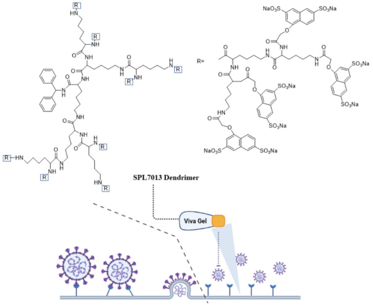
Dendrimers due to their nanoscale size and structural uniformity are attractive as a carrier for delivery of antiviral agents [138]. The branched structure of dendrimers results in spherical and symmetric nanostructures with a monodisperse size in the 2–10 nm range [139]. The dendrimer generation which is the number of branches or the number of repeated synthetic cycles, controls the size of dendrimers [139]. The chemical groups on the dendrimer surface can be functionalized to produce polycationic or polyanionic dendrimers [139,140]. Peptides and drug molecules can be conjugated to dendrimers via cleavable linkages to form functional dendrimers with controlled biological activity [141]. Antiviral activity of dendrimers is attributed to their multivalency, symmetrical and monodisperse molecular structure, and the presence of multiple functional groups on the dendrimer surface. The dendrimer-based antiviral agents act mechanistically by interfering with the interaction of virus with the host cell [142]. As a result, antiviral activity depends on dendrimer molecular weight and type of functional groups present on the dendrimer surface [142]. Among different types, peptide-conjugated dendrimers show excellent antiviral activity against many virus types. A sulfonated derivative of polylysine dendrimer with a benzhydrylamine core was used to prevent infection against intravaginal herpes simplex virus (HSV) and simian immunodeficiency virus/human immunodeficiency virus (SIV/HIV) chimeric viruses [143,144]. In vitro studies with monkey kidney epithelial cells (Vero) demonstrated that this dendrimer completely prevented adsorption and penetration of the virus in host cells and inhibited DNA synthesis in the infected cells [144]. Figure 10 shows the chemical structure and mechanism of action of an FDA approved VivaGel® dendrimer (SPL7013), which is used in preventing genital herpes (HSV-2) and HIV. The VivaGel® is composed of anionic G4-poly(L-lysine) dendrimer with a benzylhydramine-amide-lysine core with 32 sodium (1-naphthyleneyl-3,6-disulphonic acid)-oxyacetamide functional groups [145]. The functional groups on the dendrimer surface act to prevent infection by blocking the virus from binding to host cells [145].
Figure 10.
Chemical structure and mechanism of action of VivaGel® as an antiviral agent. The image was created by BioRender software.
Paull and coworkers evaluated the antiviral activity of SPL7013 dendrimer against SARS-CoV-2 [146]. Compared to polyanionic biopolymers such as io-ta-carrageenan and heparin (as linear sulphated molecules), the branched sulfonated structure of SPL7013 dendrimer inhibited SARS-CoV-2 infection in Vero E6 cells early in the replication cycle by blocking viral entry to the cells. In another study, a new class of dendrimers with tryptophan (Trp) functional groups on the surface were synthe-sized and their antiviral activity was evaluated against HIV and enterovirus-71 (EV71) [147]. Dendrimers with different surface amino acid groups including aromatic and non-aromatic, decarboxylated Trp (tryptamine), and methylated Trp were synthe-sized to study the relation between amino acid structure and antiviral activity [147]. According to the results, Trp-conjugated dendrimers act mechanistically by interacting with cell surface glycoproteins to interfere with virus-cell interaction early in the HIV replication cycle prior to viral entry in the host cell [147]. The type of peptide and its structure (aliphatic versus aromatic) affected antiviral activity of the dendrimer. Dendrimers conjugated with aromatic amino acids like tryptophan showed antiviral activity against both HIV and EV71 whereas dendrimers conjugated with aliphatic amino acids like alanine were inactive. The natural peptide LL-37 which is a member of cathelicidin family of peptides show antiviral activity against the respiratory syn-cytial virus (RSV), which is a common lower respiratory tract pathogen in children [148]. However, LL-37 peptide has limited stability in physiological medium. In a re-cent study, linear (SA-35) and hyperbranched (dendrimeric, LTP) cationic peptides from the helical region of LL-37 peptide were synthesized and their antiviral activity were compared with the natural peptide [149]. The results showed that the linear and hyperbranched peptides had higher antiviral activity as compared to the natural pep-tide which was attributed to destabilization of the viral envelope and competitive in-hibition of virus attachment/fusion with the host cell.
A range of polycationic and polyanionic dendrimers have been synthesized and evaluated against viral infections [150]. The antiviral activity of these dendrimers is due to electrostatic interaction between the viral envelope proteins and the den-drimer’s ionic functional groups [151]. In one study, polycationic dendrimers with N-alkylated 4,4′-bipyridinium units of varying charge density were synthesized and evaluated with respect to inhibition of HIV-1 [152]. Results showed that the inhibition of HIV-1 in MT-4 host T cells by the dendrimers was mediated by CXCR4 coreceptor and was independent of charge density of dendrimers. In another study, a polyanionic carbosilane dendrimer (PCD), designed to inhibit HIV-1 infection, was used to prevent viral infections causing sexually transmitted diseases [153]. The PCD dendrimer inhib-ited herpes simplex virus-2 (HSV-2) infection by reducing plaque formation in HSV-2 infected Vero cells, which was attributed to direct binding between the sulfonate groups on the dendrimer surface and glycoprotein B of the virus [154]. In another study, a PCD consisting of a polyphenolic core with 24 sulfonate groups on the den-drimer surface was shown to possess antiviral activity against hepatitis C virus (HCV) at low and high concentrations [154]. In another study, PCDs with surface sulfonate or naphthalene sulfonate functional groups prevented not only HIV-1 infection by inter-fering with cell-virus fusion but also inhibited cell-to-cell transmission [138]. List of studies on antiviral activity of dendrimers against different viruses are presented in Table 6.
Table 6.
Antiviral activity of dendrimers and their mechanisms of action.
| Dendrimer Type | Name | Virus | Cell Line | Mechanism of Action | Ref. |
|---|---|---|---|---|---|
| Polyanionic | Phenyldicarboxylic acid (BRI6195), Naphthyldisulfonic acid (BRI2923) | HIV-1 | MT-4 | Inhibit viral binding and replication | [142] |
| Polyanionic | SPL-2999 | HSV-1, HSV-2 | Vero | Inhibit late stages of viral replication | [144] |
| Polyanionic | carbosilane dendrimer G2-STE16 | HIV-1 | TZM.bl | Inhibit viral entry | [151] |
| Polyanionic | G2-S16 | HIV | PBMC | Inhibit viral replication by blocking gp120/CD4/CCR5 interaction | [155] |
| Polyanionic | G3-S16 and G2-NF16 | HIV-1 | HeLa P4.R5MAGI, TZM.bl | Inhibit viral replication by blocking gp120/CD4/CCR5 interaction | [138] |
| Polycationic | Viologen | HIV-1 | MT-4 cells | Block viral entry | [152] |
3.2. Sialyl-Based Polymers
The sialylated glycans in the protective mucin layer of the nasal cavity disrupts the entry of influenza virus (IFV) particles to host cells by complexation with viral hemagglutinin (HA) [156]. The mucin barrier is the first line of defense in the immune system that traps and clears the viral particles upon mucin turnover, as shown in Figure 11 [157]. Although sialic acid (SA) and derivatives of SA have a strong affinity for the viral HA, the monovalent interaction between HA and SA is relatively weak [158]. Therefore, polymers modified with SA that can overcome the low interaction energy by complexation with multiple SA groups can effectively inhibit IFV infections [159]. In one study, the antiviral activity of a multivalent polymer conjugate consisting of poly (methyl vinyl ether-alt-maleic anhydride) modified with thiosialoside against PR8 influenza strain was comparable to those of Zanamivir® and Oseltamivir® drugs, as measured by growth inhibition assay [160]. The antiviral activity was attributed to multivalent binding of the modified polymer to neuraminidase on the viral particles to inhibit enzymatic activity, leading to viral particle aggregation and inhibition of virus-cell fusion. These results imply that polymers functionalized with monomeric sialoside groups which possess neuraminidase inhibitory activity can serve as an antiviral scaffold against early and late stages of influenza infection.
Figure 11.
Schematic representation for the clearance of influenza virus by the mucin layer or mucin-mimetic glycol-conjugated proteins prior to uptake by epithelial cells. The image was created by BioRender.com software.
Glycopolymers based on sialyl oligosaccharides with a range of chain lengths and sugar densities were synthesized by reversible addition-fragmentation chain transfer polymerization (RAFT) for interaction with hemagglutinin on the surface of influenza virus [161]. The extent of interaction and molecular specificity of the copolymers against different types of influenza virus depended on sugar units (6′- versus 3′-sialyllactose), sugar density, and polymer chain length [161]. In another study, tri-arm star glycopolymers with homogenous number of arms consisting of inert spacers and glycomonomer segments were synthesized by a core-first approach. The star glycopolymers had three arms with each arm having a degree of polymerization ranging from 30 to 100 to match the sugar-binding pockets of hemagglutinin on influenza virus [162]. The inert core segment was based on poly(N,N-dimethylacrylamide) (PDMA) whereas the arms were made of 6′-Sialyllactose glycomonomer which served as a natural ligand for HA binding in human influenza virus [162]. The use of arms with different degrees of polymerization allowed tunning the ligand-receptor interaction to the desired strength for surface HA in influenza virus. In another study, a polymer brush consisting of side chains ending in α-2,6-linked sialic acid (SA) was synthesized by protection-group-free, ring-opening metathesis polymerization (ROMP) to enhance multivalent interaction as determined by hemagglutination binding [163]. In this work, the anomeric hydroxyl group of 6′-sialyllatose in the oligosaccharide was activated and converted to an azide by nucleophilic substitution to form an azido oligosaccharide. Next, the azido oligosaccharide was reacted with α-norbornenyl, ω-acetylene terminated PEG, prepared from amine-terminated hydroxyl PEG, using a copper-catalyzed click reaction. Then, the α-norbornenyl terminated oligosaccharide PEG was polymerized by ROMP to produce a brush polymer with oligosaccharide side chains terminated with SA [163]. According to the results, brush polymers with high degree of polymerization, high SA density, and long side chains produced strongest antiviral activity as measured by hemagglutination inhibition assay [163]. These brush polymers are useful as a model system to study the antiviral properties of native mucins with the passage of influenza A virus (IAV) through the epithelial mucin barrier as measured by the strength of interaction between the viral HA and the sialyloligosaccharides of mucin (Figure 11) [164]. List of studies on antiviral activity of sialylated polymers are presented in Table 7.
Table 7.
List of studies on antiviral activity of sialyl-based polymers with their mechanism of action.
| Sialyl-Based Polymers | Virus | Cell Line | Mechanism of Action | Ref. |
|---|---|---|---|---|
| Amide-sialoside protein conjugates | IAV | H1N1, H3N2, H9N2 | Binding to the virus and inhibiting replication | [157] |
| Thiosialoside-modified poly (methyl vinyl ether-alt-maleic anhydride) | IAV | MDCK | inhibiting both early attachment and late release during viral infection | [160] |
| silyl oligosaccharides modified glycopolymers | IAV | H1N1 | Binding with hemagglutinin on viral surface | [161] |
| Bioinspired brush polymers containing α-2,6-linked sialic acid | IAV | H1N1 | binding hemagglutination on viral surface | [163] |
4. Conclusions
Polymers provide enormous opportunity to tailor antiviral activity via specific interaction with glycoproteins on the viral surface by varying molecular weight, type and degree of functionality, sequence distribution, extent of branching, and molecular architecture. Polymers can be used to increase bioavailability and therapeutic effectiveness of antiviral agents and reduce their undesired side effects. Natural biopolymers with multiple functional groups like carrageenan, chitosan, and fucoidan are used directly to bind and form irreversible complexes with envelope glycoproteins on the viral surface to block virus-host cell interaction, fusion, and entry to the host cell. Biopolymers are also used to mobilize the immune response against the invading virus by stimulating the activation of macrophages and natural killer cells. Natural nucleic acid polymers are used as antiviral agents after phosphorothioation to render them amphipathic and stabilization against nuclease degradation. Polymers deactivate the virus before cell internalization by inhibiting virus-host cell binding or after cell internalization by blocking virus replication. Synthetic dendrimers due to their symmetry, monodisperse molecular structure, and multifunctionality bind with high affinity to envelope glycoproteins on the viral surface to block virus-host cell interaction and fusion. Brush polymers modified with sialic acid groups bind with high affinity to viral hemagglutinin to form complexes that block viral infection. Research results have demonstrated that natural and synthetic polymers are useful as broad-spectrum antiviral agents to fight against many different types of viral infections. Further preclinical research in animal models and clinical trials are needed to validate the antiviral activity of polymers to fight new viral infections in the absence of vaccines.
Abbreviations
| CXCR4 | chemokine receptor type 4 |
| ERK | extracellular signal-regulated kinase |
| gB | glycoprotein B |
| H1N1 | novel influenza A virus |
| HBV | hepatitis B virus |
| HCoV-OC43 | human coronavirus OC43 |
| HCV | hepatitis C virus |
| HDV | hepatitis delta virus |
| HIV | human immunodeficiency virus |
| hMPV | human metapneumovirus |
| HPV | human papillomavirus |
| HRV | human rhinovirus |
| HSV-1 | herpes simplex virus Type 1 |
| HSV-2 | herpes simplex virus Type 2 |
| IAV | influenza A virus |
| IC50 | Half maximal inhibitory concentration |
| NAPs | nucleic acid polymers |
| PBMC | primary peripheral blood mononuclear cells |
| PCD | polyanionic carbosilane dendrimers |
| PG | 2,3-O-disulfated-1,1,4-poly-L-guluronic acid |
| RSV | respiratory virus |
| SARS-CoV-2 | severe acute respiratory syndrome coronavirus 2 |
Author Contributions
Conceptualization, A.A., A.B. and E.J.; methodology, A.A. and A.B.; formal analysis, A.A., A.B., V.R., S.S. and E.J.; resources, E.J.; data curation, A.A., A.B., V.R., S.S. and E.J.; writing—original draft preparation, A.A. and A.B. and E.J; writing—review and editing, E.J.; visualization, A.A. and A.B.; supervision, E.J. All authors have read and agreed to the published version of the manuscript.
Funding
This research received no external funding.
Institutional Review Board Statement
Not applicable.
Informed Consent Statement
Not applicable.
Data Availability Statement
Not applicable.
Conflicts of Interest
The authors declare no conflict of interest.
Footnotes
Publisher’s Note: MDPI stays neutral with regard to jurisdictional claims in published maps and institutional affiliations.
References
- 1.Zhang N., Wang L., Deng X., Liang R., Su M., He C., Hu L., Su Y., Ren J., Yu F. Recent advances in the detection of respiratory virus infection in humans. J. Med. Virol. 2020;92:408–417. doi: 10.1002/jmv.25674. [DOI] [PMC free article] [PubMed] [Google Scholar]
- 2.Gannon O., Antonsson A., Bennett I., Saunders N. Viral infections and breast cancer—A current perspective. Cancer Lett. 2018;420:182–189. doi: 10.1016/j.canlet.2018.01.076. [DOI] [PubMed] [Google Scholar]
- 3.Feldmann H., Jones S., Klenk H.D., Schnittler H.J. Ebola virus: From discovery to vaccine. Nat. Rev. Immunol. 2003;3:677–685. doi: 10.1038/nri1154. [DOI] [PubMed] [Google Scholar]
- 4.Sullivan N., Yang Z.-Y., Nabel G.J. Ebola virus pathogenesis: Implications for vaccines and therapies. J. Virol. 2003;77:9733–9737. doi: 10.1128/JVI.77.18.9733-9737.2003. [DOI] [PMC free article] [PubMed] [Google Scholar]
- 5.Colson P., La Scola B., Levasseur A., Caetano-Anolles G., Raoult D. Mimivirus: Leading the way in the discovery of giant viruses of amoebae. Nat. Rev. Microbiol. 2017;15:243–254. doi: 10.1038/nrmicro.2016.197. [DOI] [PMC free article] [PubMed] [Google Scholar]
- 6.Villanueva R.A., Rouillé Y., Dubuisson J. Interactions between virus proteins and host cell membranes during the viral life cycle. Int. Rev. Cytol. 2005;245:171–244. doi: 10.1016/S0074-7696(05)45006-8. [DOI] [PMC free article] [PubMed] [Google Scholar]
- 7.Choi Y., Bowman J.W., Jung J.U. Autophagy during viral infection—a double-edged sword. Nat. Rev. Microbiol. 2018;16:341–354. doi: 10.1038/s41579-018-0003-6. [DOI] [PMC free article] [PubMed] [Google Scholar]
- 8.Ricci S., Francisci D., Longo V., Delfavero A. Central nervous-system side-effects of antiviral drugs. Int. J. Clin. Pharmacol. Ther. Toxicol. 1988;26:400–408. [PubMed] [Google Scholar]
- 9.Chaudhuri S., Symons J.A., Deval J. Innovation and trends in the development and approval of antiviral medicines: 1987–2017 and beyond. Antiviral Res. 2018;155:76–88. doi: 10.1016/j.antiviral.2018.05.005. [DOI] [PMC free article] [PubMed] [Google Scholar]
- 10.Zareifopoulos N., Lagadinou M., Karela A., Kyriakopoulou O., Velissaris D. Neuropsychiatric effects of antiviral drugs. Cureus. 2020;12:e9536. doi: 10.7759/cureus.9536. [DOI] [PMC free article] [PubMed] [Google Scholar]
- 11.Chun H., Yeom M., Kim H.-O., Lim J.-W., Na W., Park G., Park C., Kang A., Yun D., Kim J. Efficient antiviral co-delivery using polymersomes by controlling the surface density of cell-targeting groups for influenza A virus treatment. Polym. Chem. 2018;9:2116–2123. doi: 10.1039/C8PY00116B. [DOI] [Google Scholar]
- 12.Sun Y.Z., Gong L.D., Yin Y., Zhang L., Sun Q.M., Feng K., Cui Y.M., Zhang Q., Zhang X.H., Deng X.L., et al. A gradient pH-sensitive polymer-based antiviral strategy via viroporin-induced membrane acidification. Adv. Mater. 2022;2:e2109580. doi: 10.1002/adma.202109580. [DOI] [PubMed] [Google Scholar]
- 13.Soares D.C.F., Poletto F., Eberhardt M.J., Domingues S.C., De Sousa F.B., Tebaldi M.L. Polymer-hybrid nanosystems for antiviral applications: Current advances. Biomed. Pharmacother. 2022;146:112249. doi: 10.1016/j.biopha.2021.112249. [DOI] [PubMed] [Google Scholar]
- 14.Chen P.Y., Lang J.Y., Zhou Y.L., Khlyustova A., Zhang Z.Y., Ma X.J., Liu S., Cheng Y.F., Yang R. An imidazolium-based zwitterionic polymer for antiviral and antibacterial dual functional coatings. Sci. Adv. 2022;8:eab18812. doi: 10.1126/sciadv.abl8812. [DOI] [PMC free article] [PubMed] [Google Scholar]
- 15.Ordon M., Zdanowicz M., Nawrotek P., Stachurska X., Mizielinska M. Polyethylene films containing plant extracts in the polymer matrix as antibacterial and antiviral materials. Int. J. Mol. Sci. 2021;22:13438. doi: 10.3390/ijms222413438. [DOI] [PMC free article] [PubMed] [Google Scholar]
- 16.Nasri N., Rusli A., Teramoto N., Jaafar M., Ishak K.M.K., Shafiq M.D., Hamid Z.A.A. Past and current progress in the development of antiviral/antimicrobial polymer coating towards COVID-19 prevention: A Review. Polymers. 2021;13:4234. doi: 10.3390/polym13234234. [DOI] [PMC free article] [PubMed] [Google Scholar]
- 17.Li J.H., Wang W.Z., Jiang R., Guo C.C. Antiviral electrospun polymer composites: Recent advances and opportunities for tackling COVID-19. Front. Mater. 2021;8:773205. doi: 10.3389/fmats.2021.773205. [DOI] [Google Scholar]
- 18.Leong J.Y., Shi D.R., Tan J.P.K., Yang C., Yang S.C., Wang Y.M., Ngow Y.S., Kng J., Balakrishnan N., Peng S.Q., et al. Potent antiviral and antimicrobial polymers as safe and effective disinfectants for the prevention of infections. Adv. Healthc. Mater. 2021:2101898. doi: 10.1002/adhm.202101898. [DOI] [PubMed] [Google Scholar]
- 19.Kuroki A., Tay J., Lee G.H., Yang Y.Y. Broad-Spectrum Antiviral Peptides and Polymers. Adv. Healthc. Mater. 2021;10:e2101898. doi: 10.1002/adhm.202101113. [DOI] [PubMed] [Google Scholar]
- 20.Steinman N.Y., Hu T., Dombrovsky A., Reches M., Domb A.J. Antiviral polymers based on N-halamine polyurea. Biomacromolecules. 2021;22:4357–4364. doi: 10.1021/acs.biomac.1c00920. [DOI] [PubMed] [Google Scholar]
- 21.Yadavalli T., Mallick S., Patel P., Koganti R., Shukla D., Date A.A. Pharmaceutically acceptable carboxylic acid-terminated polymers show activity and selectivity against HSV-1 and HSV-2 and synergy with antiviral drugs. ACS Infect. Dis. 2020;6:2926–2937. doi: 10.1021/acsinfecdis.0c00368. [DOI] [PMC free article] [PubMed] [Google Scholar]
- 22.Bianculli R.H., Mase J.D., Schulz M.D. Antiviral polymers: Past approaches and future possibilities. Macromolecules. 2020;53:9158–9186. doi: 10.1021/acs.macromol.0c01273. [DOI] [Google Scholar]
- 23.Soria-Martinez L., Bauer S., Giesler M., Schelhaas S., Materlik J., Janus K., Pierzyna P., Becker M., Snyder N.L., Hartmann L. Prophylactic antiviral activity of sulfated glycomimetic oligomers and polymers. J. Am. Chem. Soc. 2020;142:5252–5265. doi: 10.1021/jacs.9b13484. [DOI] [PubMed] [Google Scholar]
- 24.Roner M.R., Carraher C.E., Miller L., Mosca F., Slawek P., Haky J.E., Frank J. Organotin polymers as antiviral agents including inhibition of Zika and Vaccinia viruses. J. Inorg. Organomet. Polym. Mater. 2020;30:684–694. doi: 10.1007/s10904-019-01250-9. [DOI] [Google Scholar]
- 25.Gunther S.C., Maier J.D., Vetter J., Podvalnyy N., Khanzhin N., Hennet T., Stertz S. Antiviral potential of 3′-sialyllactose- and 6′-sialyllactose-conjugated dendritic polymers against human and avian influenza viruses. Sci. Rep. 2020;10:768. doi: 10.1038/s41598-020-57608-4. [DOI] [PMC free article] [PubMed] [Google Scholar]
- 26.Fox M.E., Szoka F.C., Fréchet J.M. Soluble polymer carriers for the treatment of cancer: The importance of molecular architecture. Acc. Chem. Res. 2009;42:1141–1151. doi: 10.1021/ar900035f. [DOI] [PMC free article] [PubMed] [Google Scholar]
- 27.Wang Y., Canady T.D., Zhou Z., Tang Y., Price D.N., Bear D.G., Chi E.Y., Schanze K.S., Whitten D.G. Cationic phenylene ethynylene polymers and oligomers exhibit efficient antiviral activity. ACS Appl. Mater. Interf. 2011;3:2209–2214. doi: 10.1021/am200575y. [DOI] [PubMed] [Google Scholar]
- 28.Liu F., Chen Q., Liu C., Ao Q., Tian X., Fan J., Tong H., Wang X. Natural polymers for organ 3D bioprinting. Polymers. 2018;10:1278. doi: 10.3390/polym10111278. [DOI] [PMC free article] [PubMed] [Google Scholar]
- 29.Rakowska P.D., Tiddia M., Faruqui N., Bankier C., Pei Y., Pollard A.J., Zhang J., Gilmore I.S. Antiviral surfaces and coatings and their mechanisms of action. Commun. Mater. 2021;2:1–19. doi: 10.1038/s43246-021-00153-y. [DOI] [Google Scholar]
- 30.Randazzo W., Fabra M.J., Falcó I., López-Rubio A., Sánche G. Polymers and biopolymers with antiviral activity: Potential applications for improving food safety. Comp. Rev. Food Sci. Food Saf. 2018;17:754–768. doi: 10.1111/1541-4337.12349. [DOI] [PubMed] [Google Scholar]
- 31.Xie J.-H., Jin M.-L., Morris G.A., Zha X.-Q., Chen H.-Q., Yi Y., Li J.-E., Wang Z.-J., Gao J., Nie S.-P. Advances on bioactive polysaccharides from medicinal plants. Crit. Rev. Food Sci. Nutr. 2016;56((Suppl. S1)):S60–S84. doi: 10.1080/10408398.2015.1069255. [DOI] [PubMed] [Google Scholar]
- 32.Dwek R.A. Glycobiology: Toward understanding the function of sugars. Chem. Rev. 1996;96:683–720. doi: 10.1021/cr940283b. [DOI] [PubMed] [Google Scholar]
- 33.Xie L., Shen M., Hong Y., Ye H., Huang L., Xie J. Chemical modifications of polysaccharides and their anti-tumor activities. Carbohydr. Polym. 2020;229:115436. doi: 10.1016/j.carbpol.2019.115436. [DOI] [PubMed] [Google Scholar]
- 34.Pawar H.A., Kamat S.R., Choudhary P.D. An overview of natural polysaccharides as biological macromolecules: Their chemical modifications and pharmaceutical applications. Biol. Med. 2015;7:1. doi: 10.4172/0974-8369.1000224. [DOI] [Google Scholar]
- 35.Jiang J.-L., Zhang W.-Z., Ni W.-X., Shao J.-W. Insight on structure-property relationships of carrageenan from marine red algal: A review. Carbohydr. Polym. 2021;257:117642. doi: 10.1016/j.carbpol.2021.117642. [DOI] [PubMed] [Google Scholar]
- 36.Khayrova A., Lopatin S., Varlamov V. Obtaining chitin, chitosan and their melanin complexes from insects. Int. J. Biol. Macromol. 2021;167:1319–1328. doi: 10.1016/j.ijbiomac.2020.11.086. [DOI] [PubMed] [Google Scholar]
- 37.Oliveira C., Neves N.M., Reis R.L., Martins A., Silva T.H. A review on fucoidan antitumor strategies: From a biological active agent to a structural component of fucoidan-based systems. Carbohydr. Polym. 2020;239:116131. doi: 10.1016/j.carbpol.2020.116131. [DOI] [PubMed] [Google Scholar]
- 38.Darge H.F., Andrgie A.T., Tsai H.-C., Lai J.-Y. Polysaccharide and polypeptide based injectable thermo-sensitive hydrogels for local biomedical applications. Int. J. Biol. Macromol. 2019;133:545–563. doi: 10.1016/j.ijbiomac.2019.04.131. [DOI] [PubMed] [Google Scholar]
- 39.Gupta N., Jangid A.K., Pooja D., Kulhari H. Inulin: A novel and stretchy polysaccharide tool for biomedical and nutritional applications. Int. J. Biol. Macromol. 2019;132:852–863. doi: 10.1016/j.ijbiomac.2019.03.188. [DOI] [PubMed] [Google Scholar]
- 40.Shi Q.M., Wang A.J., Lu Z.H., Qin C.J., Hu J., Yin J. Overview on the antiviral activities and mechanisms of marine polysaccharides from seaweeds. Carbohydr. Res. 2017;453–454:1–9. doi: 10.1016/j.carres.2017.10.020. [DOI] [PubMed] [Google Scholar]
- 41.Lu W.J., Yang Z.F., Chen J., Wang D., Zhang Y. Recent advances in antiviral activities and potential mechanisms of sulfated polysaccharides. Carbohydr. Polym. 2021;272:118526. doi: 10.1016/j.carbpol.2021.118526. [DOI] [PubMed] [Google Scholar]
- 42.Li Z.H., Cui B., Liu X.W., Wang L.C., Xian Q.J., Lu Z.X., Liu S.T., Cao Y.G., Zhao Y.R. Virucidal activity and the antiviral mechanism of acidic polysaccharides against Enterovirus 71 infection in vitro. Microbiol. Immunol. 2020;64:189–201. doi: 10.1111/1348-0421.12763. [DOI] [PubMed] [Google Scholar]
- 43.Zhao C., Gao L., Wang C., Liu B., Jin Y., Xing Z. Structural characterization and antiviral activity of a novel heteropolysaccharide isolated from Grifola frondosa against enterovirus 71. Carbohydr. Polym. 2016;144:382–389. doi: 10.1016/j.carbpol.2015.12.005. [DOI] [PubMed] [Google Scholar]
- 44.Leibbrandt A., Meier C., König-Schuster M., Weinmüllner R., Kalthoff D., Pflugfelder B., Graf P., Frank-Gehrke B., Beer M., Fazekas T. Iota-carrageenan is a potent inhibitor of influenza A virus infection. PLoS ONE. 2010;5:e14320. doi: 10.1371/journal.pone.0014320. [DOI] [PMC free article] [PubMed] [Google Scholar]
- 45.Buck C.B., Thompson C.D., Roberts J.N., Müller M., Lowy D.R., Schiller J.T. Carrageenan is a potent inhibitor of papillomavirus infection. PLoS Pathog. 2006;2:e69. doi: 10.1371/journal.ppat.0020069. [DOI] [PMC free article] [PubMed] [Google Scholar]
- 46.Chen Y., Luo Q., Li S., Li C., Liao S., Yang X., Zhou R., Zhu Y., Teng L., Chen H. Antiviral activity against porcine epidemic diarrhea virus of Pogostemon cablin polysaccharide. J. Ethnopharmacol. 2020;259:113009. doi: 10.1016/j.jep.2020.113009. [DOI] [PubMed] [Google Scholar]
- 47.Liang Y.-Y., Li K.-W., Niu F.-J., Li Y., Wei H.-C., Dai Y.-L., Wang Y.-Y., Zhou C.-Z., Wan X.-H. Salvia plebeia R. Br. polysaccharides (SPP) against RSV (respiratory syncytial virus) infection: Antiviral effect and mechanisms of action. Biomed. Pharmacother. 2021;141:111843. doi: 10.1016/j.biopha.2021.111843. [DOI] [PubMed] [Google Scholar]
- 48.Wang T., Wang X., Zhuo Y., Si C., Yang L., Meng L., Zhu B. Antiviral activity of a polysaccharide from Radix Isatidis (Isatis indigotica Fortune) against hepatitis B virus (HBV) in vitro via activation of JAK/STAT signal pathway. J. Ethnopharmacol. 2020;257:112782. doi: 10.1016/j.jep.2020.112782. [DOI] [PubMed] [Google Scholar]
- 49.Chen X.Y., Han W.W., Wang G.X., Zhao X. Application prospect of polysaccharides in the development of anti-novel coronavirus drugs and vaccines. Int. J. Biol. Macromol. 2020;164:331–343. doi: 10.1016/j.ijbiomac.2020.07.106. [DOI] [PMC free article] [PubMed] [Google Scholar]
- 50.Yu C., Wei K., Liu L., Yang S., Hu L., Zhao P., Meng X., Shao M., Wang C., Zhu L. Taishan Pinus massoniana pollen polysaccharide inhibits subgroup J avian leucosis virus infection by directly blocking virus infection and improving immunity. Sci. Rep. 2017;7:1–14. doi: 10.1038/srep44353. [DOI] [PMC free article] [PubMed] [Google Scholar]
- 51.Carbone D.A., Pellone P., Lubritto C., Ciniglia C. Evaluation of microalgae antiviral activity and their bioactive compounds. Antibiotics. 2021;10:746. doi: 10.3390/antibiotics10060746. [DOI] [PMC free article] [PubMed] [Google Scholar]
- 52.Sanniyasi E., Venkatasubramanian G., Anbalagan M.M., Raj P.P., Gopal R.K. In vitro anti-HIV-1 activity of the bioactive compound extracted and purified from two different marine macroalgae (seaweeds) (Dictyota bartayesiana J.V.Lamouroux and Turbinaria decurrens Bory) Sci. Rep. 2019;9:12185. doi: 10.1038/s41598-019-47917-8. [DOI] [PMC free article] [PubMed] [Google Scholar]
- 53.Pereira L., Critchley A.T. The COVID 19 novel coronavirus pandemic 2020: Seaweeds to the rescue? Why does substantial, supporting research about the antiviral properties of seaweed polysaccharides seem to go unrecognized by the pharmaceutical community in these desperate times? J. Appl. Phycol. 2020;32:1875–1877. doi: 10.1007/s10811-020-02143-y. [DOI] [PMC free article] [PubMed] [Google Scholar]
- 54.Alam M.A., Parra-Saldivar R., Bilal M., Afroze C.A., Ahmed M.N., Iqbal H.M.N., Xu J.L. Algae-derived bioactive molecules for the potential treatment of SARS-CoV-2. Molecules. 2021;26:2134. doi: 10.3390/molecules26082134. [DOI] [PMC free article] [PubMed] [Google Scholar]
- 55.Andrew M., Jayaraman G. Marine sulfated polysaccharides as potential antiviral drug candidates to treat corona virus disease (COVID-19) Carbohydr. Res. 2021;505:108326. doi: 10.1016/j.carres.2021.108326. [DOI] [PMC free article] [PubMed] [Google Scholar]
- 56.Wang W., Wu J., Zhang X., Hao C., Zhao X., Jiao G., Shan X., Tai W., Yu G. Inhibition of influenza A virus infection by fucoidan targeting viral neuraminidase and cellular EGFR pathway. Sci. Rep. 2017;7:1–14. doi: 10.1038/srep40760. [DOI] [PMC free article] [PubMed] [Google Scholar]
- 57.Sun T., Zhang X., Miao Y., Zhou Y., Shi J., Yan M., Chen A. Studies on antiviral and immuno-regulation activity of low molecular weight fucoidan from Laminaria japonica. J. Ocean Univ. China. 2018;17:705–711. doi: 10.1007/s11802-018-3794-1. [DOI] [Google Scholar]
- 58.Kalitnik A.A., Byankina Barabanova A., Nagorskaya V., Reunov A., Glazunov V., Solov’eva T., Yermak I. Low molecular weight derivatives of different carrageenan types and their antiviral activity. J. Appl. Phycol. 2013;25:65–72. doi: 10.1007/s10811-012-9839-8. [DOI] [Google Scholar]
- 59.Aga M.B., Dar A.H., Nayik G.A., Panesar P.S., Allai F., Khan S.A., Shams R., Kennedy J.F., Altaf A. Recent insights into carrageenan-based bio-nanocomposite polymers in food applications: A review. Int. J. Biol. Macromol. 2021;192:197–209. doi: 10.1016/j.ijbiomac.2021.09.212. [DOI] [PubMed] [Google Scholar]
- 60.Zia K.M., Tabasum S., Nasif M., Sultan N., Aslam N., Noreen A., Zuber M. A review on synthesis, properties and applications of natural polymer based carrageenan blends and composites. Int. J. Biol. Macromol. 2017;96:282–301. doi: 10.1016/j.ijbiomac.2016.11.095. [DOI] [PubMed] [Google Scholar]
- 61.He X., Fang J., Guo Q., Wang M., Li Y., Meng Y., Huang L. Advances in antiviral polysaccharides derived from edible and medicinal plants and mushrooms. Carbohydr. Polym. 2020;229:115548. doi: 10.1016/j.carbpol.2019.115548. [DOI] [PubMed] [Google Scholar]
- 62.Hans N., Malik A., Naik S. Antiviral activity of sulfated polysaccharides from marine algae and its application in combating COVID-19: Mini review. Bioresour. Technol. Rep. 2020;13:100623. doi: 10.1016/j.biteb.2020.100623. [DOI] [PMC free article] [PubMed] [Google Scholar]
- 63.Ana P., Nathalie B., Gilles B., Daniel R., Tomás M.-S., Yolanda F.-P. Anti-Herpes simplex virus (HSV-1) activity and antioxidant capacity of carrageenan-rich enzymatic extracts from Solieria filiformis (Gigartinales, Rhodophyta) Int. J. Biol. Macromol. 2021;168:322–330. doi: 10.1016/j.ijbiomac.2020.12.064. [DOI] [PubMed] [Google Scholar]
- 64.Talarico L.B., Damonte E.B. Interference in dengue virus adsorption and uncoating by carrageenans. Virology. 2007;363:473–485. doi: 10.1016/j.virol.2007.01.043. [DOI] [PubMed] [Google Scholar]
- 65.Kolender A.A., Pujol C.A., Damonte E.B., Cerezo A.S., Matulewicz M. Sulfation of kappa-carrageenan and antiviral activity. An. Asoc. Quim. Argent. 1998;86:304–311. [Google Scholar]
- 66.Baba M., Snoeck R., Pauwels R., De Clercq E. Sulfated polysaccharides are potent and selective inhibitors of various enveloped viruses, including herpes simplex virus, cytomegalovirus, vesicular stomatitis virus, and human immunodeficiency virus. Antimicrob. Agents Chemother. 1988;32:1742–1745. doi: 10.1128/AAC.32.11.1742. [DOI] [PMC free article] [PubMed] [Google Scholar]
- 67.Morokutti-Kurz M., König-Schuster M., Koller C., Graf C., Graf P., Kirchoff N., Reutterer B., Seifert J.-M., Unger H., Grassauer A. The intranasal application of Zanamivir and carrageenan is synergistically active against influenza A virus in the murine model. PLoS ONE. 2015;10:e0128794. doi: 10.1371/journal.pone.0128794. [DOI] [PMC free article] [PubMed] [Google Scholar]
- 68.Morokutti-Kurz M., Fröba M., Graf P., Große M., Grassauer A., Auth J., Schubert U., Prieschl-Grassauer E. Iota-carrageenan neutralizes SARS-CoV-2 and inhibits viral replication in vitro. PLoS ONE. 2021;16:e0237480. doi: 10.1371/journal.pone.0237480. [DOI] [PMC free article] [PubMed] [Google Scholar]
- 69.Witvrouw M., De Clercq E. Sulfated polysaccharides extracted from sea algae as potential antiviral drugs. Gen. Pharmacol. Vasc. Sys. 1997;29:497–511. doi: 10.1016/S0306-3623(96)00563-0. [DOI] [PubMed] [Google Scholar]
- 70.Ghosh T., Chattopadhyay K., Marschall M., Karmakar P., Mandal P., Ray B. Focus on antivirally active sulfated polysaccharides: From structure–activity analysis to clinical evaluation. Glycobiology. 2009;19:2–15. doi: 10.1093/glycob/cwn092. [DOI] [PubMed] [Google Scholar]
- 71.Piccini L.E., Carro A.C., Quintana V.M., Damonte E.B. Antibody-independent and dependent infection of human myeloid cells with dengue virus is inhibited by carrageenan. Virus Res. 2020;290:198150. doi: 10.1016/j.virusres.2020.198150. [DOI] [PubMed] [Google Scholar]
- 72.Vissani A., Galdo Novo S., Ciancia M., Zabal O.A., Thiry E., Bratanich A.C., Barrandeguy M.E. Effects of lambda-carrageenan on equid herpesvirus 3 in vitro. J. Equine Vet. Sci. 2016;39:S61–S62. doi: 10.1016/j.jevs.2016.02.133. [DOI] [Google Scholar]
- 73.Ciejka J., Botwina P., Nowakowska M., Szczubiałka K., Pyrc K. Synthetic sulfonated derivatives of poly (allylamine hydrochloride) as inhibitors of human metapneumovirus. PLoS ONE. 2019;14:e0214646. doi: 10.1371/journal.pone.0214646. [DOI] [PMC free article] [PubMed] [Google Scholar]
- 74.Koenighofer M., Lion T., Bodenteich A., Prieschl-Grassauer E., Grassauer A., Unger H., Mueller C.A., Fazekas T. Carrageenan nasal spray in virus confirmed common cold: Individual patient data analysis of two randomized controlled trials. Multidiscip. Respir. Med. 2014;9:1–12. doi: 10.4081/mrm.2014.392. [DOI] [PMC free article] [PubMed] [Google Scholar]
- 75.Morokutti-Kurz M., Graf C., Prieschl-Grassauer E. Amylmetacresol/2, 4-dichlorobenzyl alcohol, hexylresorcinol, or carrageenan lozenges as active treatments for sore throat. Int. J. Gen. Med. 2017;10:53. doi: 10.2147/IJGM.S120665. [DOI] [PMC free article] [PubMed] [Google Scholar]
- 76.Buck C.B., Cheng N., Thompson C.D., Lowy D.R., Steven A.C., Schiller J.T., Trus B.L. Arrangement of L2 within the papillomavirus capsid. J. Virol. 2008;82:5190–5197. doi: 10.1128/JVI.02726-07. [DOI] [PMC free article] [PubMed] [Google Scholar]
- 77.Rodríguez A., Kleinbeck K., Mizenina O., Kizima L., Levendosky K., Jean-Pierre N., Villegas G., Ford B.E., Cooney M.L., Teleshova N. In vitro and in vivo evaluation of two carrageenan-based formulations to prevent HPV acquisition. Antivir. Res. 2014;108:88–93. doi: 10.1016/j.antiviral.2014.05.018. [DOI] [PMC free article] [PubMed] [Google Scholar]
- 78.Jang Y., Shin H., Lee M.K., Kwon O.S., Shin J.S., Kim Y.-i., Kim C.W., Lee H.-R., Kim M. Antiviral activity of lambda-carrageenan against influenza viruses and severe acute respiratory syndrome coronavirus 2. Sci. Rep. 2021;11:1–12. doi: 10.1038/s41598-020-80896-9. [DOI] [PMC free article] [PubMed] [Google Scholar]
- 79.Schütz D., Conzelmann C., Fois G., Groß R., Weil T., Wettstein L., Stenger S., Zelikin A., Hoffmann T.K., Frick M. Carrageenan containing over-the-counter nasal and oral sprays inhibit SARS-CoV-2 infection of airway epithelial cultures. Am. J. Physiol. Lung Cell. Mol. Physiol. 2021;320:L750–L756. doi: 10.1152/ajplung.00552.2020. [DOI] [PMC free article] [PubMed] [Google Scholar]
- 80.Parhi R. Drug delivery applications of chitin and chitosan: A review. Environ. Chem. Lett. 2020;18:577–594. doi: 10.1007/s10311-020-00963-5. [DOI] [Google Scholar]
- 81.Riaz Rajoka M.S., Mehwish H.M., Wu Y., Zhao L., Arfat Y., Majeed K., Anwaar S. Chitin/chitosan derivatives and their interactions with microorganisms: A comprehensive review and future perspectives. Crit. Rev. Biotechnol. 2020;40:365–379. doi: 10.1080/07388551.2020.1713719. [DOI] [PubMed] [Google Scholar]
- 82.Shamshina J.L., Berton P., Rogers R.D. Advances in functional chitin materials: A review. ACS Sustain. Chem. Eng. 2019;7:6444–6457. doi: 10.1021/acssuschemeng.8b06372. [DOI] [Google Scholar]
- 83.Safarzadeh M., Sadeghi S., Azizi M., Rastegari-Pouyani M., Pouriran R., Hoseini M.H.M. Chitin and chitosan as tools to combat COVID-19: A triple approach. Int. J. Biol. Macromol. 2021;183:235–244. doi: 10.1016/j.ijbiomac.2021.04.157. [DOI] [PMC free article] [PubMed] [Google Scholar]
- 84.Jana B., Chatterjee A., Roy D., Ghorai S., Pan D., Pramanik S.K., Chakraborty N., Ganguly J. Chitosan/benzyloxy-benzaldehyde modified ZnO nano template having optimized and distinct antiviral potency to human cytomegalovirus. Carbohydr. Polym. 2022;278:118965. doi: 10.1016/j.carbpol.2021.118965. [DOI] [PubMed] [Google Scholar]
- 85.Davydova V., Nagorskaya V., Gorbach V., Kalitnik A., Reunov A., Solov’Eva T., Ermak I. Chitosan antiviral activity: Dependence on structure and depolymerization method. Appl. Biochem. Microbiol. 2011;47:103–108. doi: 10.1134/S0003683811010042. [DOI] [PubMed] [Google Scholar]
- 86.Chirkov S. The antiviral activity of chitosan. Appl. Biochem. Microbiol. 2002;38:1–8. doi: 10.1023/A:1013206517442. [DOI] [PubMed] [Google Scholar]
- 87.Sharma N., Modak C., Singh P.K., Kumar R., Khatri D., Singh S.B. Underscoring the immense potential of chitosan in fighting a wide spectrum of viruses: A plausible molecule against SARS-CoV-2? Int. J. Biol. Macromol. 2021;179:33–44. doi: 10.1016/j.ijbiomac.2021.02.090. [DOI] [PMC free article] [PubMed] [Google Scholar]
- 88.Nishimura S.-I., Kai H., Shinada K., Yoshida T., Tokura S., Kurita K., Nakashima H., Yamamoto N., Uryu T. Regioselective syntheses of sulfated polysaccharides: Specific anti-HIV-1 activity of novel chitin sulfates. Carbohydr. Res. 1998;306:427–433. doi: 10.1016/S0008-6215(97)10081-7. [DOI] [PubMed] [Google Scholar]
- 89.Shibata Y., Foster L.A., Metzger W.J., Myrvik Q.N. Alveolar macrophage priming by intravenous administration of chitin particles, polymers of N-acetyl-D-glucosamine, in mice. Infect. Immunity. 1997;65:1734–1741. doi: 10.1128/iai.65.5.1734-1741.1997. [DOI] [PMC free article] [PubMed] [Google Scholar]
- 90.Sosa M.A.G., Fazely F., Koch J.A., Vercellotti S.V., Ruprecht R.M. N-Carboxymethylchitosan-N, O-sulfate as an anti-HIV-1 agent. Biochem. Biophys. Res. Commun. 1991;174:489–496. doi: 10.1016/0006-291X(91)91443-G. [DOI] [PubMed] [Google Scholar]
- 91.Ishihara C., Yoshimatsu K., Tsuji M., Arikawa J., Saiki I., Tokura S., Azuma I. Anti-viral activity of sulfated chitin derivatives against Friend murine leukaemia and herpes simplex type-1 viruses. Vaccine. 1993;11:670–674. doi: 10.1016/0264-410X(93)90315-O. [DOI] [PubMed] [Google Scholar]
- 92.Hassan M.I., Mohamed A.F., Taher F.A., Kamel M.R. Antimicrobial activities of chitosan nanoparticles prepared from Lucilia cuprina maggots (Diptera: Calliphoridae) J. Egypt. Soc. Parasitol. 2016;46:563–570. [PubMed] [Google Scholar]
- 93.Mallakpour S., Azadi E., Hussain C.M. Chitosan, alginate, hyaluronic acid, gums, and β-glucan as potent adjuvants and vaccine delivery systems for viral threats including SARS-CoV-2: A review. Int. J. Biol. Macromol. 2021;182:1931–1940. doi: 10.1016/j.ijbiomac.2021.05.155. [DOI] [PMC free article] [PubMed] [Google Scholar]
- 94.Loutfy S.A., Elberry M.H., Farroh K.Y., Mohamed H.T., Mohamed A.A., Mohamed E.B., Faraag A.H.I., Mousa S.A. Antiviral activity of chitosan nanoparticles encapsulating curcumin against hepatitis C virus genotype 4a in human hepatoma cell lines. Int. J. Nanomed. 2020;15:2699. doi: 10.2147/IJN.S241702. [DOI] [PMC free article] [PubMed] [Google Scholar] [Retracted]
- 95.Wu D., Ensinas A., Verrier B., Primard C., Cuvillier A., Champier G., Paul S., Delair T. Zinc-stabilized chitosan-chondroitin sulfate nanocomplexes for HIV-1 infection inhibition application. Mol. Pharmaceut. 2016;13:3279–3291. doi: 10.1021/acs.molpharmaceut.6b00568. [DOI] [PubMed] [Google Scholar]
- 96.Milewska A., Chi Y., Szczepanski A., Barreto-Duran E., Dabrowska A., Botwina P., Obloza M., Liu K., Liu D., Guo X. HTCC as a polymeric inhibitor of SARS-CoV-2 and MERS-CoV. J. Virol. 2021;95:e01622-20. doi: 10.1128/JVI.01622-20. [DOI] [PMC free article] [PubMed] [Google Scholar]
- 97.Pyrć K., Milewska A., Duran E.B., Botwina P., Lopes R., Arenas-Pinto A., Badr M., Mellor R., Kalber T.L., Fernandes-Reyes D. SARS-CoV-2 inhibition in human airway epithelial cells using a mucoadhesive, amphiphilic chitosan that may serve as an anti-viral nasal spray. BioRxiv. 2021;11:20012. doi: 10.1038/s41598-021-99404-8. [DOI] [PMC free article] [PubMed] [Google Scholar]
- 98.Russo E., Gaglianone N., Baldassari S., Parodi B., Cafaggi S., Zibana C., Donalisio M., Cagno V., Lembo D., Caviglioli G. Preparation, characterization and in vitro antiviral activity evaluation of foscarnet-chitosan nanoparticles. Colloids Surf. B Biointerf. 2014;118:117–125. doi: 10.1016/j.colsurfb.2014.03.037. [DOI] [PubMed] [Google Scholar]
- 99.Mori Y., Ono T., Miyahira Y., Nguyen V.Q., Matsui T., Ishihara M. Antiviral activity of silver nanoparticle/chitosan composites against H1N1 influenza A virus. Nanoscale Res. Lett. 2013;8:1–6. doi: 10.1186/1556-276X-8-93. [DOI] [PMC free article] [PubMed] [Google Scholar]
- 100.Sofy A.R., Hmed A.A., Abd El Haliem N.F., Zein M.A.-E., Elshaarawy R.F. Polyphosphonium-oligochitosans decorated with nanosilver as new prospective inhibitors for common human enteric viruses. Carbohydr. Polym. 2019;226:115261. doi: 10.1016/j.carbpol.2019.115261. [DOI] [PubMed] [Google Scholar]
- 101.Gao Y., Liu W., Wang W., Zhang X., Zhao X. The inhibitory effects and mechanisms of 3, 6-O-sulfated chitosan against human papillomavirus infection. Carbohydr. Polym. 2018;198:329–338. doi: 10.1016/j.carbpol.2018.06.096. [DOI] [PubMed] [Google Scholar]
- 102.Zayed A., Ulber R. Fucoidans: Downstream processes and recent applications. Mar. Drugs. 2020;18:170. doi: 10.3390/md18030170. [DOI] [PMC free article] [PubMed] [Google Scholar]
- 103.Wu L., Sun J., Su X., Yu Q., Yu Q., Zhang P. A review about the development of fucoidan in antitumor activity: Progress and challenges. Carbohydr. Polym. 2016;154:96–111. doi: 10.1016/j.carbpol.2016.08.005. [DOI] [PubMed] [Google Scholar]
- 104.Thuy T.T.T., Ly B.M., Van T.T.T., Van Quang N., Tu H.C., Zheng Y., Seguin-Devaux C., Mi B., Ai U. Anti-HIV activity of fucoidans from three brown seaweed species. Carbohydr. Polym. 2015;115:122–128. doi: 10.1016/j.carbpol.2014.08.068. [DOI] [PubMed] [Google Scholar]
- 105.Cao Y.-G., Hao Y., Li Z.-H., Liu S.-T., Wang L.-X. Antiviral activity of polysaccharide extract from Laminaria japonica against respiratory syncytial virus. Biomed. Pharmacother. 2016;84:1705–1710. doi: 10.1016/j.biopha.2016.10.082. [DOI] [PubMed] [Google Scholar]
- 106.Trejo-Avila L.M., Elizondo-Gonzalez R., Rodriguez-Santillan P., Aguilar-Briseño J.A., Ricque-Marie D., Rodriguez-Padilla C., Cruz-Suarez L.E. Innocuity and anti-Newcastle-virus-activity of Cladosiphon okamuranus fucoidan in chicken embryos. Poult. Sci. 2016;95:2795–2802. doi: 10.3382/ps/pew201. [DOI] [PubMed] [Google Scholar]
- 107.Diaz-Resendiz K.J.G., Toledo-Ibarra G.A., Ruiz-Manzano R., Perez D.A.G., Covantes-Rosales C.E., Benitez-Trinidad A.B., Ramirez-Ibarra K.M., Escobedo A.T.H., Gonzalez-Navarro I., Ventura-Ramon G.H., et al. Ex vivo treatment with fucoidan of mononuclear cells from SARS-CoV-2 infected patients. Int. J. Environ. Health Res. 2021:1–19. doi: 10.1080/09603123.2021.1982875. [DOI] [PubMed] [Google Scholar]
- 108.Sanjeewa K.K.A., Fernando I.P.S., Kim E.-A., Ahn G., Jee Y., Jeon Y.-J. Anti-inflammatory activity of a sulfated polysaccharide isolated from an enzymatic digest of brown seaweed Sargassum horneri in RAW 264.7 cells. Nutr. Res. Pract. 2017;11:3. doi: 10.4162/nrp.2017.11.1.3. [DOI] [PMC free article] [PubMed] [Google Scholar]
- 109.Amin M.L., Mawad D., Dokos S., Koshy P., Martens P.J., Sorrell C.C. Immunomodulatory properties of photopolymerizable fucoidan and carrageenans. Carbohydr. Polym. 2020;230:115691. doi: 10.1016/j.carbpol.2019.115691. [DOI] [PubMed] [Google Scholar]
- 110.Rasin A.B., Silchenko A.S., Kusaykin M.I., Malyarenko O.S., Zueva A.O., Kalinovsky A.I., Airong J., Surits V.V., Ermakova S.P. Enzymatic transformation and anti-tumor activity of Sargassum horneri fucoidan. Carbohydr. Polym. 2020;246:116635. doi: 10.1016/j.carbpol.2020.116635. [DOI] [PubMed] [Google Scholar]
- 111.Dai Y.-L., Jiang Y.-F., Lee H.G., Jeon Y.-J., Kang M.-C. Characterization and screening of anti-tumor activity of fucoidan from acid-processed hijiki (Hizikia fusiforme) Int. J. Biol. Macromol. 2019;139:170–180. doi: 10.1016/j.ijbiomac.2019.07.119. [DOI] [PubMed] [Google Scholar]
- 112.Ponce N.M.A., Flores M.L., Pujol C.A., Becerra M.B., Navarro D.A., Córdoba O., Damonte E.B., Stortz C.A. Fucoidans from the phaeophyta Scytosiphon lomentaria: Chemical analysis and antiviral activity of the galactofucan component. Carbohydr. Res. 2019;478:18–24. doi: 10.1016/j.carres.2019.04.004. [DOI] [PubMed] [Google Scholar]
- 113.Sun Q.L., Li Y., Ni L.Q., Li Y.X., Cui Y.S., Jiang S.L., Xie E.Y., Du J., Deng F., Dong C.X. Structural characterization and antiviral activity of two fucoidans from the brown algae Sargassum henslowianum. Carbohydr. Polym. 2020;229:115487. doi: 10.1016/j.carbpol.2019.115487. [DOI] [PubMed] [Google Scholar]
- 114.Li Y., Hu H., Zhou Q., Ao Y., Xiao C., Wan J., Wan Y., Xu H., Li Z., Yang X. α-Amylase-and redox-responsive nanoparticles for tumor-targeted drug delivery. ACS Appl. Mater. Interf. 2017;9:19215–19230. doi: 10.1021/acsami.7b04066. [DOI] [PubMed] [Google Scholar]
- 115.Elizondo-Gonzalez R., Cruz-Suarez L.E., Ricque-Marie D., Mendoza-Gamboa E., Rodriguez-Padilla C., Trejo-Avila L.M. In vitro characterization of the antiviral activity of fucoidan from Cladosiphon okamuranus against Newcastle Disease Virus. Virol. J. 2012;9:1–9. doi: 10.1186/1743-422X-9-307. [DOI] [PMC free article] [PubMed] [Google Scholar]
- 116.Lee J.-B., Hayashi K., Hashimoto M., Nakano T., Hayashi T. Novel antiviral fucoidan from sporophyll of Undaria pinnatifida (Mekabu) Chem. Pharmaceut. Bull. 2004;52:1091–1094. doi: 10.1248/cpb.52.1091. [DOI] [PubMed] [Google Scholar]
- 117.Krylova N.V., Ermakova S.P., Lavrov V.F., Leneva I.A., Kompanets G.G., Iunikhina O.V., Nosik M.N., Ebralidze L.K., Falynskova I.N., Silchenko A.S. The comparative analysis of antiviral activity of native and modified fucoidans from brown algae Fucus evanescens in vitro and in vivo. Mar. Drugs. 2020;18:224. doi: 10.3390/md18040224. [DOI] [PMC free article] [PubMed] [Google Scholar]
- 118.Vaillant A. Nucleic acid polymers: Broad spectrum antiviral activity, antiviral mechanisms and optimization for the treatment of hepatitis B and hepatitis D infection. Antivir. Res. 2016;133:32–40. doi: 10.1016/j.antiviral.2016.07.004. [DOI] [PubMed] [Google Scholar]
- 119.Vaillant A., Juteau J.-M., Lu H., Liu S., Lackman-Smith C., Ptak R., Jiang S. Phosphorothioate oligonucleotides inhibit human immunodeficiency virus type 1 fusion by blocking gp41 core formation. Antimicrob. Agents Chemother. 2006;50:1393–1401. doi: 10.1128/AAC.50.4.1393-1401.2006. [DOI] [PMC free article] [PubMed] [Google Scholar]
- 120.Guzman E.M., Cheshenko N., Shende V., Keller M.J., Goyette N., Juteau J.-M., Boivin G., Vaillant A., Herold B.C. Amphipathic DNA polymers are candidate vaginal microbicides and block herpes simplex virus binding, entry and viral gene expression. Antivir. Ther. 2007;12:1147. doi: 10.1177/135965350701200810. [DOI] [PubMed] [Google Scholar]
- 121.Lee A.M., Rojek J.M., Gundersen A., Ströher U., Juteau J.-M., Vaillant A., Kunz S. Inhibition of cellular entry of lymphocytic choriomeningitis virus by amphipathic DNA polymers. Virology. 2008;372:107–117. doi: 10.1016/j.virol.2007.10.016. [DOI] [PMC free article] [PubMed] [Google Scholar]
- 122.Bernstein D.I., Goyette N., Cardin R., Kern E.R., Boivin G., Ireland J., Juteau J.-M., Vaillant A. Amphipathic DNA polymers exhibit antiherpetic activity in vitro and in vivo. Antimicrob. Agents Chemother. 2008;52:2727–2733. doi: 10.1128/AAC.00279-08. [DOI] [PMC free article] [PubMed] [Google Scholar]
- 123.Cardin R.D., Bravo F.J., Sewell A.P., Cummins J., Flamand L., Juteau J.-M., Bernstein D.I., Vaillant A. Amphipathic DNA polymers exhibit antiviral activity against systemic murine Cytomegalovirus infection. Virol. J. 2009;6:1–14. doi: 10.1186/1743-422X-6-214. [DOI] [PMC free article] [PubMed] [Google Scholar]
- 124.Basu A., Kanda T., Beyene A., Saito K., Meyer K., Ray R. Sulfated homologues of heparin inhibit hepatitis C virus entry into mammalian cells. J. Virol. 2007;81:3933–3941. doi: 10.1128/JVI.02622-06. [DOI] [PMC free article] [PubMed] [Google Scholar]
- 125.Chang K.-S., Jiang J., Cai Z., Luo G. Human apolipoprotein e is required for infectivity and production of hepatitis C virus in cell culture. J. Virol. 2007;81:13783–13793. doi: 10.1128/JVI.01091-07. [DOI] [PMC free article] [PubMed] [Google Scholar]
- 126.Noordeen F., Vaillant A., Jilbert A.R. Nucleic acid polymers inhibit duck hepatitis B virus infection in vitro. Antimicrob. Agents Chemother. 2013;57:5291–5298. doi: 10.1128/AAC.01003-13. [DOI] [PMC free article] [PubMed] [Google Scholar]
- 127.Al-Mahtab M., Bazinet M., Vaillant A. Safety and efficacy of nucleic acid polymers in monotherapy and combined with immunotherapy in treatment-naive Bangladeshi patients with HBeAg+ chronic hepatitis B infection. PLoS ONE. 2016;11:e0156667. doi: 10.1371/journal.pone.0156667. [DOI] [PMC free article] [PubMed] [Google Scholar]
- 128.Noordeen F., Scougall C.A., Grosse A., Qiao Q., Ajilian B.B., Reaiche-Miller G., Finnie J., Werner M., Broering R., Schlaak J.F. Therapeutic antiviral effect of the nucleic acid polymer REP 2055 against persistent duck hepatitis B virus infection. PLoS ONE. 2015;10:e0140909. doi: 10.1371/journal.pone.0140909. [DOI] [PMC free article] [PubMed] [Google Scholar]
- 129.Bazinet M., Pantea V., Placinta G., Moscalu I., Cebotarescu V., Cojuhari L., Jimbei P., Iarovoi L., Smesnoi V., Musteata T., et al. Safety and efficacy of 48 weeks REP 2139 or REP 2165, tenofovir disoproxil, and pegylated interferon alfa-2a in patients with chronic HBV infection naive to nucleos(t)ide therapy. Gastroenterology. 2020;158:2180–2194. doi: 10.1053/j.gastro.2020.02.058. [DOI] [PubMed] [Google Scholar]
- 130.Blanchet M., Sinnathamby V., Vaillant A., Labonté P. Inhibition of HBsAg secretion by nucleic acid polymers in HepG2. 2.15 cells. Antivir. Res. 2019;164:97–105. doi: 10.1016/j.antiviral.2019.02.009. [DOI] [PubMed] [Google Scholar]
- 131.Blome S., Staubach C., Henke J., Carlson J., Beer M. Classical swine fever—An updated review. Viruses. 2017;9:86. doi: 10.3390/v9040086. [DOI] [PMC free article] [PubMed] [Google Scholar]
- 132.Zhang H., Zhang H., Demirer G.S., González-Grandío E., Fan C., Landry M.P. Engineering DNA nanostructures for siRNA delivery in plants. Nat. Protoc. 2020;15:3064–3087. doi: 10.1038/s41596-020-0370-0. [DOI] [PMC free article] [PubMed] [Google Scholar]
- 133.Kleiboeker S.B. Swine fever: Classical swine fever and African swine fever. Vet. Clin. Food Anim. Pract. 2002;18:431–451. doi: 10.1016/S0749-0720(02)00028-2. [DOI] [PubMed] [Google Scholar]
- 134.Dong H., Song G., Ma D., Wang T., Jing S., Yang H., Tao Y., Tang Y., Shi Y., Dai Z. Improved antiviral activity of classical swine fever virus-targeted siRNA by tetrahedral framework nucleic acid-enhanced delivery. ACS Appl. Mater. Interf. 2021;13:29416–29423. doi: 10.1021/acsami.1c08143. [DOI] [PubMed] [Google Scholar]
- 135.Woo J., Seo H., Na Y., Choi S., Kim S., Choi W.I., Park M.H., Sung D. Facile synthesis and coating of aqueous antifouling polymers for inhibiting pathogenic bacterial adhesion on medical devices. Prog. Org. Coat. 2020;147:10. doi: 10.1016/j.porgcoat.2020.105772. [DOI] [Google Scholar]
- 136.Kouhi M., Prabhakaran M.P., Ramakrishna S. Edible polymers: An insight into its application in food, biomedicine and cosmetics. Trends Food Sci. Technol. 2020;103:248–263. doi: 10.1016/j.tifs.2020.05.025. [DOI] [Google Scholar]
- 137.Kenawy E.-R., Worley S., Broughton R. The chemistry and applications of antimicrobial polymers: A state-of-the-art review. Biomacromolecules. 2007;8:1359–1384. doi: 10.1021/bm061150q. [DOI] [PubMed] [Google Scholar]
- 138.Vacas-Córdoba E., Maly M., De la Mata F.J., Gomez R., Pion M., Muñoz-Fernández M.Á. Antiviral mechanism of polyanionic carbosilane dendrimers against HIV-1. Int. J. Nanomed. 2016;11:1281. doi: 10.2147/IJN.S96352. [DOI] [PMC free article] [PubMed] [Google Scholar]
- 139.Kim Y., Park E.J., Na D.H. Recent progress in dendrimer-based nanomedicine development. Arch. Pharm. Res. 2018;41:571–582. doi: 10.1007/s12272-018-1008-4. [DOI] [PubMed] [Google Scholar]
- 140.Caminade A.M., Turrin C.O., Laurent R., Rebout C., Majoral J.P. Phosphorus dendritic architectures: Polyanionic and polycationic derivatives. Polym. Int. 2006;55:1155–1160. doi: 10.1002/pi.1929. [DOI] [Google Scholar]
- 141.Dias A.P., da Silva Santos S., da Silva J.V., Parise-Filho R., Ferreira E.I., El Seoud O., Giarolla J. Dendrimers in the context of nanomedicine. Int. J. Pharmaceut. 2020;573:118814. doi: 10.1016/j.ijpharm.2019.118814. [DOI] [PubMed] [Google Scholar]
- 142.Witvrouw M., Fikkert V., Pluymers W., Matthews B., Mardel K., Schols D., Raff J., Debyser Z., De Clercq E., Holan G. Polyanionic (i.e., polysulfonate) dendrimers can inhibit the replication of human immunodeficiency virus by interfering with both virus adsorption and later steps (reverse transcriptase/integrase) in the virus replicative cycle. Mol. Pharmacol. 2000;58:1100–1108. doi: 10.1124/mol.58.5.1100. [DOI] [PubMed] [Google Scholar]
- 143.Bernstein D., Stanberry L., Sacks S., Ayisi N., Gong Y., Ireland J., Mumper R., Holan G., Matthews B., McCarthy T. Evaluations of unformulated and formulated dendrimer-based microbicide candidates in mouse and guinea pig models of genital herpes. Antimicrob. Agents Chemother. 2003;47:3784–3788. doi: 10.1128/AAC.47.12.3784-3788.2003. [DOI] [PMC free article] [PubMed] [Google Scholar]
- 144.Gong Y., Matthews B., Cheung D., Tam T., Gadawski I., Leung D., Holan G., Raff J., Sacks S. Evidence of dual sites of action of dendrimers: SPL-2999 inhibits both virus entry and late stages of herpes simplex virus replication. Antivir. Res. 2002;55:319–329. doi: 10.1016/S0166-3542(02)00054-2. [DOI] [PubMed] [Google Scholar]
- 145.Mumper R.J., Bell M.A., Worthen D.R., Cone R.A., Lewis G.R., Paull J.R., Moench T.R. Formulating a sulfonated antiviral dendrimer in a vaginal microbicidal gel having dual mechanisms of action. Drug Dev. Ind. Pharm. 2009;35:515–524. doi: 10.1080/03639040802488097. [DOI] [PMC free article] [PubMed] [Google Scholar]
- 146.Paull J.R.A., Heery G.P., Bobardt M.D., Castellarnau A., Luscombe C.A., Fairley J.K., Gallay P.A. Virucidal and antiviral activity of astodrimer sodium against SARS-CoV-2 in vitro. Antivir. Res. 2021;191:105089. doi: 10.1016/j.antiviral.2021.105089. [DOI] [PMC free article] [PubMed] [Google Scholar]
- 147.Martínez-Gualda B., Sun L., Rivero-Buceta E., Flores A., Quesada E., Balzarini J., Noppen S., Liekens S., Schols D., Neyts J. Structure-activity relationship studies on a Trp dendrimer with dual activities against HIV and enterovirus A71. Modifications on the amino acid. Antivir. Res. 2017;139:32–40. doi: 10.1016/j.antiviral.2016.12.010. [DOI] [PubMed] [Google Scholar]
- 148.Anderson L.J., Dormitzer P.R., Nokes D.J., Rappuoli R., Roca A., Graham B.S. Strategic priorities for respiratory syncytial virus (RSV) vaccine development. Vaccine. 2013;31((Suppl. S2)):B209–B215. doi: 10.1016/j.vaccine.2012.11.106. [DOI] [PMC free article] [PubMed] [Google Scholar]
- 149.Kozhikhova K.V., Shilovskiy I.P., Shatilov A.A., Timofeeva A.V., Turetskiy E.A., Vishniakova L.I., Nikolskii A.A., Barvinskaya E.D., Karthikeyan S., Smirnov V.V. Linear and dendrimeric antiviral peptides: Design, chemical synthesis and activity against human respiratory syncytial virus. J. Mater. Chem. B. 2020;8:2607–2617. doi: 10.1039/C9TB02485A. [DOI] [PubMed] [Google Scholar]
- 150.Ortega M.A., Guzman Merino A., Fraile-Martinez O., Recio-Ruiz J., Pekarek L., Guijarro L.G., Garcia-Honduvilla N., Alvarez-Mon M., Bujan J., Garcia-Gallego S. Dendrimers and dendritic materials: From laboratory to medical practice in infectious diseases. Pharmaceutics. 2020;12:874. doi: 10.3390/pharmaceutics12090874. [DOI] [PMC free article] [PubMed] [Google Scholar]
- 151.Sepulveda-Crespo D., Lorente R., Leal M., Gomez R., Francisco J., Jiménez J.L., Muñoz-Fernández M.Á. Synergistic activity profile of carbosilane dendrimer G2-STE16 in combination with other dendrimers and antiretrovirals as topical anti-HIV-1 microbicide. Nanomedicine. 2014;10:609–618. doi: 10.1016/j.nano.2013.10.002. [DOI] [PubMed] [Google Scholar]
- 152.Asaftei S., Huskens D., Schols D. HIV-1 X4 activities of polycationic “viologen” based dendrimers by interaction with the chemokine receptor CXCR4: Study of structure–activity relationship. J. Med. Chem. 2012;55:10405–10413. doi: 10.1021/jm301337y. [DOI] [PubMed] [Google Scholar]
- 153.Relaño-Rodríguez I., Muñoz-Fernández M.Á. Emergence of nanotechnology to fight HIV sexual transmission: The trip of G2-S16 polyanionic carbosilane dendrimer to possible pre-clinical trials. Int. J. Mol. Sci. 2020;21:9403. doi: 10.3390/ijms21249403. [DOI] [PMC free article] [PubMed] [Google Scholar]
- 154.Sepúlveda-Crespo D., Jiménez J.L., Gómez R., De La Mata F.J., Majano P.L., Muñoz-Fernández M.Á., Gastaminza P. Polyanionic carbosilane dendrimers prevent hepatitis C virus infection in cell culture. Nanomedicine. 2017;13:49–58. doi: 10.1016/j.nano.2016.08.018. [DOI] [PubMed] [Google Scholar]
- 155.Chonco L., Pion M., Vacas E., Rasines B., Maly M., Serramia M., Lopez-Fernandez L., De la Mata J., Alvarez S., Gomez R. Carbosilane dendrimer nanotechnology outlines of the broad HIV blocker profile. J. Contr. Rel. 2012;161:949–958. doi: 10.1016/j.jconrel.2012.04.050. [DOI] [PubMed] [Google Scholar]
- 156.Ogata M., Umemura S., Sugiyama N., Kuwano N., Koizumi A., Sawada T., Yanase M., Takaha T., Kadokawa J.-I., Usui T. Synthesis of multivalent sialyllactosamine-carrying glyco-nanoparticles with high affinity to the human influenza virus hemagglutinin. Carbohydr. Polym. 2016;153:96–104. doi: 10.1016/j.carbpol.2016.07.083. [DOI] [PubMed] [Google Scholar]
- 157.Zhong M., Yu Y., Song J.-Q., Jia T.-W., Liu A.-Y., Zhao T.-F., He H.-J., Yang M.-B., Zhang W.-X., Yang Y. Amide-sialoside protein conjugates as neomucin bioshields prevent influenza virus infection. Carbohydr. Res. 2020;495:108088. doi: 10.1016/j.carres.2020.108088. [DOI] [PubMed] [Google Scholar]
- 158.Stencel-Baerenwald J.E., Reiss K., Reiter D.M., Stehle T., Dermody T.S. The sweet spot: Defining virus–sialic acid interactions. Nat. Rev. Microbiol. 2014;12:739–749. doi: 10.1038/nrmicro3346. [DOI] [PMC free article] [PubMed] [Google Scholar]
- 159.Gulati S., Smith D.F., Cummings R.D., Couch R.B., Griesemer S.B., George K.S., Webster R.G., Air G.M. Human H3N2 influenza viruses isolated from 1968 to 2012 show varying preference for receptor substructures with no apparent consequences for disease or spread. PLoS ONE. 2013;8:e66325. doi: 10.1371/journal.pone.0066325. [DOI] [PMC free article] [PubMed] [Google Scholar]
- 160.Yu Y., Qin H.-J., Shi X.-X., Song J.-Q., Zhou J.-P., Yu P., Fan Z.-C., Zhong M., Yang Y. Thiosialoside-decorated polymers use a two-step mechanism to inhibit both early and late stages of influenza virus infection. Eur. J. Med. Chem. 2020;199:112357. doi: 10.1016/j.ejmech.2020.112357. [DOI] [PubMed] [Google Scholar]
- 161.Nagao M., Fujiwara Y., Matsubara T., Hoshino Y., Sato T., Miura Y. Design of glycopolymers carrying sialyl oligosaccharides for controlling the interaction with the influenza virus. Biomacromolecules. 2017;18:4385–4392. doi: 10.1021/acs.biomac.7b01426. [DOI] [PubMed] [Google Scholar]
- 162.Nagao M., Matsubara T., Hoshino Y., Sato T., Miura Y. Topological design of star glycopolymers for controlling the interaction with the influenza virus. Bioconj. Chem. 2019;30:1192–1198. doi: 10.1021/acs.bioconjchem.9b00134. [DOI] [PubMed] [Google Scholar]
- 163.Tang S., Puryear W.B., Seifried B.M., Dong X., Runstadler J.A., Ribbeck K., Olsen B.D. Antiviral agents from multivalent presentation of sialyl oligosaccharides on brush polymers. ACS Macro Lett. 2016;5:413–418. doi: 10.1021/acsmacrolett.5b00917. [DOI] [PubMed] [Google Scholar]
- 164.Matrosovich M.N., Gambaryan A.S., Teneberg S., Piskarev V.E., Yamnikova S.S., Lvov D.K., Robertson J.S., Karlsson K.A. Avian influenza a viruses differ from human viruses by recognition of sialyloligosaccharides and gangliosides and by a higher conservation of the HA receptor-binding site. Virology. 1997;233:224–234. doi: 10.1006/viro.1997.8580. [DOI] [PubMed] [Google Scholar]
Associated Data
This section collects any data citations, data availability statements, or supplementary materials included in this article.
Data Availability Statement
Not applicable.


