

Abstract
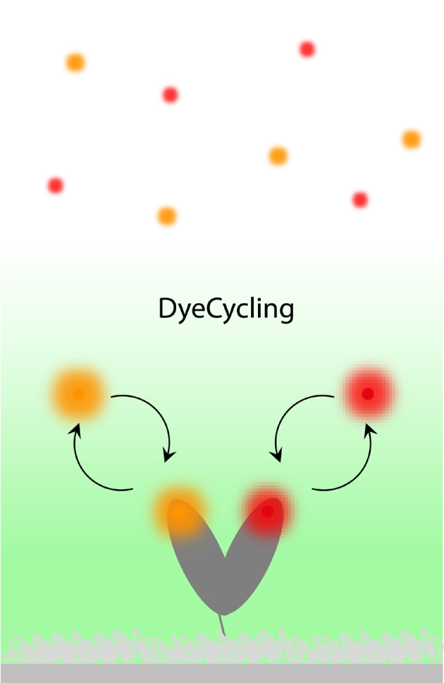
Biomolecular systems, such as proteins, crucially rely on dynamic processes at the nanoscale. Detecting biomolecular nanodynamics is therefore key to obtaining a mechanistic understanding of the energies and molecular driving forces that control biomolecular systems. Single-molecule fluorescence resonance energy transfer (smFRET) is a powerful technique to observe in real-time how a single biomolecule proceeds through its functional cycle involving a sequence of distinct structural states. Currently, this technique is fundamentally limited by irreversible photobleaching, causing the untimely end of the experiment and thus, a narrow temporal bandwidth of ≤ 3 orders of magnitude. Here, we introduce “DyeCycling”, a measurement scheme with which we aim to break the photobleaching limit in smFRET. We introduce the concept of spontaneous dye replacement by simulations, and as an experimental proof-of-concept, we demonstrate the intermittent observation of a single biomolecule for one hour with a time resolution of milliseconds. Theoretically, DyeCycling can provide > 100-fold more information per single molecule than conventional smFRET. We discuss the experimental implementation of DyeCycling, its current and fundamental limitations, and specific biological use cases. Given its general simplicity and versatility, DyeCycling has the potential to revolutionize the field of time-resolved smFRET, where it may serve to unravel a wealth of biomolecular dynamics by bridging from milliseconds to the hour range.
Electronic Supplementary Material
Supplementary material is available for this article at 10.1007/s12274-022-4420-5 and is accessible for authorized users.
Keywords: biomolecular dynamics, single-molecule fluorescence resonance energy transfer (smFRET), photobleaching, conformational changes, single-molecule kinetics
Electronic Supplementary Material
Can DyeCycling break the photobleaching limit in single-molecule FRET?
Acknowledgements
We thank Chirlmin Joo, Johannes Hohlbein, Mattia Fontana, and Abbas Jabermoradi for helpful discussions before and during this project. We thank John Philippi for support with electronic triggering. We thank Johannes Hohlbein, Mattia Fontana, Katarzyna Tych, Mahipal Ganji, and David Dulin for helpful comments on the manuscript. Parts of the analysis code used herein were co-developed in Thorsten Hugel’s lab by previous lab members (including SS). We are thankful for this contribution.
References
- [1].California Institute of Technology. The Feynman Lectures on Physics [Online]. https://www.feynmanlectures.caltech.edu (accessed Jan 31, 2022).
- [2].Hellenkamp B, Schmid S, Doroshenko O, Opanasyuk O, Kühnemuth R, Adariani S R, Ambrose B, Aznauryan M, Barth A, Birkedal V, et al. Precision and accuracy of single-molecule FRET measurements—A multi-laboratory benchmark study. Nat. Methods. 2018;15:669–676. doi: 10.1038/s41592-018-0085-0. [DOI] [PMC free article] [PubMed] [Google Scholar]
- [3].Lerner E, Cordes T, Ingargiola A, Alhadid Y, Chung S, Michalet X, Weiss S. Toward dynamic structural biology: Two decades of single-molecule förster resonance energy transfer. Science. 2018;359:eaan1133. doi: 10.1126/science.aan1133. [DOI] [PMC free article] [PubMed] [Google Scholar]
- [4].Lerner E, Barth A, Hendrix J, Ambrose B, Birkedal V, Blanchard S C, Börner R, Chung H S, Cordes T, Craggs T D, et al. FRET-based dynamic structural biology: Challenges, perspectives and an appeal for open-science practices. eLife. 2021;10:e60416. doi: 10.7554/eLife.60416. [DOI] [PMC free article] [PubMed] [Google Scholar]
- [5].Hohng S, Wilson T J, Tan E, Clegg R M, Lilley D M J, Ha T. Conformational flexibility of four-way junctions in RNA. J. Mol. Biol. 2004;336:69–79. doi: 10.1016/j.jmb.2003.12.014. [DOI] [PubMed] [Google Scholar]
- [6].Joo C, McKinney S A, Nakamura M, Rasnik I, Myong S, Ha T. Real-time observation of RecA filament dynamics with single monomer resolution. Cell. 2006;126:515–527. doi: 10.1016/j.cell.2006.06.042. [DOI] [PubMed] [Google Scholar]
- [7].Feldman M B, Terry D S, Altman R B, Blanchard S C. Aminoglycoside activity observed on single pre-translocation ribosome complexes. Nat. Chem. Biol. 2010;6:54–62. doi: 10.1038/nchembio.274. [DOI] [PMC free article] [PubMed] [Google Scholar]
- [8].Ruer M, Krainer G, Gröger P, Schlierf M. ATPase and protease domain movements in the bacterial AAA+ protease FtsH are driven by thermal fluctuations. J. Mol. Biol. 2018;430:4592–4602. doi: 10.1016/j.jmb.2018.07.023. [DOI] [PubMed] [Google Scholar]
- [9].Kilic S, Felekyan S, Doroshenko O, Boichenko I, Dimura M, Vardanyan H, Bryan L C, Arya G, Seidel C A M, Fierz B. Single-molecule FRET reveals multiscale chromatin dynamics modulated by HP1α. Nat. Commun. 2018;9:235. doi: 10.1038/s41467-017-02619-5. [DOI] [PMC free article] [PubMed] [Google Scholar]
- [10].Zosel F, Mercadante D, Nettels D, Schuler B. A proline switch explains kinetic heterogeneity in a coupled folding and binding reaction. Nat. Commun. 2018;9:3332. doi: 10.1038/s41467-018-05725-0. [DOI] [PMC free article] [PubMed] [Google Scholar]
- [11].Choi J, Marks J, Zhang J J, Chen D H, Wang J F, Vázquez-Laslop N, Mankin A S, Puglisi J D. Dynamics of the context-specific translation arrest by chloramphenicol and linezolid. Nat. Chem. Biol. 2020;16:310–317. doi: 10.1038/s41589-019-0423-2. [DOI] [PMC free article] [PubMed] [Google Scholar]
- [12].Schmid S, Hugel T. Controlling protein function by fine-tuning conformational flexibility. eLife. 2020;9:e57180. doi: 10.7554/eLife.57180. [DOI] [PMC free article] [PubMed] [Google Scholar]
- [13].Bauer B W, Davidson I F, Canena D, Wutz G, Tang W, Litos G, Horn S, Hinterdorfer P, Peters J M. Cohesin mediates DNA loop extrusion by a “swing and clamp” mechanism. Cell. 2021;184:5448–5464. doi: 10.1016/j.cell.2021.09.016. [DOI] [PMC free article] [PubMed] [Google Scholar]
- [14].Mazumder A, Wang A N, Uhm H, Ebright R H, Kapanidis A N. RNA polymerase clamp conformational dynamics: Long-lived states and modulation by crowding, cations, and nonspecific DNA binding. Nucleic Acids Res. 2021;49:2790–2802. doi: 10.1093/nar/gkab074. [DOI] [PMC free article] [PubMed] [Google Scholar]
- [15].Teilum K, Olsen J G, Kragelund B B. Functional aspects of protein flexibility. Cell. Mol. Life Sci. 2009;66:2231–2247. doi: 10.1007/s00018-009-0014-6. [DOI] [PMC free article] [PubMed] [Google Scholar]
- [16].Burley S K, Bhikadiya C, Bi C X, Bittrich S, Chen L, Crichlow G V, Duarte J M, Dutta S, Fayazi M, Feng Z K, et al. RCSB protein data bank: Celebrating 50 years of the PDB with new tools for understanding and visualizing biological macromolecules in 3D. Protein Sci. 2022;31:187–208. doi: 10.1002/pro.4213. [DOI] [PMC free article] [PubMed] [Google Scholar]
- [17].Jumper J, Evans R, Pritzel A, Green T, Figurnov M, Ronneberger O, Tunyasuvunakool K, Bates R, Žídek A, Potapenko A, et al. Highly accurate protein structure prediction with AlphaFold. Nature. 2021;596:583–589. doi: 10.1038/s41586-021-03819-2. [DOI] [PMC free article] [PubMed] [Google Scholar]
- [18].Varadi M, Anyango S, Deshpande M, Nair S, Natassia C, Yordanova G, Yuan D, Stroe O, Wood G, Laydon A, et al. AlphaFold protein structure database: Massively expanding the structural coverage of protein-sequence space with high-accuracy models. Nucleic Acids Res. 2022;50:D439–D444. doi: 10.1093/nar/gkab1061. [DOI] [PMC free article] [PubMed] [Google Scholar]
- [19].Vallat B, Webb B, Westbrook J D, Sali A, Berman H M. Development of a prototype system for archiving integrative/hybrid structure models of biological macromolecules. Structure. 2018;26:894–904. doi: 10.1016/j.str.2018.03.011. [DOI] [PMC free article] [PubMed] [Google Scholar]
- [20].Henzler-Wildman K A, Lei M, Thai V, Kerns S J, Karplus M, Kern D. A hierarchy of timescales in protein dynamics is linked to enzyme catalysis. Nature. 2007;450:913–916. doi: 10.1038/nature06407. [DOI] [PubMed] [Google Scholar]
- [21].Hellenkamp B, Wortmann P, Kandzia F, Zacharias M, Hugel T. Multidomain structure and correlated dynamics determined by self-consistent FRET networks. Nat. Methods. 2017;14:174–180. doi: 10.1038/nmeth.4081. [DOI] [PMC free article] [PubMed] [Google Scholar]
- [22].Geeves M A, Holmes K C. Structural mechanism of muscle contraction. Annu. Rev. Biochem. 1999;68:687–728. doi: 10.1146/annurev.biochem.68.1.687. [DOI] [PubMed] [Google Scholar]
- [23].Kodera N, Yamamoto D, Ishikawa R, Ando T. Video imaging of walking myosin V by high-speed atomic force microscopy. Nature. 2010;468:72–76. doi: 10.1038/nature09450. [DOI] [PubMed] [Google Scholar]
- [24].Ernst O P, Lodowski D T, Elstner M, Hegemann P, Brown L S, Kandori H. Microbial and animal rhodopsins: Structures, functions, and molecular mechanisms. Chem. Rev. 2014;114:126–163. doi: 10.1021/cr4003769. [DOI] [PMC free article] [PubMed] [Google Scholar]
- [25].Fields R D, Burnstock G. Purinergic signalling in neuron—glia interactions. Nat. Rev. Neurosci. 2006;7:423–436. doi: 10.1038/nrn1928. [DOI] [PMC free article] [PubMed] [Google Scholar]
- [26].Henzler-Wildman K, Kern D. Dynamic personalities of proteins. Nature. 2007;450:964–972. doi: 10.1038/nature06522. [DOI] [PubMed] [Google Scholar]
- [27].Ode H, Nakashima M, Kitamura S, Sugiura W, Sato H. Molecular dynamics simulation in virus research. Front. Microbiol. 2012;3:258. doi: 10.3389/fmicb.2012.00258. [DOI] [PMC free article] [PubMed] [Google Scholar]
- [28].Mitra K, Frank J. Ribosome dynamics: Insights from atomic structure modeling into cryo-electron microscopy maps. Annu. Rev. Biophys. Biomol. Struct. 2006;35:299–317. doi: 10.1146/annurev.biophys.35.040405.101950. [DOI] [PubMed] [Google Scholar]
- [29].Tan J Z, Verschueren K H G, Anand K, Shen J H, Yang M J, Xu Y C, Rao Z H, Bigalke J, Heisen B, Mesters J R, et al. pH-dependent conformational flexibility of the SARS-CoV main proteinase (Mpro) dimer: Molecular dynamics simulations and multiple X-ray structure analyses. J. Mol. Biol. 2005;354:25–40. doi: 10.1016/j.jmb.2005.09.012. [DOI] [PMC free article] [PubMed] [Google Scholar]
- [30].Santoso Y, Joyce C M, Potapova O, Le Reste L, Hohlbein J, Torella J P, Grindley N D F, Kapanidis A N. Conformational transitions in DNA polymerase I revealed by single-molecule FRET. Proc. Natl. Acad. Sci. USA. 2010;107:715–720. doi: 10.1073/pnas.0910909107. [DOI] [PMC free article] [PubMed] [Google Scholar]
- [31].Nüesch M F, Ivanovć M T, Claude J B, Nettels D, Best R B, Wenger J, Schuler B. Single-molecule detection of ultrafast biomolecular dynamics with nanophotonics. J. Am. Chem. Soc. 2022;144:52–56. doi: 10.1021/jacs.1c09387. [DOI] [PubMed] [Google Scholar]
- [32].McKinney S A, Joo C, Ha T. Analysis of single-molecule FRET trajectories using hidden markov modeling. Biophys. J. 2006;91:1941–1951. doi: 10.1529/biophysj.106.082487. [DOI] [PMC free article] [PubMed] [Google Scholar]
- [33].Schmid S, Götz M, Hugel T. Single-molecule analysis beyond dwell times: Demonstration and assessment in and out of equilibrium. Biophys. J. 2016;111:1375–1384. doi: 10.1016/j.bpj.2016.08.023. [DOI] [PMC free article] [PubMed] [Google Scholar]
- [34].Götz, M.; Barth, A.; Bohr, S. S. R.; Börner, R.; Chen, J. X.; Cordes, T.; Erie, D. A.; Gebhardt, C.; Hadzic, M. C. A. S.; Hamilton, G. L. et al. Inferring kinetic rate constants from single-molecule FRET trajectories-a blind benchmark of kinetic analysis tools. 2021, bioRxiv 2021.11.23.469671. https://www.biorxiv.org/content/10.1101/2021.11.23.469671v2.article-info (accessed Jan 5, 2022). [DOI] [PMC free article] [PubMed]
- [35].Miller H, Zhou Z K, Shepherd J, Wollman A J M, Leake M C. Single-molecule techniques in biophysics: A review of the progress in methods and applications. Rep. Prog. Phys. 2018;81:024601. doi: 10.1088/1361-6633/aa8a02. [DOI] [PubMed] [Google Scholar]
- [36].Okumus B, Wilson T J, Lilley D M J, Ha T. Vesicle encapsulation studies reveal that single molecule ribozyme heterogeneities are intrinsic. Biophys. J. 2004;87:2798–2806. doi: 10.1529/biophysj.104.045971. [DOI] [PMC free article] [PubMed] [Google Scholar]
- [37].Ha T, Tinnefeld P. Photophysics of fluorescent probes for single-molecule biophysics and super-resolution imaging. Annu. Rev. Phys. Chem. 2012;63:595–617. doi: 10.1146/annurev-physchem-032210-103340. [DOI] [PMC free article] [PubMed] [Google Scholar]
- [38].Schmid S, Hugel T. Efficient use of single molecule time traces to resolve kinetic rates, models and uncertainties. J. Chem. Phys. 2018;148:123312. doi: 10.1063/1.5006604. [DOI] [PMC free article] [PubMed] [Google Scholar]
- [39].Nettels D, Hoffmann A, Schuler B. Unfolded protein and peptide dynamics investigated with single-molecule FRET and correlation spectroscopy from picoseconds to seconds. J. Phys. Chem. B. 2008;112:6137–6146. doi: 10.1021/jp076971j. [DOI] [PubMed] [Google Scholar]
- [40].Juette M F, Terry D S, Wasserman M R, Altman R B, Zhou Z, Zhao H, Blanchard S C. Single-molecule imaging of non-equilibrium molecular ensembles on the millisecond timescale. Nat. Methods. 2016;13:341–344. doi: 10.1038/nmeth.3769. [DOI] [PMC free article] [PubMed] [Google Scholar]
- [41].Zheng Q S, Juette M F, Jockusch S, Wasserman M R, Zhou Z, Altman R B, Blanchard S C. Ultra-stable organic fluorophores for single-molecule research. Chem. Soc. Rev. 2014;43:1044–1056. doi: 10.1039/C3CS60237K. [DOI] [PMC free article] [PubMed] [Google Scholar]
- [42].Zheng Q S, Lavis L D. Development of photostable fluorophores for molecular imaging. Curr. Opin. Chem. Biol. 2017;39:32–38. doi: 10.1016/j.cbpa.2017.04.017. [DOI] [PubMed] [Google Scholar]
- [43].Isselstein M, Zhang L, Glembockyte V, Brix O, Cosa G, Tinnefeld P, Cordes T. Self-healing dyes-keeping the promise. J. Phys. Chem. Lett. 2020;11:4462–4480. doi: 10.1021/acs.jpclett.9b03833. [DOI] [PubMed] [Google Scholar]
- [44].Grimm J B, Xie L Q, Casler J C, Patel R, Tkachuk A N, Falco N, Choi H, Lippincott-Schwartz J, Brown T A, Glick B S, et al. A general method to improve fluorophores using deuterated auxochromes. JACS Au. 2021;1:690–696. doi: 10.1021/jacsau.1c00006. [DOI] [PMC free article] [PubMed] [Google Scholar]
- [45].Smit J H, Van Der Velde J H M, Huang J Y, Trauschke V, Henrikus S S, Chen S, Eleftheriadis N, Warszawik E M, Herrmann A, Cordes T. On the impact of competing intra- and intermolecular triplet-state quenching on photobleaching and photoswitching kinetics of organic fluorophores. Phys. Chem. Chem. Phys. 2019;21:3721–3733. doi: 10.1039/C8CP05063E. [DOI] [PubMed] [Google Scholar]
- [46].Frauenfelder H, Sligar S G, Wolynes P G. The energy landscapes and motions of proteins. Science. 1991;254:1598–1603. doi: 10.1126/science.1749933. [DOI] [PubMed] [Google Scholar]
- [47].Hyeon C, Lee J, Yoon J, Hohng S, Thirumalai D. Hidden complexity in the isomerization dynamics of Holliday junctions. Nat. Chem. 2012;4:907–914. doi: 10.1038/nchem.1463. [DOI] [PubMed] [Google Scholar]
- [48].Liu B, Baskin R J, Kowalczykowski S C. DNA unwinding heterogeneity by RecBCD results from static molecules able to equilibrate. Nature. 2013;500:482–485. doi: 10.1038/nature12333. [DOI] [PMC free article] [PubMed] [Google Scholar]
- [49].Zhuang X W, Kim H, Pereira M J B, Babcock H P, Walter N G, Chu S. Correlating structural dynamics and function in single ribozyme molecules. Science. 2002;296:1473–1476. doi: 10.1126/science.1069013. [DOI] [PubMed] [Google Scholar]
- [50].Solomatin S V, Greenfeld M, Chu S, Herschlag D. Multiple native states reveal persistent ruggedness of an RNA folding landscape. Nature. 2010;463:681–684. doi: 10.1038/nature08717. [DOI] [PMC free article] [PubMed] [Google Scholar]
- [51].Ditzler M A, Rueda D, Mo J J, Håkansson K, Walter N G. A rugged free energy landscape separates multiple functional RNA folds throughout denaturation. Nucleic Acids Res. 2008;36:7088–7099. doi: 10.1093/nar/gkn871. [DOI] [PMC free article] [PubMed] [Google Scholar]
- [52].Sharonov A, Hochstrasser R M. Wide-field subdiffraction imaging by accumulated binding of diffusing probes. Proc. Natl. Acad. Sci. USA. 2006;103:18911–18916. doi: 10.1073/pnas.0609643104. [DOI] [PMC free article] [PubMed] [Google Scholar]
- [53].Jungmann R, Steinhauer C, Scheible M, Kuzyk A, Tinnefeld P, Simmel F C. Single-molecule kinetics and super-resolution microscopy by fluorescence imaging of transient binding on DNA origami. Nano Lett. 2010;10:4756–4761. doi: 10.1021/nl103427w. [DOI] [PubMed] [Google Scholar]
- [54].Auer A, Strauss M T, Schlichthaerle T, Jungmann R. Fast, background-free DNA-PAINT imaging using FRET-based probes. Nano Lett. 2017;17:6428–6434. doi: 10.1021/acs.nanolett.7b03425. [DOI] [PubMed] [Google Scholar]
- [55].Filius M, Kim S H, Severins I, Joo C. High-resolution single-molecule FRET via DNA eXchange (FRET X) Nano Lett. 2021;21:3295–3301. doi: 10.1021/acs.nanolett.1c00725. [DOI] [PMC free article] [PubMed] [Google Scholar]
- [56].Stehr F, Stein J, Bauer J, Niederauer C, Jungmann R, Ganzinger K, Schwille P. Tracking single particles for hours via continuous DNA-mediated fluorophore exchange. Nat. Commun. 2021;12:4432. doi: 10.1038/s41467-021-24223-4. [DOI] [PMC free article] [PubMed] [Google Scholar]
- [57].Dupuis N F, Holmstrom E D, Nesbitt D J. Single-molecule kinetics reveal cation-promoted DNA duplex formation through ordering of single-stranded helices. Biophys. J. 2013;105:756–766. doi: 10.1016/j.bpj.2013.05.061. [DOI] [PMC free article] [PubMed] [Google Scholar]
- [58].Ouldridge T E, Šulc P, Romano F, Doye J P K, Louis A A. DNA hybridization kinetics: zippering, internal displacement and sequence dependence. Nucleic Acids Res. 2013;418886:8886–8895. doi: 10.1093/nar/gkt687. [DOI] [PMC free article] [PubMed] [Google Scholar]
- [59].Swenson C S, Lackey H H, Reece E J, Harris J M, Heemstra J M, Peterson E M. Evaluating the effect of ionic strength on PNA: DNA duplex formation kinetics. RSC Chem. Biol. 2021;2:1249–1256. doi: 10.1039/D1CB00025J. [DOI] [PMC free article] [PubMed] [Google Scholar]
- [60].Schueder F, Stein J, Stehr F, Auer A, Sperl B, Strauss M T, Schwille P, Jungmann R. An order of magnitude faster DNA-PAINT imaging by optimized sequence design and buffer conditions. Nat. Methods. 2019;16:1101–1104. doi: 10.1038/s41592-019-0584-7. [DOI] [PubMed] [Google Scholar]
- [61].Andrews R. DNA hybridisation kinetics using single-molecule fluorescence imaging. Essays Biochem. 2021;65:27–36. doi: 10.1042/EBC20200040. [DOI] [PMC free article] [PubMed] [Google Scholar]
- [62].Madsen M, Gothelf K V. Chemistries for DNA nanotechnology. Chem. Rev. 2019;119:6384–6458. doi: 10.1021/acs.chemrev.8b00570. [DOI] [PubMed] [Google Scholar]
- [63].Egholm M, Buchardt O, Christensen L, Behrens C, Freier S M, Driver D A, Berg R H, Kim S K, Norden B, Nielsen P E. PNA hybridizes to complementary oligonucleotides obeying the Watson—Crick hydrogen-bonding rules. Nature. 1993;365:566–568. doi: 10.1038/365566a0. [DOI] [PubMed] [Google Scholar]
- [64].Saarbach J, Sabale P M, Winssinger N. Peptide nucleic acid (PNA) and its applications in chemical biology, diagnostics, and therapeutics. Curr. Opin. Chem. Biol. 2019;52:112–124. doi: 10.1016/j.cbpa.2019.06.006. [DOI] [PubMed] [Google Scholar]
- [65].Gavins G C, Gröger K, Bartoschek M D, Wolf P, Beck-Sickinger A G, Bultmann S, Seitz O. Live cell PNA labelling enables erasable fluorescence imaging of membrane proteins. Nat. Chem. 2021;13:15–23. doi: 10.1038/s41557-020-00584-z. [DOI] [PubMed] [Google Scholar]
- [66].Biomers. net. PNA Oligomers [Online], https://www.biomers.net/en/products/PNA_Oligomers.html (accessed Jan 31, 2022).
- [67].Panagene. Custom PNA oligonucleotide synthesis [Online]. http://www.panagene.com/_ENG/html/ (accessed Jan 31, 2022).
- [68].Wiita A P, Ainavarapu S R K, Huang H H, Fernandez J M. Force-dependent chemical kinetics of disulfide bond reduction observed with single-molecule techniques. Proc. Natl. Acad. Sci. USA. 2006;103:7222–7227. doi: 10.1073/pnas.0511035103. [DOI] [PMC free article] [PubMed] [Google Scholar]
- [69].Vincent S, Subramanian S, Vollmer F. Optoplasmonic characterisation of reversible disulfide interactions at single thiol sites in the attomolar regime. Nat. Commun. 2020;11:2043. doi: 10.1038/s41467-020-15822-8. [DOI] [PMC free article] [PubMed] [Google Scholar]
- [70].Lotze J, Reinhardt U, Seitz O, Beck-Sickinger A G. Peptidetags for site-specific protein labelling in vitro and in vivo. Mol. Biosyst. 2016;12:1731–1745. doi: 10.1039/C6MB00023A. [DOI] [PubMed] [Google Scholar]
- [71].Knecht S, Ricklin D, Eberle A N, Ernst B. Oligohis-tags: Mechanisms of binding to Ni2+-NTA surfaces. J. Mol. Recognit. 2009;22:270–279. doi: 10.1002/jmr.941. [DOI] [PubMed] [Google Scholar]
- [72].Day J W, Kim C H, Smider V V, Schultz P G. Identification of metal ion binding peptides containing unnatural amino acids by phage display. Bioorganic Med. Chem. Lett. 2013;23:2598–2600. doi: 10.1016/j.bmcl.2013.02.106. [DOI] [PubMed] [Google Scholar]
- [73].Ryu Y, Schultz P G. Efficient incorporation of unnatural amino acids into proteins in Escherichia coli. Nat. Methods. 2006;3:263–265. doi: 10.1038/nmeth864. [DOI] [PubMed] [Google Scholar]
- [74].Koehler C, Sauter P F, Wawryszyn M, Girona G E, Gupta K, Landry J J M, Fritz M H Y, Radic K, Hoffmann J E, Chen Z A, et al. Genetic code expansion for multiprotein complex engineering. Nat. Methods. 2016;13:997–1000. doi: 10.1038/nmeth.4032. [DOI] [PubMed] [Google Scholar]
- [75].Scinto S L, Bilodeau D A, Hincapie R, Lee W, Nguyen S S, Xu M H, am Ende C W, Finn M G, Lang K, Lin Q, et al. Bioorthogonal chemistry. Nat. Rev. Methods Prim. 2021;1:30. doi: 10.1038/s43586-021-00028-z. [DOI] [PMC free article] [PubMed] [Google Scholar]
- [76].Sindbert S, Kalinin S, Nguyen H, Kienzler A, Clima L, Bannwarth W, Appel B, Müller S, Seidel C A M. Accurate distance determination of nucleic acids via förster resonance energy transfer: Implications of dye linker length and rigidity. J. Am. Chem. Soc. 2011;133:2463–2480. doi: 10.1021/ja105725e. [DOI] [PubMed] [Google Scholar]
- [77].Roy R, Hohng S, Ha T. A practical guide to single-molecule FRET. Nat. Methods. 2008;5:507–516. doi: 10.1038/nmeth.1208. [DOI] [PMC free article] [PubMed] [Google Scholar]
- [78].Eid J, Fehr A, Gray J, Luong K, Lyle J, Otto G, Peluso P, Rank D, Baybayan P, Bettman B, et al. Real-time DNA sequencing from single polymerase molecules. Science. 2009;323:133–138. doi: 10.1126/science.1162986. [DOI] [PubMed] [Google Scholar]
- [79].Levene H J, Korlach J, Turner S W, Foquet M, Craighead H G, Webb W W. Zero-mode waveguides for single-molecule analysis at high concentrations. Science. 2003;299:682–686. doi: 10.1126/science.1079700. [DOI] [PubMed] [Google Scholar]
- [80].Klughammer N, Dekker C. Palladium zero-mode waveguides for optical single-molecule detection with nanopores. Nanotechnology. 2021;32:18LT01. doi: 10.1088/1361-6528/abd976. [DOI] [PubMed] [Google Scholar]
- [81].Jeffet J, Ionescu A, Michaeli Y, Torchinsky D, Perlson E, Craggs T D, Ebenstein Y. Multimodal single-molecule microscopy with continuously controlled spectral resolution. Biophys. Rep. 2021;1:100013. doi: 10.1016/j.bpr.2021.100013. [DOI] [PMC free article] [PubMed] [Google Scholar]
- [82].Farhangdoust F, Cheng F, Liang W T, Liu Y M, Wanunu M. Rapid identification of DNA fragments through direct sequencing with electro-optical zero-mode waveguides. Adv. Mater. 2022;34:e2108479. doi: 10.1002/adma.202108479. [DOI] [PMC free article] [PubMed] [Google Scholar]
- [83].McKinney S A, Déclais A C, Lilley D M J, Ha T. Structural dynamics of individual Holliday junctions. Nat. Struct. Biol. 2003;10:93–97. doi: 10.1038/nsb883. [DOI] [PubMed] [Google Scholar]
- [84].Samiee K T, Moran-Mirabal J M, Cheung Y K, Craighead H G. Zero mode waveguides for single-molecule spectroscopy on lipid membranes. Biophys. J. 2006;90:3288–3299. doi: 10.1529/biophysj.105.072819. [DOI] [PMC free article] [PubMed] [Google Scholar]
- [85].Levitus M, Ranjit S. Cyanine dyes in biophysical research: The photophysics of polymethine fluorescent dyes in biomolecular environments. Q. Rev. Biophys. 2011;44:123–151. doi: 10.1017/S0033583510000247. [DOI] [PubMed] [Google Scholar]
- [86].Chandradoss, S. D.; Haagsma, A. C.; Lee, Y. K.; Hwang, J. H.; Nam, J. M.; Joo, C. Surface passivation for single-molecule protein studies. J. Vis. Exp.2014, 50549. [DOI] [PMC free article] [PubMed]
- [87].Boukobza E, Sonnenfeld A, Haran G. Immobilization in surface-tethered lipid vesicles as a new tool for single biomolecule spectroscopy. J. Phys. Chem. B. 2001;105:12165–12170. doi: 10.1021/jp012016x. [DOI] [Google Scholar]
- [88].Mets Ü, Rigler R. Submillisecond detection of single rhodamine molecules in water. J. Fluoresc. 1994;4:259–264. doi: 10.1007/BF01878461. [DOI] [PubMed] [Google Scholar]
- [89].Wayment J R, Harris J M. Biotin-avidin binding kinetics measured by single-molecule imaging. Anal. Chem. 2009;81:336–342. doi: 10.1021/ac801818t. [DOI] [PubMed] [Google Scholar]
- [90].Joo C, McKinney S A, Lilley D M J, Ha T. Exploring rare conformational species and ionic effects in DNA Holliday junctions using single-molecule spectroscopy. J. Mol. Biol. 2004;341:739–751. doi: 10.1016/j.jmb.2004.06.024. [DOI] [PubMed] [Google Scholar]
- [91].Evans G W, Hohlbein J, Craggs T, Aigrain L, Kapanidis A N. Real-time single-molecule studies of the motions of DNA polymerase fingers illuminate DNA synthesis mechanisms. Nucleic Acids Res. 2015;43:5998–6008. doi: 10.1093/nar/gkv547. [DOI] [PMC free article] [PubMed] [Google Scholar]
- [92].Rasnik I, McKinney S A, Ha T. Nonblinking and long-lasting single-molecule fluorescence imaging. Nat. Methods. 2006;3:891–893. doi: 10.1038/nmeth934. [DOI] [PubMed] [Google Scholar]
- [93].Farooq S, Hohlbein J. Camera-based single-molecule FRET detection with improved time resolution. Phys. Chem. Chem. Phys. 2015;17:27862–27872. doi: 10.1039/C5CP04137F. [DOI] [PubMed] [Google Scholar]
- [94].Kapanidis A N, Lee N K, Laurence T A, Doose S, Margeat E, Weiss S. Fluorescence-aided molecule sorting: Analysis of structure and interactions by alternating-laser excitation of single molecules. Proc. Natl. Acad. Sci. USA. 2004;101:8936–8941. doi: 10.1073/pnas.0401690101. [DOI] [PMC free article] [PubMed] [Google Scholar]
- [95].Laine R F, Tosheva K L, Gustafsson N, Gray R D M, Almada P, Albrecht D, Risa G T, Hurtig F, Lindås A C, Baum B, et al. NanoJ: A high-performance open-source superresolution microscopy toolbox. J. Phys. D:Appl. Phys. 2019;52:163001. doi: 10.1088/1361-6463/ab0261. [DOI] [PMC free article] [PubMed] [Google Scholar]
- [96].Ryu J K, Rah S H, Janissen R, Kerssemakers J W J, Bonato A, Michieletto D, Dekker C. Condensin extrudes DNA loops in steps up to hundreds of base pairs that are generated by ATP binding events. Nucleic Acids Res. 2022;50:820–832. doi: 10.1093/nar/gkab1268. [DOI] [PMC free article] [PubMed] [Google Scholar]
- [97].Barnes C O, Calero M, Malik I, Graham B W, Spahr H, Lin G W, Cohen A E, Brown I S, Zhang Q M, Pullara F, et al. Crystal structure of a transcribing RNA polymerase II complex reveals a complete transcription bubble. Mol. Cell. 2015;59:258–269. doi: 10.1016/j.molcel.2015.06.034. [DOI] [PMC free article] [PubMed] [Google Scholar]
- [98].Lerner E, Chung S, Allen B L, Wang S, Lee J, Lu S W, Grimaud L W, Ingargiola A, Michalet X, Alhadid Y, et al. Backtracked and paused transcription initiation intermediate of Escherichia coli RNA polymerase. Proc. Natl. Acad. Sci. USA. 2016;113:E6562–E6571. doi: 10.1073/pnas.1605038113. [DOI] [PMC free article] [PubMed] [Google Scholar]
- [99].Dulin D, Bauer D L V, Malinen A M, Bakermans J J W, Kaller M, Morichaud Z, Petushkov I, Depken M, Brodolin K, Kulbachinskiy A, et al. Pausing controls branching between productive and non-productive pathways during initial transcription in bacteria. Nat. Commun. 2018;9:1478. doi: 10.1038/s41467-018-03902-9. [DOI] [PMC free article] [PubMed] [Google Scholar]
- [100].Mazumder A, Kapanidis A N. Recent advances in understanding σ70-dependent transcription initiation mechanisms. J. Mol. Biol. 2019;431:3947–3959. doi: 10.1016/j.jmb.2019.04.046. [DOI] [PMC free article] [PubMed] [Google Scholar]
- [101].Landick R. Transcriptional pausing as a mediator of bacterial gene regulation. Annu. Rev. Microbiol. 2021;75:291–314. doi: 10.1146/annurev-micro-051721-043826. [DOI] [PubMed] [Google Scholar]
- [102].Janissen R, Eslami-Mossallam B, Artsimovitch I, Depken M, Dekker N H. A unifying mechanistic model of bacterial transcription with three interconnected pause states and non-diffusive backtrack recovery. Biophys. J. 2020;118:543a. doi: 10.1016/j.bpj.2019.11.2974. [DOI] [Google Scholar]
- [103].Jamiolkowski R M, Chen C L, Cooperman B S, Goldman Y E. tRNA fluctuations observed on stalled ribosomes are suppressed during ongoing protein synthesis. Biophys. J. 2017;113:2326–2335. doi: 10.1016/j.bpj.2017.08.052. [DOI] [PMC free article] [PubMed] [Google Scholar]
- [104].Morse J C, Girodat D, Burnett B J, Holm M, Altman R B, Sanbonmatsu K Y, Wieden H J, Blanchard S C. Elongation factor-Tu can repetitively engage aminoacyl-tRNA within the ribosome during the proofreading stage of tRNA selection. Proc. Natl. Acad. Sci. USA. 2020;117:3610–3620. doi: 10.1073/pnas.1904469117. [DOI] [PMC free article] [PubMed] [Google Scholar]
- [105].Lapointe C P, Grosely R, Johnson A G, Wang J F, Fernández I S, Puglisi J D. Dynamic competition between SARS-CoV-2 NSP1 and mRNA on the human ribosome inhibits translation initiation. Proc. Natl. Acad. Sci. USA. 2021;118:e2017715118. doi: 10.1073/pnas.2017715118. [DOI] [PMC free article] [PubMed] [Google Scholar]
- [106].Aunins T R, Marsh K A, Subramanya G, Uprichard S L, Perelson A S, Chatterjee A. Intracellular hepatitis C virus modeling predicts infection dynamics and viral protein mechanisms. J. Virol. 2018;92:e02098–17. doi: 10.1128/JVI.02098-17. [DOI] [PMC free article] [PubMed] [Google Scholar]
- [107].Yu J, Xiao J, Ren X J, Lao K Q, Xie X S. Probing gene expression in live cells, one protein molecule at a time. Science. 2006;311:1600–1603. doi: 10.1126/science.1119623. [DOI] [PubMed] [Google Scholar]
- [108].Biebl M M, Buchner J. Structure, function, and regulation of the Hsp90 machinery. Cold Spring Harb. Perspect. Biol. 2019;11:a034017. doi: 10.1101/cshperspect.a034017. [DOI] [PMC free article] [PubMed] [Google Scholar]
- [109].Taipale M, Jarosz D F, Lindquist S. HSP90 at the hub of protein homeostasis: Emerging mechanistic insights. Nat. Rev. Mol. Cell Biol. 2010;11:515–528. doi: 10.1038/nrm2918. [DOI] [PubMed] [Google Scholar]
- [110].Picard, D. Curated list of Hsp90 Interactors [Online]. https://www.picard.ch/downloads/Hsp90interactors.pdf (accessed Jan 31, 2022).
- [111].Whitesell L, Lindquist S L. Hsp90 and the chaperoning of cancer. Nat. Rev. Cancer. 2005;5:761–772. doi: 10.1038/nrc1716. [DOI] [PubMed] [Google Scholar]
- [112].Blagg B S J, Kerr T D. Hsp90 inhibitors: Small molecules that transform the Hsp90 protein folding machinery into a catalyst for protein degradation. Med. Res. Rev. 2006;26:310–338. doi: 10.1002/med.20052. [DOI] [PubMed] [Google Scholar]
- [113].Burlison J A, Neckers L, Smith A B, Maxwell A, Blagg B S J. Novobiocin: Redesigning a DNA gyrase inhibitor for selective inhibition of Hsp90. J. Am. Chem. Soc. 2006;128:15529–15536. doi: 10.1021/ja065793p. [DOI] [PubMed] [Google Scholar]
- [114].Joshi S, Wang T, Araujo T L S, Sharma S, Brodsky J L, Chiosis G. Adapting to stress—chaperome networks in cancer. Nat. Rev. Cancer. 2018;18:562–575. doi: 10.1038/s41568-018-0020-9. [DOI] [PMC free article] [PubMed] [Google Scholar]
- [115].Mishra S J, Liu W Y, Beebe K, Banerjee M, Kent C N, Munthali V, Koren J, III, Taylor J A, III, Neckers L M, Holzbeierlein J, et al. The development of Hsp90β-selective inhibitors to overcome detriments associated with pan-Hsp90 inhibition. J. Med. Chem. 2021;64:1545–1557. doi: 10.1021/acs.jmedchem.0c01700. [DOI] [PMC free article] [PubMed] [Google Scholar]
- [116].Pearl L H. Review: The HSP90 molecular chaperone—An enigmatic ATPase. Biopolymers. 2016;105:594–607. doi: 10.1002/bip.22835. [DOI] [PMC free article] [PubMed] [Google Scholar]
- [117].Wang R Y R, Noddings C M, Kirschke E, Myasnikov A G, Johnson J L, Agard D A. Structure of Hsp90-Hsp70-Hop-GR reveals the Hsp90 client-loading mechanism. Nature. 2022;601:460–464. doi: 10.1038/s41586-021-04252-1. [DOI] [PMC free article] [PubMed] [Google Scholar]
- [118].Noddings C M, Wang R Y R, Johnson J L, Agard D A. Structure of Hsp90-p23-GR reveals the Hsp90 client-remodelling mechanism. Nature. 2022;601:465–469. doi: 10.1038/s41586-021-04236-1. [DOI] [PMC free article] [PubMed] [Google Scholar]
- [119].Bhattacharya K, Weidenauer L, Luengo T M, Pieters E C, Echeverría P C, Bernasconi L, Wider D, Sadian Y, Koopman M B, Villemin M, et al. The Hsp70-Hsp90 co-chaperone Hop/Stip1 shifts the proteostatic balance from folding towards degradation. Nat. Commun. 2020;11:5975. doi: 10.1038/s41467-020-19783-w. [DOI] [PMC free article] [PubMed] [Google Scholar]
- [120].Luengo T M, Kityk R, Mayer M P, Rüdiger S G D. Hsp90 breaks the deadlock of the Hsp70 chaperone system. Mol. Cell. 2018;70:545–552. doi: 10.1016/j.molcel.2018.03.028. [DOI] [PubMed] [Google Scholar]
- [121].Mickler M, Hessling M, Ratzke C, Buchner J, Hugel T. The large conformational changes of Hsp90 are only weakly coupled to ATP hydrolysis. Nat. Struct. Mol. Biol. 2009;16:281–286. doi: 10.1038/nsmb.1557. [DOI] [PubMed] [Google Scholar]
- [122].Uhlmann F. SMC complexes: From DNA to chromosomes. Nat. Rev. Mol. Cell Biol. 2016;17:399–412. doi: 10.1038/nrm.2016.30. [DOI] [PubMed] [Google Scholar]
- [123].Rowley M J, Corces V G. Organizational principles of 3D genome architecture. Nat. Rev. Genet. 2018;19:789–800. doi: 10.1038/s41576-018-0060-8. [DOI] [PMC free article] [PubMed] [Google Scholar]
- [124].Ganji M, Shaltiel I A, Bisht S, Kim E, Kalichava A, Haering C H, Dekker C. Real-time imaging of DNA loop extrusion by condensin. Science. 2018;360:102–105. doi: 10.1126/science.aar7831. [DOI] [PMC free article] [PubMed] [Google Scholar]
- [125].Kim E, Kerssemakers J, Shaltiel I A, Haering C H, Dekker C. DNA-loop extruding condensin complexes can traverse one another. Nature. 2020;579:438–442. doi: 10.1038/s41586-020-2067-5. [DOI] [PubMed] [Google Scholar]
- [126].Higashi T L, Pobegalov G, Tang M Z, Molodtsov M I, Uhlmann F. A brownian ratchet model for DNA loop extrusion by the cohesin complex. eLife. 2021;10:e67530. doi: 10.7554/eLife.67530. [DOI] [PMC free article] [PubMed] [Google Scholar]
- [127].Goodsell, D. PDB Molecule of the Month: RNA Polymerase [Online]. https://pdb101.rcsb.org/motm/40 (accessed Jan 27, 2022).
- [128].Rundlet E J, Holm M, Schacherl M, Natchiar S K, Altman R B, Spahn C M T, Myasnikov A G, Blanchard S C. Structural basis of early translocation events on the ribosome. Nature. 2021;595:741–745. doi: 10.1038/s41586-021-03713-x. [DOI] [PMC free article] [PubMed] [Google Scholar]
Associated Data
This section collects any data citations, data availability statements, or supplementary materials included in this article.
Supplementary Materials
Can DyeCycling break the photobleaching limit in single-molecule FRET?