Abstract
Background. FGFR1/2/3 fusions have been reported infrequently in aNSCLC, including as a rare, acquired resistance mechanism following treatment with EGFR TKIs. Data regarding their prevalence and therapeutic implications are limited. Methods. The Guardant Health (GH) electronic database (ED) was evaluated for cases of aNSCLC and FGFR2/3 fusions; FGFR2/3 fusion prevalence with and without a co-existing EGFR mutation was assessed. The ED of Tel-Aviv Sourasky Medical Center (TASMC, June 2020–June 2021) was evaluated for cases of aNSCLC and de novo FGFR1/2/3 fusions. Patients with EGFR mutant aNSCLC progressing on EGFR TKIs and developing an FGFR1/2/3 fusion were selected from the ED of Davidoff Cancer Center (DCC) and Oncology Department, Bnei-Zion hospital (BZ) (April 2014–April 2021). Clinicopathological characteristics, systemic therapies, and outcomes were assessed. Results. In the GH ED (n = 57,445), the prevalence of FGFR2 and FGFR3 fusions were 0.02% and 0.26%, respectively. FGFR3-TACC3 fusion predominated (91.5%). In 23.8% of cases, FGFR2/3 fusions co-existed with EGFR sensitizing mutations (exon 19 del, 64.1%; L858R, 33.3%, L861Q, 2.6%). Among samples with concurrent FGFR fusions and EGFR sensitizing mutations, 41.0% also included EGFR resistant mutations. In TASMC (n = 161), 1 case of de novo FGFR3-TACC3 fusion was detected (prevalence, 0.62%). Of three patients from DCC and BZ with FGFR3-TACC3 fusions following progression on EGFR TKIs, two received EGFR TKI plus erdafitinib, an FGFR TKI, with clinical benefit duration of 13.0 and 6.0 months, respectively. Conclusions. Over 23% of FGFR2/3 fusions in aNSCLC may be associated with acquired resistance following treatment with EGFR TKIs. In this clinical scenario, a combination of EGFR TKIs and FGFR TKIs represents a promising treatment strategy.
Keywords: FGFR, FGFR fusion, acquired resistance to EGFR TKIs, lung cancer, liquid biopsy, ctDNA
1. Introduction
Epidermal growth factor receptor tyrosine kinase inhibitors (EGFR TKIs) represent an extremely effective therapy option for a distinct yet substantial subset of patients with advanced non-small lung cancer (aNSCLC) harboring EGFR activating mutations. Recently, they have also demonstrated a benefit in the adjuvant setting [1]. Unfortunately, the development of acquired resistance during EGFR TKI treatment is inevitable [2]. The mechanisms of acquired resistance to EGFR TKIs are complex and heterogeneous and often involve the co-activation of several molecular pathways. The most common resistant mechanisms involve the development of secondary resistant mutations in the EGFR kinase domain, such as EGFR T790M in approximately half of the tumors following treatment with first- or second-generation EGFR TKIs or EGFR C797S following therapy with osimertinib, which occurs in 10–26% of cases [3,4]. Among the “oncogene kinase switch”-resistant mechanisms are tyrosine-protein kinase Met (cMET)-amplification, which occurs in 5–50% of cases, phosphatidylinositol-4,5-bisphosphate 3-kinase (PIK3CA) mutations (in 4–11% of cases), Kirsten rat sarcoma viral oncogene homolog (KRAS) mutations (in 2–8% of cases), v-raf murine sarcoma viral oncogene homolog B1 (BRAF) mutations (5–8% of cases) [5,6,7,8,9], receptor tyrosine kinase (RTK) fusions (e.g., rearranged during transfection (RET)—coiled-coil domain-containing protein 6 (CCDC6), anaplastic lymphoma kinase (ALK)—echinoderm microtubule-associated protein-like 4 (EML4), and fibroblast growth factor receptor 3 (FGFR3)—transforming acidic coiled-coil–containing protein 3 (TACC3) fusions (which occur in about 1% of cases each) [10,11]. Ou et al. reported five cases of FGFR3-TACC3 fusion co-existing with an activating EGFR mutation in the setting of acquired resistance to EGFR TKIs in a large database of aNSCLC tissue samples analyzed by a comprehensive next-generation sequencing platform [12]. Acquisition of tumor tissue is more challenging in the clinical scenario of acquired resistance following EGFR TKIs treatment, given the spatial and temporal heterogeneity of resistance mechanisms and the need to subject a patient to additional invasive procedure(s) to acquire a timely tissue specimen reflective of the genomic landscape at the time of progression. This is the clinical setup in which liquid biopsy may be of value, eliminating the need for repeated tissue biopsy and allowing for the detection of molecular resistance mechanisms.
Based on the preclinical data, FGFR tyrosine kinase inhibition (FGFR TKIs) leads to G2/M cell cycle blockade and cell proliferation arrest [13]. Two different FGFR TKIs were recently approved by the United States Food and Drug Administration Agency (US FDA) for clinical use in tumors harboring FGFR alterations: pemigatinib, for the treatment of advanced intrahepatic cholangiocarcinoma, and erdafitinib, for the treatment of refractory urothelial cancers [13,14]. The clinical data regarding FGFR TKIs as a treatment for FGFR-rearranged aNSCLC is limited. Twenty-four patients with aNSCLC (eight of those with an FGFR fusion) were included in a phase 1 trial of erdafitinib in patients with advanced or refractory solid tumors. The objective response rate with erdafitinib in 21 evaluable aNSCLC patients was 5% [15]. Additionally, one patient with an EGFR mutant aNSCLC that developed an FGFR3-TACC3 fusion following treatment with osimertinib has been reported. The patient received a combination of erdafitinib and osimertinib; the treatment was well tolerated, and a partial response was achieved [16].
We aimed to further explore the prevalence and therapeutic implications of FGFR fusions in aNSCLC, mainly focusing on the acquired resistance setting following treatment with EGFR TKIs.
2. Materials and Methods
2.1. Guardant Health (GH) Electronic Database (ED)
FGFR2/3 fusion frequency and EGFR mutation co-occurrence in plasma samples were calculated from the ED of Guardant360 CDx and Guardant360 LDT version 2.11 (genomic results from 57445 patients). Guardant360 is not designed to detect FGFR1 fusions. The database included results of plasma samples with successfully extracted circulating tumor deoxyribonucleic acid (ctDNA) from individuals with aNSCLC undergoing testing as part of their routine care. For the purposes of this analysis, all samples were de-identified, and corresponding clinical treatment history was not available.
Guardant360 is a Clinical Laboratory Improvement Amendments (CLIA)-certified, College of American Pathologists (CAP)-accredited, New York State Department of Health–approved laboratory, and FDA-approved assay [17,18]. The assay detects single-nucleotide variants (SNV), insertions and deletions (indels), fusions, and copy number variations in 74 genes with a reportable range of ≥0.04%, ≥0.02%, ≥0.04%, and ≥2.12 copies, respectively, as well as microsatellite instability. ctDNA was extracted from whole blood collected in 10-mL Streck tubes. After double ultracentrifugation, 5–30 ng of ctDNA was isolated from plasma for digital sequencing. After the isolation of ctDNA by hybrid capture, the assay was performed using molecular barcoding and proprietary bioinformatics algorithms with massively parallel sequencing on an Illumina Hi-Seq 2500 platform in a single laboratory (Guardant Health; Redwood City, CA, USA) [17,18].
2.2. Tel-Aviv Sourasky Medical Center (TASMC) ED
The frequency of de novo FGFR1/2/3 fusion and EGFR mutation co-occurrence in the tumor tissue specimens were determined using Tel-Aviv Sourasky Medical Center (TASMC) clinicopathological ED. TASMC represents a referral center for upfront tumor molecular testing. The mentioned database comprised of tumor samples from 161 aNSCLC patients collected in June 2020–June 2021. NGS was done using Archer® VariantPlex® and FusionPlex® Comprehensive Thyroid and Lung kit. Briefly, total nucleic acid (DNA and ribonucleic acid [RNA]) was extracted using the QIAamp DNA formalin-fixed paraffin-embedded (FFPE) Tissue kit (Qiagen Inc., Hilden, Germany). Libraries were generated using the Archer® VariantPlex® Thyroid and Lung kit and the Archer FusionPlex® Lung panels (Archer, Boulder, CO, USA). Libraries were run on MiniSeq platform. Binary alignment map (BAM) files were uploaded into the Archer data analysis pipeline (Archer™ analysis software version).
2.3. Patients with FGFR1/2/3 Fusion as an Acquired Resistance Mechanism Following Treatment with EGFR TKIs
Patients with an EGFR mutant aNSCLC progressing on EGFR TKIs and developing an FGFR1/2/3 fusion (detected either in the ctDNA or in the tumor tissue specimen) were selected from the ED of Davidoff Cancer Center (DCC) and the Oncology Department of Bnei-Zion hospital (BZ) (April 2014–April 2021, n = 3). Clinicopathological patients’ characteristics and systemic therapy outcomes were assessed. The response assessment was done either using computer tomography (CT) or fluorodeoxyglucose positron emission tomography/computer tomography (FDG-PET/CT) every 8–12 weeks.
2.4. Ethical Aspects
Institutional review board approval was received before study initiation in TASMC ED. No patient-identifying data was included in the central data collection.
3. Results
3.1. FGFR2/3 Fusion Frequency in GH Electronic Database
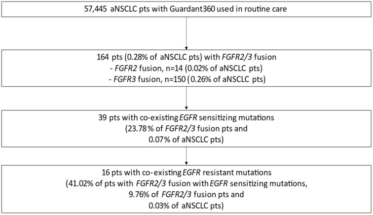
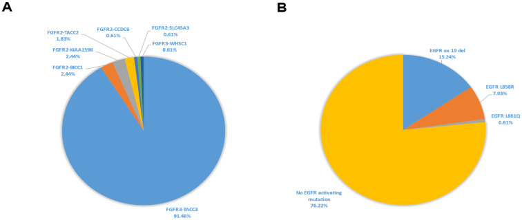
This analysis was done on the validated dataset in GH ED and comprised genomic data from 57,445 patients with aNSCLC. A total of 164 (0.28% of all aNSCLC cases) carried either FGFR2 or FGFR3 fusion; the prevalence of FGFR2 and FGFR3 fusions were 0.02% and 0.26%, respectively (Figure 1). Approximately 50% of tumors with FGFR rearrangements were adenocarcinoma, 38% were squamous cell carcinoma, and the remaining cases were of either mixed or unknown histology (Table 1). Amongst all FGFR fusion subtypes, FGFR3-TACC3 fusions were the most abundant (91.46% of cases); other fusion partners were TACC2, Bicaudal C homolog 1 (BICC1), shootin-1 (SHTN1, also called KIAA1598), CCDC6, solute carrier family 45 member 3 (SLC45A3), and Wolf-Hirschhorn syndrome candidate gene-1 (WHSC1) (Figure 2A, Table 1). The median FGFR mutant allele frequency (MAF) was 0.5%.
Figure 1.
Prevalence of FGFR2/3 fusions and co-existing EGFR sensitizing and resistant mutations in aNSCLC in the GH electronic database. Abbreviations: aNSCLC—advanced non-small cell lung cancer; EGFR—epidermal growth factor receptor; FGFR—fibroblast growth factor receptor; GH—Guardant Health; pts—patients.
Table 1.
FGFR2/3 fusion subtype prevalence and distribution across different histological non-small cell lung cancer subtypes in the Guardant Health electronic database.
| FGFR Fusion Type | Large-Cell Carcinoma | ADC | Adeno-Squamous Carcinoma | SCC | NSCLC NOS Carcinoma |
Total |
|---|---|---|---|---|---|---|
| FGFR2-BICC1 | 4 | 4 | ||||
| FGFR2-CCDC6 | 1 | 1 | ||||
| FGFR2-KIAA1598 | 3 | 1 | 4 | |||
| FGFR2-SLC45A3 | 1 | 1 | ||||
| FGFR2-TACC2 | 2 | 1 | 3 | |||
| FGFR3-TACC3 | 4 | 71 | 1 | 61 | 13 | 150 |
| FGFR3-WHSC1 | 1 | 1 | ||||
| Total | 4 | 81 | 1 | 62 | 16 | 164 |
Abbreviations: ADC—adenocarcinoma; FGFR—fibroblast growth factor receptor; NSCLC NOS—non-small cell lung cancer non otherwise specified carcinoma; SCC—squamous cell carcinoma.
Figure 2.
FGFR2/3 fusion subtypes distribution (A) and prevalence of co-existing EGFR sensitizing mutations (B) in aNSCLC in the GH electronic database. Abbreviations: aNSCLC—advanced non-small cell lung cancer; EGFR—epidermal growth factor receptor; FGFR—fibroblast growth factor receptor; GH—Guardant Health.
Out of all the 164 patients with FGFR2/3 fusions, 39 (23.78%) patients (0.07% of all aNSCLC cases) had concurrent EGFR sensitizing mutations (Figure 1 and Figure 2B). In samples with FGFR2/3 fusions with co-existing EGFR mutations, subtypes of the EGFR sensitizing mutation were as follows: exon 19 del—64.1%, L8585R—33.3%, and L861Q—2.6% (Figure 2B, Table 2). Among the 39 patients with FGFR2/3 fusions and concurrent EGFR sensitizing mutations, co-existing EGFR resistance mutations (T790M and/or C797X) were observed in 16 (41.02%) patients (0.03% of all aNSCLC cases) (Figure 1). Those EGFR resistant mutation types were as follows: T790M—20.5%, C797X—2.6%, and both T790M and C797X—17.9% (Table 2).
Table 2.
EGFR mutation types co-occurring with FGFR2/3 fusions in the Guardant Health electronic database.
| FGFR Fusion Type | EGFR Sensitizing Mutation Type | EGFR Resistance Mutation Type | ||||||
|---|---|---|---|---|---|---|---|---|
| Exon 19 Deletion, n (%) |
L858R, n (%) |
L861Q, n (%) |
Total, n (%) |
T790M, n (%) |
C797X, n (%) |
T790M and C797X, n (%) |
None, n (%) |
|
| FGFR2-CCDC6 | 1 (2.6) | 0 (0) | 0 (0) | 1 (2.6) | 1 (2.6) | 0 (0) | 0 (0) | 0 (0) |
| FGFR2-KIAA1598 | 1 (2.6) | 1 (2.6) | 0 (0) | 2 (5.1) | 1 (2.6) | 0 (0) | 0 (0) | 1 (2.6) |
| FGFR2-TACC2 | 0 (0) | 1 (2.6) | 0 (0) | 1 (2.6) | 1 (2.6) | 0 (0) | 0 (0) | 0 (0) |
| FGFR3-TACC3 | 23 (58.9) | 11 (28.2) | 1 (2.6) | 35 (89.7) | 5 (12.8) | 1 (2.6) | 7 (17.9) | 22 (56.4) |
| Total | 25 (64.1) | 13 (33.3) | 1 (2.6) | 39 (100) | 8 (20.5) | 1 (2.6) | 7 (17.9) | 23 (59.0) |
Abbreviations: EGFR—epidermal growth factor receptor; FGFR—fibroblast growth factor receptor.
3.2. FGFR1/2/3 Fusion Frequency in TASMC Electronic Database
The TASMC ED included 161 cases with aNSCLC, among which there was one case of de novo FGFR3-TACC3 fusion (prevalence—0.62%). This tumor did not harbor a co-existing EGFR mutation.
3.3. Case-Series of Patients with FGFR3-TACC3 Fusion as an Acquired Resistance Mechanism Following Treatment with EGFR TKIs
Three patients with EGFR mutant aNSCLC progressing on EGFR TKIs and developing an FGFR3-TACC3 fusion were identified in the ED of DCC and BZ. In all the three cases, FGFR3-TACC3 fusion was detected by Guardant360 performed at radiological progression on osimertinib. Clinicopathological patient characteristics are summarized in Table 3.
Table 3.
Demographic and clinico-pathological characteristics of patients with EGFR mutant aNSCLC progressing on EGFR TKIs and developing an FGFR3-TACC3 fusion. All FGFR3-TACC3 fusions were detected by Guardant 360.
| Case Number | Sex | Age, Years | Tumor Histology | Smoking History | EGFR Mutation Subtype | Treatment History before FGFR3-TACC3 Fusion Diagnosis: Agent (PFS, mo) | FGFR3-TACC3 Fusion MAF, % | Concurrent Alterations, MAF, % |
|---|---|---|---|---|---|---|---|---|
| #1 | F | 59 | ADC | Never- smoker |
L858R | Gefitinib (7 mo), osimertinib (13 mo), carboplatin/pemetrexed (6 mo) | 0.3 |
EGFR L858R, 33.4, PIK3CA E545K, 47.5, CCND1 amplification, CDK4 amplification, KRAS amplification, MYC amplification |
| #2 | M | 84 | ADC | Never- smoker |
E746_A750del | Osimertinib (11 mo) | 0.04 |
EGFR E746_A750del, 1.3, TP53 Y163C, 0.4 |
| #3 | F | 63 | ADC | Never- smoker |
L747_A750delinsP | Gefitinib (52 mo), osimertinib (14 mo) | 0.07 | Gardant360: EGFR L747_A750delinsP, 0.5, PIK3CA V344G, 1.3 Tempus xT: EGFR L747_A750delinsP, 14.4, EGFR p. C797S, 3.6, PIK3CA V344G, 15.9 |
Abbreviations: ADC—adenocarcinoma; EGFR—epidermal growth factor receptor; F—female; FGFR—fibroblast growth factor receptor; M—male; MAF—mutant allele frequency; mo—months; PFS—progression-free survival.
3.4. Clinical Case #1
The first patient was a 59-year-old never-smoking female diagnosed with lung adenocarcinoma harboring an EGFR L858R substitution on exon 21 (verified by polymerase chain reaction [PCR] in the tumor specimen) with bilateral lung metastases, liver metastases, and a left-sided malignant pleural effusion. Gefitinib was initiated, and a partial response was achieved. Seven months later, upon disease progression, droplet digital PCR (ddPCR) of the plasma was performed and revealed the presence of an EGFR T790M mutation. Gefitinib was replaced by osimertinib with a partial response lasting 13 months. Upon radiological disease progression in the lungs and pleura, treatment was changed to platinum and pemetrexed, with disease stabilization for an additional 6 months. Eventually, following a disease progression, blood was collected for Guardant360 testing, which revealed an EGFR L858R mutation (MAF 33.4%), PIK3CA E545K mutation (MAF 47.5%), FGFR3-TACC3 fusion (MAF 0.3%), cyclin D1 (CCND1) amplification, cyclin-dependent kinase 4 (CDK4) amplification, KRAS amplification, and Myelocytomatosis viral oncogene homolog (MYC) amplification. The patient did not receive any subsequent systemic therapy and died of aNSCLC 3 months thereafter.
3.5. Clinical Case #2
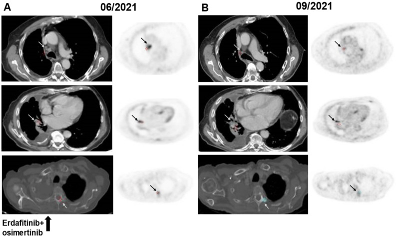
The second patient is an 84-year-old never-smoking male who was diagnosed with advanced-stage lung adenocarcinoma, a malignant right-sided pleural effusion, and bone metastases. PCR of the pleural fluid cell-block revealed the presence of an EGFR exon 19 deletion (E746_A750del). Osimertinib 80 mg once daily was initiated, and a partial response was achieved. Eleven months after treatment initiation, the disease progressed with the development of new mediastinal lymphadenopathy and recurrent right-sided pleural effusion necessitating the insertion of a drain. At that point, Guardant360 testing was performed and, in addition to detecting the original sensitizing mutation EGFR E746_A750del (MAF 1.3%), identified a novel FGFR3-TACC3 fusion (MAF 0.04%) along with tumor protein P53 (TP53) Y163C (MAF 0.4%). Erdafitinib 8 mg was added to osimertinib 80 mg daily, resulting in mediastinal lymph node shrinkage, stabilization of the lung metastasis and pleural effusion, and calcification of lytic bone metastases. All metastatic sites showed a reduction in fluorodeoxyglucose (FDG)-avidity in the FDG-PET/CT (Figure 3). The treatment was well-tolerated; no laboratory abnormalities were seen. The patient continued with combination TKI treatment with disease stabilization lasting for 6 months since treatment initiation.
Figure 3.
FDG-PET/CT images before (A) and during (B) therapy with osimertinib + erdafitinib in a patient with an EGFR mutated aNSCLC and FGFR3-TACC3 fusion following progression on osimertinib. Shrinkage of a retro-caval lymph node with a reduction in FDG-avidity, stable lung metastasis with a reduction in FDG-avidity, calcification of a D3 lytic bone metastasis with a reduction in FDG-avidity (gray and black arrows). Abbreviations: aNSCLC—advanced non-small cell lung cancer; EGFR—epidermal growth factor receptor; FDG—fluorodeoxyglucose; FGFR3-TACC3—fibroblast growth factor receptor 3-transforming acidic coiled-coil-containing protein 3; PET/CT—positron emission tomography/computer tomography.
3.6. Clinical Case #3
The third patient is a 63-year-old never-smoking female who was diagnosed with advanced-stage lung adenocarcinoma harboring an EGFR L747_A750delinsP mutation (exon 19 deletion). She was initially treated with gefitinib, with partial response lasting 52 months. Upon radiological disease progression, treatment was changed to osimertinib following the detection of an EGFR T790M mutation by ddPCR. Eleven months later, disease spread to the peritoneum (omental cake) and was confirmed by peritoneal biopsy. NGS (Tempus xT) was performed on the tissue specimen and revealed an EGFR L747_A750delinsP (MAF 14.4%), EGFR C797S (MAF 3.6%), and PIK3CA V344G (MAF 15.9%). Guardant360 performed on plasma collected at the same time revealed the presence of an FGFR3-TACC3 fusion (MAF 0.07%) in addition to the previously described molecular alterations (EGFR L747_A750delinsP, PIK3CA V344G). Treatment with erdafitinb 9 mg and gefitinb 250 mg daily was initiated with clinical improvement and radiological disease stabilization. Four months later, a solitary brain metastasis was detected. Stereotactic radiosurgery was conducted, and combined systemic treatment was continued. Due to isolated asymptomatic grade 3 alanine transaminase (ALT) elevation, the erdafitinib dose was reduced to 4 mg daily. Nine months after the initiation of the combined TKI treatment, the disease progressed systemically and intracranially, and systemic treatment was changed to carboplatin with pemetrexed.
4. Discussion
Analysis of FGFR2/3 fusions in aNSCLC patients in the GH electronic database, the largest analysis yet reported on the subject, revealed that FGFR2/3 fusions co-exist with EGFR sensitizing mutations in 24% of cases, and in 41% of those cases, concurrent EGFR resistant mutations are also seen. This observation suggests that FGFR2/3 fusions may represent a rare but clinically important acquired molecular resistance mechanism following treatment with EGFR TKIs in EGFR mutant aNSCLC. Moreover, in the presented clinical series, two out of three patients with an FGFR3-TACC3 fusion, as a confirmed acquired molecular resistance mechanism to EGFR TKIs, derived clinical benefit from adding an FGFR TKI, erdafitinib, to EGFR TKI.
According to the GH electronic database, the prevalence of FGFR2/3 fusions in liquid biopsy specimens from patients with aNSCLC was 0.28%, whereas the analysis of TASMC electronic database demonstrated FGFR fusions prevalence in tumor tissue specimens of 0.62%. Both results are in line with previous reports on FGFR2/3 fusions prevalence ranging between 0.2% and 1.3% when assessed by comprehensive molecular profiling. For instance, Qin et al. [19] reported FGFR fusions retaining the kinase domain in 0.2% (52 of 26.054 NSCLC cases), including 37 FGFR3-TACC3 fusions, two FGFR2 fusions, one FGFR1 fusion (all previously reported), and 12 novel FGFR1, FGFR2, FGFR3, and FGFR4 fusions. Wang et al. reported a FGFR fusion prevalence of 1.3% in a cohort of 1.328 patients with NSCLC, amd the majority of cases (15 of 17) were FGFR3-TACC3 fusions [20]. Additionally, Capelletti et al. reported a FGFR3-TACC3 fusions prevalence of 0.5% in a cohort of 576 patients with lung adenocarcinoma [21]. The differences in FGFR rearrangements prevalence may result from the techniques used for the assessment of fusions in different assays. Specifically, the technical limitations of DNA-based NGS for detection of fusions may lead to a slightly lower prevalence of FGFR fusions compared to assays that assess for gene rearrangements using RNA or protein. On the other hand, tissue-based NGS databases are mainly composed of samples from treatment-naive patients, as opposed to plasma-based NGS databases which are enriched for patients who have had multiple lines of systemic treatment, hence increasing the probability of finding FGFR fusions representing an acquired resistance mechanism.
Amongst all the FGFR fusion subtypes, FGFR3-TACC3 predominated; this subtype represented 91% of all FGFR fusions in the GH electronic database. This observation is in line with the previously reported data regarding the frequency of FGFR3-TACC3 fusions in FGFR-rearranged NSCLC (77–88%) [19,20].
In the GH electronic database, FGFR2/3 fusions were more frequently seen in lung adenocarcinomas as compared to other histological lung cancer subtypes. The previously reported data, on the other hand, points to the two most common NSCLC histological subtypes (adenocarcinoma and squamous cell carcinoma) as the main source of FGFR fusions [19,20]. Additionally, in the GH database, about one-quarter of patients with FGFR fusions had co-existing EGFR sensitizing mutations. Nearly half of those also harbored one of the EGFR resistant mutations, suggesting possible tumor progression on 1st or 2nd-generation EGFR TKI before sample acquisition. The higher frequency of adenocarcinoma-associated FGFR fusions and the higher frequency of co-existing EGFR sensitizing and EGFR resistant mutations in the GH electronic database may both reflect the higher proportion of cases with acquired resistance to targeted treatments included in the GH database, which, in turn, reflects real-world referral patterns for liquid biopsy testing after disease progression on prior therapies.
The acquisition of FGFR3-TACC3 fusion has been described as a molecular resistance mechanism following treatment with EGFR TKIs in EGFR mutant aNSCLC [12,22]. We observed that 0.06% of aNSCLC samples in the GH database had FGFR3-TACC3 fusion with a concurrent EGFR mutation, similar to the results reported by Ou et al. (0.03%; 5 of 17,319 aNSCLC cases) using either tumor tissue or plasma as source material [12], but somewhat greater than those reported by Quin et al. (0.007%) using tumor tissue only [19].
It is difficult to estimate the exact prevalence of acquired FGFR fusion following treatment with EGFR TKIs in EGFR mutant aNSCLC because most large molecular databases do not include testing and/or treatment histories. In the absence of such information in the GH database, we assumed that in cases with both EGFR sensitizing mutation and FGFR fusion, the former represented the original oncogenic driver. The reported prevalence is based on the following assumptions: first, the co-existing EGFR resistant mutations reflect the underlying molecular resistant mechanism, and then, all the tumors developing an FGFR rearrangement following the progression on EGFR TKIs retain the original EGFR mutation, which may or may not be true. We also acknowledge that most cases of FGFR fusions in GH ED occurred without a concomitant EGFR mutation, and therefore, in most cases, FGFR fusion might represent a driver by itself.
The clinical data on FGFR TKIs as a treatment of de novo FGFR-aberrated aNSCLC, as opposed to intrahepatic cholangiocarcinoma and urothelial carcinoma, is limited to 24 cases treated with erdafitinib in a phase 1 trial [15]. The objective response rate with erdafitinib in 21 evaluable aNSCLC patients was 5%, efficacy in 8 patients with de novo FGFR-rearrangements has not been reported [15]. In the clinical scenario of acquired FGFR- fusions following treatment with osimertinib in EGFR mutant aNSCLC, one patient achieved a partial response with the combination of erdafitinib and osimertinib has been previously reported [16], and our case-series adds to the reported data.
This study highlights the important role of ctDNA testing as a sensitive and non-invasive approach for the detection of emerging and potentially actionable genomic alterations that may develop in aNSCLC after treatment with targeted therapies. Recently, the International Association for the Study of Lung Cancer (IASLC) updated its liquid biopsy guideline and recommended the use of ctDNA as the first method (“plasma first”) to discover molecular resistance mechanisms—due to the simplicity and high patient advocacy [23]. Such testing may help to identify actionable alterations, such as FGFR3 fusions that inform effective targeted therapeutic options.
5. Conclusions
In conclusion, although FGFR fusions represent a rare molecular event in the setting of acquired resistance to EGFR TKIs, its finding enables an emerging treatment strategy of combining EGFR TKIs with FGFR TKIs. Such combinations warrant a prospective evaluation.
Acknowledgments
Authors would like to acknowledge contribution by Angie Thompson and Lesli Kiedrowski (both employees of Guardant Health) for data analysis and interpretation. Israeli Lung Cancer Group, ISCORT: Elizabeth Dudnik, Chair of the Israeli Lung Cancer Group; Ari Raphael, Hovav Nechushtan, Nir Peled and Abed Agbarya—members of the Israeli Lung Cancer Group.
Abbreviations
ADC—adenocarcinoma; ALK—anaplastic lymphoma kinase; ALT—alanine transaminase; (a) NSCLC—(advanced) non-small lung cancer; BAM—binary alignment map; BICC1—Bicaudal C homolog 1; BRAF—v-raf murine sarcoma viral oncogene homolog B1; BZ—Bnei-Zion; CAP—college of American pathologists; CCDC6—coiled-coil domain-containing protein 6; CCND1—cyclin D1; CDK4—cyclin-dependent kinase 4; CLIA—Clinical Laboratory Improvement Amendments; cMET—tyrosine-protein kinase Met; (ct)DNA—(circulating tumor) deoxyribonucleic acid; CT—computer tomography; DCC—Davidoff Cancer Center; (dd) PCR—(droplet digital) polymerase chain reaction; ED—electronic database; EGFR—epidermal growth factor receptor; EML4—echinoderm microtubule-associated protein-like 4; F—female; FDG—fluorodeoxyglucose; (FDG)-PET/CT—fluorodeoxyglucose positron emission tomography/computer tomography; FFPE—formalin-fixed paraffin-embedded; FGFR—fibroblast growth factor receptor; GH—Guardant Health; IASLC—International Association for the Study of Lung Cancer; indels—insertions and deletions; KRAS—Kirsten rat sarcoma viral oncogene homolog; M—male; MAF—mutant allele frequency; mo—months; MYC—Myelocytomatosis viral oncogene homolog; NGS—next generation sequencing; NOS—non-small cell lung cancer non otherwise specified carcinoma; PFS—progression-free survival; PIK3CA—phosphatidylinositol-4,5-bisphosphate 3-kinase; pts—patients; RET—rearranged during transfection; RNA—ribonucleic acid; RT—reverse transcription; RTK—receptor tyrosine kinase; SHTN1/KIAA1598—shootin-1; SLC45A3—solute carrier family 45 member 3; SNV—single-nucleotide variants; SCC—squamous cellcarcinoma; TACC2/3—transforming acidic coiled-coil containing protein; TASMC—Tel-Aviv Sourasky medical center; TKI—tyrosine kinase inhibitor; TP53—tumor protein P53; (US) FDA—(United States) Food and Drug Administration Agency; WHSC1—Wolf-Hirschhorn syndrome candidate gene-1.
Author Contributions
Conceptualization: E.D., A.R. and A.A.; Methodology: E.D., A.R., D.H., S.J. and A.A.; Validation: D.H., S.J. and S.O.; Formal analysis: S.J. and S.O.; Investigation: A.R., E.D., S.J. and A.A.; Resources: A.R., H.N., D.H., S.J., A.A., N.P., A.O. and E.D.; Data Curation: D.H., S.J. and A.D.; Writing—Original Draft: E.D., A.R. and A.A.; Writing—Review & Editing: A.R., E.D., D.H., S.J., S.O., L.S.-G., T.B.-S., A.D., H.N., N.P., A.O. and A.A.; Visualization: A.R.; Supervision: E.D. and A.A.; Project administration: A.R. and E.D. All authors have read and agreed to the published version of the manuscript.
Institutional Review Board Statement
Institutional review board approval was received before study initiation in TASMC ED (approval number: -TLV0685-21).
Informed Consent Statement
Patient consent was waived.
Data Availability Statement
According to the GH policy, the access to the database cannot be provided.
Conflicts of Interest
Ari Raphael reported personal fees from Roche, Astra Zeneca, Merck Sharpe & Dohme, Novartis, Takeda, Elli Lilly, support for attending meetings from Bristol Myers Squibb, Roche, Boehringer Ingelheim—all outside of the submitted work. Elizabeth Dudnik reported research funding from Astra Zeneca, Boehringer Ingelheim, personal and consulting fees from Boehringer Ingelheim, Roche, Astra Zeneca, Pfizer, Merck Sharpe & Dohme, Bristol Myers Squibb, Novartis, Takeda, Sanofi, Merck Serono, Medison Pharma, Janssen Israel—all outside of the submitted work. Suyog Jain and Steve Olsen reported a relationship as an employee at Guardant Health AMEA Inc. Lior Soussan-Gutman reported a relationship as a Head of Oncotest-Rhenium, a company that provides Guardant Health services. Taly Ben-Shitrit and Addie Dvir reported a relationship as an employee at Oncotest-Rhenium, a company that provides Guardant Health services. Nir Peled reported research funding from Bristol-Myers Squibb, Eli Lilly, Foundation Medicine, Guardant Health, Merck Serono, Merck Sharpe & Dohme, Novartis, NovellusDx, Pfizer, Roche, Takeda, IP: Volatile Organic Compounds For Detecting Cell Dysplasia And Genetic Alterations Associated With Lung Cancer, WO2012023138; Breath Analysis of Pulmonary Nodules, US20130150261 A1; Apparatus for treating a target site of a body, WO/2015/059646—all outside of the submitted work. Amir Onn reported personal and consulting fees from Merck Sharpe & Dohme, Bristol Myers Squibb, Roche, Astra Zeneca, Novartis, Boehringer Ingelheim—all outside of the submitted work. Abed Agbarya reported research funding from Bristol Myers Squibb, personal and consulting fees from Bristol Myers Squibb, Roche, Pfizer, Astra Zeneca, Takeda, Novartis. Other authors declared no conflict of interest.
Funding Statement
This research did not receive any specific grant from funding agencies in the public, commercial, or not-for-profit sectors.
Footnotes
Publisher’s Note: MDPI stays neutral with regard to jurisdictional claims in published maps and institutional affiliations.
References
- 1.Wu Y.L., Tsuboi M., He J., John T., Grohe C., Majem M., Goldman J.W., Laktionov K., Kim S.W., Kato T., et al. Osimertinib in Resected EGFR-Mutated Non–Small-Cell Lung Cancer. N. Engl. J. Med. 2020;383:1711–1723. doi: 10.1056/NEJMoa2027071. [DOI] [PubMed] [Google Scholar]
- 2.Ramalingam S.S., Vansteenkiste J., Planchard D., Cho B.C., Gray J.E., Ohe Y., Zhou C., Reungwetwattana T., Cheng Y., Chewaskulyong B., et al. Overall Survival with Osimertinib in Untreated, EGFR-Mutated Advanced NSCLC. N. Engl. J. Med. 2020;382:41–50. doi: 10.1056/NEJMoa1913662. [DOI] [PubMed] [Google Scholar]
- 3.Helena A.Y., Tian S.K., Drilon A.E., Borsu L., Riely G.J., Arcila M.E., Ladanyi M. Acquired Resistance of EGFR-Mutant Lung Cancer to a T790M-Specific EGFR Inhibitor Emergence of a Third Mutation (C797S) in the EGFR Tyrosine Kinase Domain. JAMA Oncol. 2015;1:982–984. doi: 10.1001/jamaoncol.2015.1066. [DOI] [PMC free article] [PubMed] [Google Scholar]
- 4.Thress K.S., Paweletz C.P., Felip E., Cho B.C., Stetson D., Dougherty B., Lai Z., Markovets A., Vivancos A., Kuang Y., et al. Acquired EGFR C797S mutation mediates resistance to AZD9291 in non–small cell lung cancer harboring EGFR T790M. Nat. Med. 2015;21:560–562. doi: 10.1038/nm.3854. [DOI] [PMC free article] [PubMed] [Google Scholar]
- 5.Le X., Puri S., Negrao M.V., Nilsson M.B., Robichaux J., Boyle T., Hicks J.K., Lovinger K.L., Roarty E., Rinsurongkawong W., et al. Landscape of EGFR-Dependent and -Independent Resistance Mechanisms to Osimertinib and Continuation Therapy Beyond Progression in EGFR-Mutant NSCLC. Clin. Cancer Res. 2018;24:6195–6203. doi: 10.1158/1078-0432.CCR-18-1542. [DOI] [PMC free article] [PubMed] [Google Scholar]
- 6.Lin C.-C., Shih J.-Y., Yu C.-J., Ho C.-C., Liao W.-Y., Lee J.-H., Tsai T.-H., Su K.-Y., Hsieh M.-S., Chang Y.-L., et al. Outcomes in patients with non-small-cell lung cancer and acquired Thr790Met mutation treated with osimertinib: A genomic study. Lancet Respir. Med. 2018;6:107–116. doi: 10.1016/S2213-2600(17)30480-0. [DOI] [PubMed] [Google Scholar]
- 7.Oxnard G.R., Hu Y., Mileham K.F., Husain H., Costa D.B., Tracy P., Feeney N., Sholl L.M., Dahlberg S.E., Redig A.J., et al. Assessment of Resistance Mechanisms and Clinical Implications in Patients with EGFR T790M-Positive Lung Cancer and Acquired Resistance to Osimertinib. JAMA Oncol. 2018;4:1527–1534. doi: 10.1001/jamaoncol.2018.2969. [DOI] [PMC free article] [PubMed] [Google Scholar]
- 8.Piotrowska Z., Thress K.S., Mooradian M., Heist R.S., Azzoli C.G., Temel J.S., Rizzo C., Nagy R.J., Lanman R.B., Gettinger S.N., et al. MET amplification (amp) as a resistance mechanism to osimertinib. J. Clin. Oncol. 2017;35:9020. doi: 10.1200/JCO.2017.35.15_suppl.9020. [DOI] [Google Scholar]
- 9.Yang Z., Yang N., Ou Q., Xiang Y., Jiang T., Wu X., Bao H., Tong X., Wang X., Shao Y.W., et al. Investigating Novel Resistance Mechanisms to Third-Generation EGFR Tyrosine Kinase Inhibitor Osimertinib in Non-Small Cell Lung Cancer Patients. Clin. Cancer Res. 2018;24:3097–3107. doi: 10.1158/1078-0432.CCR-17-2310. [DOI] [PubMed] [Google Scholar]
- 10.Allen J.M., Schrock A.B., Erlich R.L., Miller V.A., Stephens P.J., Ross J.S., Ou S.-H.I., Ali S.M., Vafai D. Genomic Profiling of Circulating Tumor DNA in Relapsed EGFR-Mutated Lung Adenocarcinoma Reveals an Acquired FGFR3- TACC3 Fusion. Clin. Lung Cancer. 2017;18:219–222. doi: 10.1016/j.cllc.2016.12.006. [DOI] [PubMed] [Google Scholar]
- 11.Helsten T., Elkin S., Arthur E., Tomson B.N., Carter J., Kurzrock R. The FGFR Landscape in Cancer: Analysis of 4,853 Tumors by Next-Generation Sequencing. Clin. Cancer Res. 2015;22:259–267. doi: 10.1158/1078-0432.CCR-14-3212. [DOI] [PubMed] [Google Scholar]
- 12.Ou S.-H.I., Horn L., Cruz M., Vafai D., Lovly C.M., Spradlin A., Williamson M.J., Dagogo-Jack I., Johnson A., Miller V.A., et al. Emergence of FGFR3-TACC3 fusions as a potential by-pass resistance mechanism to EGFR tyrosine kinase inhibitors in EGFR mutated NSCLC patients. Lung Cancer. 2017;111:61–64. doi: 10.1016/j.lungcan.2017.07.006. [DOI] [PMC free article] [PubMed] [Google Scholar]
- 13.Soria J.-C., Strickler J.H., Govindan R., Chai S., Chan N., Quiroga-Garcia V., Bahleda R., Hierro C., Zhong B., Gonzalez M., et al. Safety and activity of the pan-fibroblast growth factor receptor (FGFR) inhibitor erdafitinib in phase 1 study patients (Pts) with molecularly selected advanced cholangiocarcinoma (CCA) J. Clin. Oncol. 2017;35:4074. doi: 10.1200/JCO.2017.35.15_suppl.4074. [DOI] [Google Scholar]
- 14.Loriot Y., Necchi A., Park S.H., Garcia-Donas J., Huddart R., Burgess E., Fleming M., Rezazadeh A., Mellado B., Varlamov S., et al. Erdafitinib in Locally Advanced or Metastatic Urothelial Carcinoma. N. Engl. J. Med. 2019;381:338–348. doi: 10.1056/NEJMoa1817323. [DOI] [PubMed] [Google Scholar]
- 15.Bahleda R., Italiano A., Hierro C., Mita A., Cervantes A., Chan N., Awad M., Calvo E., Moreno V., Govindan R., et al. Multicenter Phase I Study of Erdafitinib (JNJ-42756493), Oral Pan-Fibroblast Growth Factor Receptor Inhibitor, in Patients with Advanced or Refractory Solid Tumors. Clin. Cancer Res. 2019;25:4888–4897. doi: 10.1158/1078-0432.CCR-18-3334. [DOI] [PubMed] [Google Scholar]
- 16.Haura E.B., Hicks J.K., Boyle T.A. Erdafitinib Overcomes FGFR3-TACC3–Mediated Resistance to Osimertinib. J. Thorac. Oncol. 2020;15:e154–e156. doi: 10.1016/j.jtho.2019.12.132. [DOI] [PubMed] [Google Scholar]
- 17.Odegaard J.I., Vincent J.J., Mortimer S., Vowles J.V., Ulrich B.C., Banks K.C., Fairclough S.R., Zill O.A., Sikora M., Mokhtari R., et al. Validation of a Plasma-Based Comprehensive Cancer Genotyping Assay Utilizing Orthogonal Tissue- and Plasma-Based Methodologies. Clin. Cancer Res. 2018;24:3539–3549. doi: 10.1158/1078-0432.CCR-17-3831. [DOI] [PubMed] [Google Scholar]
- 18.Lanman R.B., Mortimer S.A., Zill O.A., Sebisanovic D., Lopez R., Blau S., Collisson E.A., Divers S.G., Hoon D., Kopetz S., et al. Analytical and Clinical Validation of a Digital Sequencing Panel for Quantitative, Highly Accurate Evaluation of Cell-Free Circulating Tumor DNA. PLoS ONE. 2015;10:e0140712. doi: 10.1371/journal.pone.0140712. [DOI] [PMC free article] [PubMed] [Google Scholar]
- 19.Qin A., Johnson A., Ross J.S., Miller V.A., Ali S.M., Schrock A.B., Gadgeel S.M. Detection of Known and Novel FGFR Fusions in Non-Small Cell Lung Cancer by Comprehensive Genomic Profiling. J. Thorac. Oncol. 2019;14:54–62. doi: 10.1016/j.jtho.2018.09.014. [DOI] [PubMed] [Google Scholar]
- 20.Wang R., Wang L., Li Y., Hu H., Shen L., Shen X., Pan Y., Ye T., Zhang Y., Luo X., et al. FGFR1/3 tyrosine kinase fusions define a unique molecular subtype of non-small cell lung cancer. Clin. Cancer Res. 2014;20:4107–4114. doi: 10.1158/1078-0432.CCR-14-0284. [DOI] [PubMed] [Google Scholar]
- 21.Capelletti M., Dodge M.E., Ercan D., Hammerman P.S., Park S.I., Kim J., Sasaki H., Jablons D.M., Lipson D., Young L., et al. Identification of recurrent FGFR3-TACC3 fusion oncogenes from lung adenocarcinoma. Clin. Cancer Res. 2014;20:6551–6558. doi: 10.1158/1078-0432.CCR-14-1337. [DOI] [PubMed] [Google Scholar]
- 22.Papadimitrakopoulou V.A., Wu Y.L., Han J.Y., Ahn M.J., Ramalingam S.S., John T., Okamoto I., Yang J.H., Bulusu K.C., Laus G.J.A.O.O., et al. Analysis of resistance mechanisms to osimertinib in patients with EGFR T790M advanced NSCLC from the AURA3 study. Ann. Oncol. 2018;29:VIII741. doi: 10.1093/annonc/mdy424.064. [DOI] [Google Scholar]
- 23.Rolfo C., Mack P.C., Scagliotti G.V., Baas P., Barlesi F., Bivona T.G., Herbst R.S., Mok T.S., Peled N., Pirker R., et al. Liquid Biopsy for Advanced Non-Small Cell Lung Cancer: A Consensus Statement from The International Association for the Study of Lung Cancer (IASLC) J. Thorac. Oncol. 2021;16:1647–1662. doi: 10.1016/j.jtho.2021.06.017. [DOI] [PubMed] [Google Scholar]
Associated Data
This section collects any data citations, data availability statements, or supplementary materials included in this article.
Data Availability Statement
According to the GH policy, the access to the database cannot be provided.


