



Abstract
Objective
Patients with liver cirrhosis (LC) commonly exhibit hypercoagulability and tend to develop thrombosis. Neutrophil extracellular traps (NETs) are associated with a variety of thrombotic conditions, but their possible value in portal vein thrombosis (PVT) is not known. We assessed whether NETs promote thrombosis and contribute to the procoagulant state in patients with LC.
Methods
The circulating levels of NETs markers (myeloperoxidase, neutrophil elastase, citrullinated histone H3) were measured in 72 patients (median age, 55 years; 48 [66.7%] men) with LC from September 2020 to February 2021. Then they were divided into two groups: patients with or without PVT. NETs procoagulant activity was assessed based on thrombin–antithrombin complex (TAT complex) and Factor X. The levels of plasma markers were determined by ELISA.
Results
There were 28 patients with PVT and 44 patients without PVT. The levels of NETs markers and hypercoagulability markers in the plasma of cirrhosis patients with PVT were significantly higher than those of cirrhosis patients without PVT (p < 0.05). Additionally, the levels of the NETs markers correlated with TAT complex and Factor X (Spearman correlation rho >0.73, p < 0.0001).
Conclusions
Neutrophil extracellular traps seem to enhance procoagulant activity in LC patients with PVT; thus, they may be a practical predictor of PVT as well as a rapid and easy‐to‐use diagnostic and treatment guide for PVT in patients with cirrhosis.
Keywords: hypercoagulability, liver cirrhosis, neutrophil extracellular traps, portal vein thrombosis
Patients with PVT had higher serum NETs markers levels compared with non‐PVT patients. Levels of MPO, NE, and CitH3 in the PVT group (n = 28) were significantly higher than those in the non‐PVT group (n = 44). Levels of MPO were positively associated with circulating levels of NE and CitH3 in PVT patients. **: p < 0.01; *: p < 0.05.

1. INTRODUCTION
Portal vein thrombosis (PVT) is characterized by thrombosis within the portal vein trunk and intrahepatic portal branches, with or without the involvement of mesenteric and splenic veins. 1 PVT is a frequent and serious complication of liver cirrhosis (LC), particularly in its advanced stages. Namely, the prevalence of PVT range between 0.6% and 28% in patients with cirrhosis. 2 Remarkably, most patients are asymptomatic at the time of PVT diagnosis. 3 However, the 2‐year survival rate of patients with PVT is reduced by 55% compared with patients without PVT due to hepatic dysfunction. 4 Therefore, there is an urgent requirement for validated biomarkers that might predict the development of PVT.
For the past few years, terms such as “thromboinflammation,” “immunothrombosis,” and “immunohemostasis” have been used to describe responses/mechanisms associated with both thrombosis and inflammation. 5 Neutrophil extracellular traps (NETs) are extracellular DNA fibers comprising histones and neutrophil antimicrobial proteins released from activated neutrophils. 6 NETs stimulate thrombosis and coagulation, which are abundant in venous,7, 8 arterial, and cancer‐associated thrombosis.9, 10 Therefore, NETs have been identified to play a major role in immune thrombosis.11, 12 Recently, several studies have suggested that NETs are potential contributors to hypercoagulability in cancers.13, 14 However, there have been no reports as to whether there is a correlation between NETs and a hypercoagulable state in LC patients with PVT. The aim of this study was to assess whether NETs are related to hypercoagulability and whether they can predict PVT formation in patients with LC.
2. MATERIALS AND METHODS
Demographic status, etiology, and clinical laboratory test data were available through the electronic medical records at the Affiliated Hospital of Qingdao University from September 2020 to February 2021. The Model of End‐Stage Liver Disease (MELD) and Child–Pugh scores were used to evaluate the severity of LC. Patients with primary or secondary hepatic malignancy, hematologic diseases, Budd–Chiari syndrome, or inflammatory diseases were excluded. On the basis of the Consensus for Management of Portal Vein Thrombosis in Liver Cirrhosis (2020, Shanghai), 1 the patients were divided into two groups, PVT group (n = 28) and non‐PVT group (n = 44). The flowchart of this study is shown in Figure 1. After obtaining informed consent from all the patients, blood samples were taken from the antebrachial vein and collected into vacuum tubes containing EDTAK2 on the day of admission to the hospital and were then centrifuged for 15 min at 1000 g. The plasma was frozen and stored at a temperature of −80°C for further analysis. Enzyme‐linked immunosorbent assays (ELISA) were used for measurement of the plasma levels of TAT complex, Factor X, MPO, NE, CitH3, endotoxin, and tissue factor (Enzyme‐linked Biotechnology Co.). The current study was approved by the Clinical Trials (NCT05012501) and Ethics Committee of the Affiliated Hospital of Qingdao University, China (QYFYWZLL26363).
FIGURE 1.
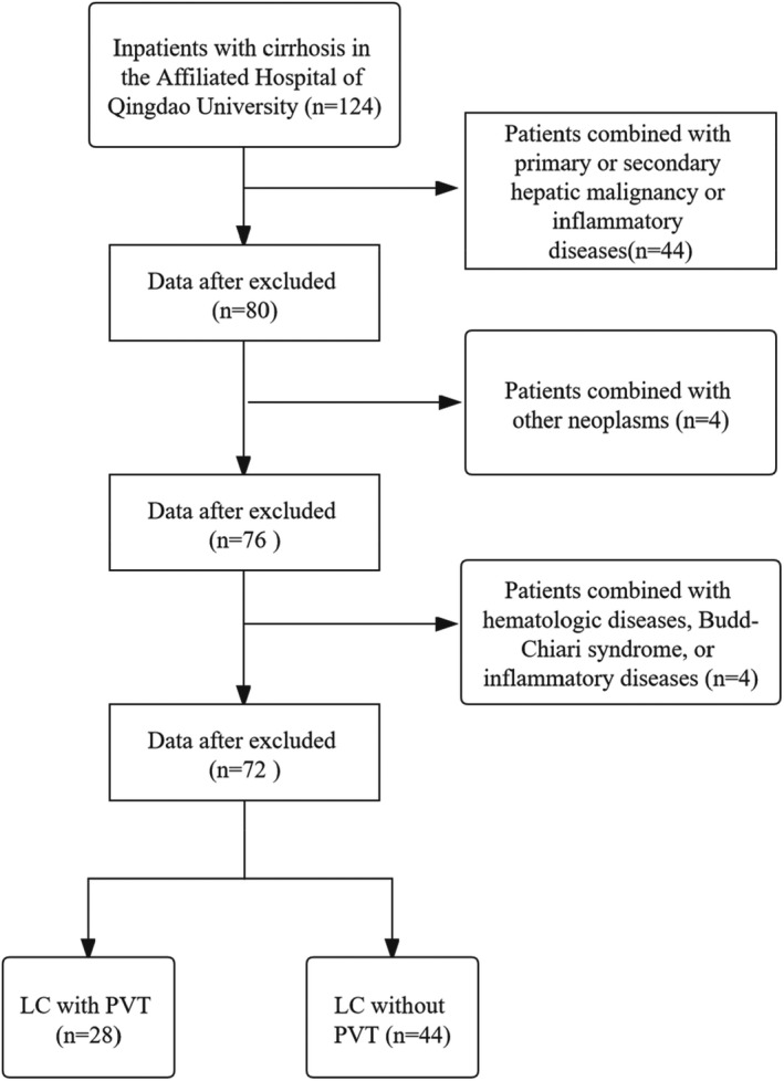
Flowchart of this study
3. STATISTICAL ANALYSIS
Quantitative variables are expressed as mean ± standard deviation, and significance of difference was determined using Student's t test. Categorical variables are expressed as frequencies and percentages, and significance of difference was determined using the χ² or Fisher's exact test. Non‐normally distributed variables are expressed as median and interquartile range, and significance of difference was determined by Mann–Whitney U test. Multivariate logistic regression analyses were carried out to identify independent risk factors for PVT. We used the cutoff points of test variables obtained from receiver operating characteristic (ROC) curves. Correlations were tested by Spearman's correlation. p value < 0.05 was considered statistically significant. All analyses were carried out using the SPSS version 24.0 (IBM inc.) and GraphPad Prism version 8.0.1 (GraphPad Software inc.).
4. RESULTS
4.1. Characteristics of patients
After screening by inclusion and exclusion criteria, 28 LC patients with PVT and 44 cirrhosis patients without PVT were chosen. The basic characteristics of the patients with PVT (n = 28) and those without PVT (n = 44) are presented in Table 1. The study participants in the PVT group had a mean age of 55.61 ± 7.01 years and 60.7% were men; in the non‐PVT cohort, the mean age was 54.43 ± 11.19 years and 70.5% were men. There were no significant differences in age, gender, or history of smoking and alcohol consumption between the two groups. According to MELD score and Child–Pugh level, liver function was worse in the non‐PVT group as shown in Table 1.
TABLE 1.
Characteristics of all patients
| PVT group (n = 28) | Non‐PVT group (n = 44) | p Value | |
|---|---|---|---|
| Age, years | 55.61 ± 7.01 | 54.43 ± 11.19 | 0.586 |
| Gender, n (%) | |||
| Male | 17 (60.7) | 31 (70.5) | 0.393 |
| Female | 11 (39.3) | 13 (29.5) | |
| BMI, kg/m2 | 24.35 ± 2.81 | 24.47 ± 3.95 | 0.891 |
| Site of PVT, n (%) | |||
| Only trunk | 7 (25) | NA | |
| Only branch | 7 (25) | NA | |
| Trunk and branches | 14 (50) | NA | |
| Smoking, n (%) | |||
| Yes | 7 (25) | 15 (34.1) | 0.414 |
| No | 21(75) | 29 (65.9) | |
| Alcohol, n (%) | |||
| Yes | 8 (28.6) | 16 (36.4) | 0.494 |
| No | 20 (71.4) | 28 (63.6) | |
| Pathology, n (%) | |||
| Hepatitis | 15 (53.6) | 16 (36.4) | 0.107 |
| Alcoholic | 4 (14.3) | 14 (31.8) | |
| Autoimmune | 4 (14.3) | 11 (25) | |
| Other | 5 (17.8) | 3 (6.8) | |
| MELD score | 8.26 (7.62, 9.56)* | 10.28 (8.33, 15.89) | 0.008 |
| Child–Pugh stage, n (%) | |||
| A | 11 (39.3) | 11 (25) | 0.319 |
| B | 15 (53.6) | 26 (59.1) | |
| C | 2 (7.1) | 7 (15.9) | |
| Diabetes mellitus, n (%) | |||
| Yes | 2 (7.1) | 11 (25) | 0.55 |
| No | 26 (92.9) | 33 (75) | |
| Endoscopic ligation, n (%) | |||
| Yes | 10 (35.7) | 10 (22.7) | 0.23 |
| No | 18 (64.3) | 34 (77.3) | |
| Endoscopic sclerotherapy, n (%) | |||
| Yes | 6 (21.4) | 11 (25) | 0.728 |
| No | 22 (78.6) | 33 (75) | |
| Splenectomy, n (%) | |||
| Yes | 7 (25) | 3 (6.8) | 0.3 |
| No | 21 (75) | 41 (93.2) | |
Indicates that there was a significant difference between cases and controls. (p < 0.05).
4.2. Systemic inflammatory markers are independent risk factors for PVT
We analyzed the laboratory data of the PVT group and non‐PVT group (Table 2). Patients with PVT had lower levels of Hb (p = 0.26), serum bilirubin (p = 0.002), aspartate aminotransferase (p = 0.038), and alanine aminotransferase (p = 0.03) than patients without PVT. Albumin level in PVT patients was significantly higher than that in non‐PVT patients (p = 0.03). The systemic inflammatory markers (NLR and PLR) in the PVT group were higher than those in the non‐PVT group (p = 0.022 and p = 0.026, respectively). Via multivariate logistic regression analyses, albumin (p = 0.005, 95% CI 1.073–1.422), D‐dimer (p < 0.0001, 95% CI 1.001–1.003), and NLR (p = 0.017, 95% CI 1.111–2.878) were found to be independently associated with PVT.
TABLE 2.
Laboratory data of patients with or without PVT
| PVT group (n = 28) | Non‐PVT group (n = 44) | Univariate (p Value) | Multivariate (p Value) | |
|---|---|---|---|---|
| Hb (g/L) | 74 (68.25, 105.75) | 93 (75.5, 116) | 0.026 | |
| PLT (109/L) | 56.5 (42, 95.5) | 63 (46.5, 103.5) | 0.556 | |
| TBIL (μmol/L) | 15.69 (12.63, 26.09) | 23.98 (17.51, 61.36) | 0.002 | |
| ALT (U/L) | 19.5 (14, 25.5) | 28 (16, 43) | 0.038 | |
| AST (U/L) | 24.5 (16.75, 31.25) | 31 (22, 48.5) | 0.03 | |
| Alb (g/L) | 34.57 ± 5.8 | 31.463 ± 5.82 | 0.03 | 0.005 |
| PT (s) | 13.65 (12.3, 14.65) | 14.2 (12.2,18.35) | 0.136 | |
| INR | 1.18 (1.095, 1.28) | 1.25 (1.11, 1.61) | 0.073 | |
| APTT (s) | 31.45 (29.18, 37.13) | 32.9 (28.85, 39.15) | 0.477 | |
| Fib (g/L) | 1.82 ± 0.78 | 1.68 ± 0.73 | 0.457 | |
| TT (s) | 18.15 (16.58, 19.33) | 18.6 (16.9, 20.55) | 0.184 | |
| Antithrombin III activity (%) | 70.71 ± 20.70 | 59.90 ± 29.95 | 0.099 | |
| D‐dimer (ng/ml) | 1095 (532.5, 2202.5) | 390 (280,680) | <0.0001 | <0.0001 |
| SII | 155.11 (87.43, 337.17) | 240.91 (132.76, 393.10) | 0.075 | |
| NLR | 2.31 (1.97, 4.27) | 1.97 (1.28, 2.65) | 0.022 | 0.026 |
| MLR | 0.35 (0.24, 0.42) | 0.31 (0.24, 0.40) | 0.267 | |
| PLR | 100.54 (76.8, 120.62) | 81.30 (45.65, 138.52) | 0.026 | |
| CRP (mg/L) | 2.05 (0.58, 11.26) | 3.13 (0.5, 8.77) | 0.949 |
Abbreviations: Alb, albumin; ALT, alanine aminotransferase; APTT, activated partial thromboplastin time; AST, aspartate aminotransferase; Fib, fibrinogen; INR, international normalized ratio; MLR, monocyte‐to‐lymphocyte ratio; NLR, neutrophil‐to‐lymphocyte ratio; PLR, platelet‐to‐lymphocyte ratio; PT, prothrombin time; SII, systemic immune‐inflammation index; TBIL, total bilirubin; TT, thrombin time. The bold values indicate the p values are significant (p < 0.05).
4.3. Circulating markers of NETs are associated with PVT
Plasma levels of MPO, NE, and CitH3 in LC patients with PVT were all significantly higher than those in non‐PVT patients (Figure 2). We used ROC curves to obtain the cutoff points and found that patients with PVT had significantly higher levels of NETs markers compared with those without PVT (Table 3), indicating that NETs were associated with PVT. The levels of circulating MPO, NE, and CitH3 in both groups showed a significant positive correlation with each other (Spearman correlation coefficient Rs = 0.8918, p < 0.0001; Rs = 0.8343, p < 0.0001; Rs = 0.9310, p < 0.0001, respectively) (Figure 3 and Table 4).
FIGURE 2.

Patients with PVT had higher serum NETs markers levels compared with non‐PVT patients. Levels of MPO (A), NE (B), and CitH3 (C) in the PVT group (n = 28) were significantly higher than those in the non‐PVT group (n = 44). **p < 0.01; *p < 0.05
TABLE 3.
NETs markers between PVT group and non‐PVT group
| PVT group (n = 28) | Non‐PVT group (n = 44) | p Value | |
|---|---|---|---|
| MPO (ng/ml) [n (%)] | |||
| <9.04 | 10 (35.7) | 30 (68.2) | 0.007 |
| ≥9.04 | 18 (53.6) | 14 (31.8) | |
| NE (ng/ml) [n (%)] | |||
| <2467.73 | 16 (57.1) | 36 (81.8) | 0.023 |
| ≥2467.73 | 12 (42.9) | 8 (18.2) | |
| CitH3 (ng/ml) [n (%)] | |||
| <68.05 | 16 (57.1) | 36 (81.8) | 0.023 |
| ≥68.05 | 12 (42.9) | 8 (18.2) | |
Abbreviations: CitH3, citrullinated histone H3; MPO, myeloperoxidase; NE, neutrophil elastase.
FIGURE 3.
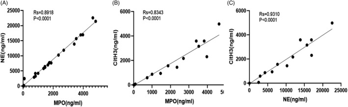
Relationships between MPO, NE, and CitH3. Levels of MPO were positively associated with circulating levels of NE and CitH3 in PVT patients (A and B). Levels of NE were positively associated with circulating levels of MPO and CitH3 in PVT patients (A and C). Levels of CitH3 were positively associated with circulating levels of MPO and NE in PVT patients (B and C)
TABLE 4.
Correlation between NETs markers and hypercoagulable state in PVT
| MPO | NE | CitH3 | |||||||
|---|---|---|---|---|---|---|---|---|---|
| Rs | p Value | 95% CI | Rs | p Value | 95% CI | Rs | p Value | 95% CI | |
| MPO | 1.00 | / | / | 0.8918 | <0.0001 | 0.8299–0.9321 | 0.8343 | <0.0001 | 0.6632–0.9225 |
| NE | 0.8918 | <0.0001 | 0.8299–0.9321 | 1.00 | / | / | 0.9310 | <0.0001 | 0.8516–0.9686 |
| CitH3 | 0.8343 | <0.0001 | 0.6632–0.9225 | 0.9310 | <0.0001 | 0.8516–0.9686 | 1.00 | / | / |
| TAT | 0.7837 | <0.0001 | 0.5725–0.8973 | 0.7903 | <0.0001 | 0.5840–0.9007 | 0.8462 | <0.0001 | 0.6786–0.9266 |
| FX | 0.7908 | <0.0001 | 0.5850–0.9009 | 0.7382 | <0.0001 | 0.4952–0.8741 | 0.7865 | <0.0001 | 0.5774–0.8988 |
| D‐dimer | 0.1041 | 0.5981 | −0.2905–0.4684 | 0.1044 | 0.5971 | −0.2902–0.4686 | 0.05724 | 0.7724 | −0.3331–0.4308 |
Abbreviations: CitH3, citrullinated histone H3; FX, Factor X; MPO, myeloperoxidase; NE, neutrophil elastase; TAT complex, thrombin‐antithrombin complex.
4.4. Circulating markers of NETs and hypercoagulability
The levels of PT, APTT, fibrinogen, TT, and antithrombin III activity were not different between patients with or without PVT (Table 2). However, D‐dimer level was significantly higher in PVT patients than in non‐PVT patients (p < 0.0001). Moreover, the plasma levels of Factor X and TAT complex were significantly higher in patients with PVT (Figure 4). The results of Spearman's correlation analyses indicated significant correlations between the levels of circulating NETs markers and TAT complex or Factor X (Figure 5 and Table 4).
FIGURE 4.

Patients with PVT exhibit hypercoagulability compared with non‐PVT patients. Levels of TAT (A) and FX (B) in the PVT group (n = 28) were significantly higher than those in the non‐PVT group (n = 44). *p < 0.05
FIGURE 5.

Relationship between NETs markers and TAT/FX. TAT complexes were positively associated with circulating levels of MPO, NE, and CitH3 in PVT patients (A–C). FX level was positively associated with circulating levels of MPO, NE, and CitH3 in PVT patients (D–F)
4.5. Circulating markers of NETs are associated with the activation of the TF/endotoxin in patients with PVT
Plasma levels of TF and endotoxin were significantly higher in PVT patients than in non‐PVT patients (p = 0.046, p = 0.045, respectively; Figure 6). Moreover, except for D‐dimer, the results showed strong correlations between the levels of circulating NETs markers, hypercoagulability markers, and TF/endotoxin (Table 5).
FIGURE 6.

Patients with PVT had higher serum TF and endotoxin levels compared with non‐PVT patients. Levels of endotoxin (A) and TF (B) in the PVT group (n = 28) were significantly higher than those in the non‐PVT group (n = 44)
TABLE 5.
Correlation between NETs markers, hypercoagulable state, and tissue factor/endotoxin in patients with PVT
| TF | Endotoxin | |||
|---|---|---|---|---|
| Rs | p Value | Rs | p Value | |
| MPO | 0.8045 | <0.0001 | 0.8910 | <0.0001 |
| NE | 0.8297 | <0.0001 | 0.9584 | <0.0001 |
| CitH3 | 0.8834 | <0.0001 | 0.9425 | <0.0001 |
| TAT | 0.9704 | <0.0001 | 0.7581 | <0.0001 |
| FX | 0.8867 | <0.0001 | 0.7203 | <0.0001 |
| D‐dimer | 0.0899 | 0.6495 | 0.0967 | 0.6247 |
Abbreviations: CitH3, citrullinated histone H3; FX, Factor X; MPO, myeloperoxidase; NE, neutrophil elastase; TAT complex, thrombin‐antithrombin complex.
5. DISCUSSION
Traditionally, it has been considered that the development of PVT in patients with liver disease primarily results from a reduction in portal blood flow and hypercoagulability. Recently, “thromboinflammation,” “immunothrombosis,” and “immunohemostasis” have been shown to be associated with both thrombosis and inflammation. 5 It has been proposed that the systemic activation of the coagulation system is a response to the dysregulation of inflammatory markers. 15 The current study showed that the systemic inflammatory marker, NLR, was an independent risk factor for PVT.
Additionally, NETs have been identified as an essential mediator of thrombosis.16, 17 Activated neutrophils activate NE, MPO, and protein‐arginine deiminase type 4 (PAD4) to catalyze citrullination of histones. Then, reactive oxygen species (ROS) promote NETosis through the release of chromatin outside the cell; a final release of DNA, histones, and other intracellular granules forms the extracellular traps. 18 NETs adhere to red blood cells, platelets, and platelet adhesion molecules, such as fibrinogen, von Willebrand factor (VWF), and fibronectin, to form a scaffold that can trigger platelet activation and blood coagulation. 19 Moreover, mainly via the activity of neutrophil serine proteases, NETs can promote both the intrinsic 20 and the extrinsic coagulation pathway. 19 In this study, we found that the levels of plasma NETs markers were significantly higher in LC patients with PVT. We also detected intrinsic and extrinsic common coagulation pathway (Factor X). The observed link with a prothrombotic state in the present study was a remarkable association between Factor X, TAT complex, and NETs markers. This finding supports a link between NETs formation and immunothrombosis, suggesting that circulating markers of NETs may be useful in clinical prognostics.
The later stage of LC is prone to form enterogenous endotoxemia to activate hepatic mononuclear macrophages releasing inflammatory factors such as TNF‐α, IL‐8, IL‐6, thereby leading to a cascade reaction of inflammatory factors. 21 Endotoxemia often triggers abundant inflammatory responses and activation of the coagulation cascade. 22 During endotoxin shock, neutrophils can lead to multiple organ dysfunction and death through overproducing proinflammatory cytokines and release excessive NETs.22, 23 In turn, NETs also play an important role in the development of endotoxemia. 24 NETs bind several proteins such as TF and induce TF expression to initiate coagulation and promote thrombin generation.25, 26 This study demonstrated that inflammatory factors could induce the formation of NETs in PVT patients through TF and endotoxin.
This study also revealed that NETs were more abundant in patients with PVT than in those non‐PVT. In addition, NET formation promoted the hypercoagulable state in patients with PVT, suggesting that NETs may be a predictor and a potential new target for the treatment of PVT. Patients with enhanced TF and endotoxin are more likely to suffer from PVT.
Our study has some strengths and limitations should be acknowledged. As far as we know, the present study is the first report on circulating NETs markers in patients with PVT. However, as a single‐center study, it lacks representativeness and a sufficient number of participants. Additionally, this is a cross‐sectional study, which cannot claim causality between NETs markers and PVT formation. Causality can be confirmed in longitudinal studies or in animal models in the future. Moreover, prospectively designed studies are necessary to confirm these results. Further research is needed to determine whether NETs can be used as PVT‐targeted therapy.
6. CONCLUSION
In our cohort, we observed a strong correlation between PVT and increased plasma levels of NETs markers. NETs may be a practical predictor of PVT and may be helpful to clinicians as a rapid and easy‐to‐use diagnostic indicator, possibly guiding the treatment of PVT in patients with cirrhosis.
CONFLICT OF INTEREST
No authors had conflict of interest.
AUTHOR CONTRIBUTIONS
Y‐yX made the major contribution to the conception and design of the study, performed the experiments, and drafted the manuscript. Y‐pJ obtained institutional review board approval. S‐cX directed the completion of experiments. TM contributed to the data acquisition. GG, Q‐hN, X‐zZ, and J‐rZ assisted in blood specimen collection. XJ revised the manuscript. XJ is the guarantor of the study. All authors read and approved the final manuscript prior to the submission.
Xing Y, Jiang Y, Xing S, et al. Neutrophil extracellular traps are associated with enhanced procoagulant activity in liver cirrhosis patients with portal vein thrombosis. J Clin Lab Anal. 2022;36:e24433. doi: 10.1002/jcla.24433
Funding information
This work was supported by Shandong provincial medicine and health science and technology development plan [grant numbers 202003030683] and the National Natural Science Foundation of Shandong Province [grant numbers ZR202103040311]
DATA AVAILABILITY STATEMENT
Raw data can be obtained by contacting the corresponding author.
REFERENCES
- 1. Hepatobiliary Disease Study Group, Chinese Society of Gastroenterology, Chinese Medical Association [Wei Fen Xie, Ji Yao Wang, Yu Lan Liu, et al] . Consensus for management of portal vein thrombosis in liver cirrhosis (2020, Shanghai). J Dig Dis. 2021;22(4):176‐186. [DOI] [PMC free article] [PubMed] [Google Scholar]
- 2. Zhan C, Prabhu V, Kang SK, et al. Comparison of non‐tumoral portal vein thrombosis management in cirrhotic patients: TIPS versus anticoagulation versus no treatment. J Clin Med. 2021;10(11):2316. [DOI] [PMC free article] [PubMed] [Google Scholar]
- 3. Senzolo M, Garcia‐Tsao G, García‐Pagán JC. Current knowledge and management of portal vein thrombosis in cirrhosis. J Hepatol. 2021;75(2):442‐453. [DOI] [PubMed] [Google Scholar]
- 4. Samant H, Asafo‐Agyei KO, Garfield K. Portal Vein Thrombosis. StatPearls Publishing; 2021. StatPearls Publishing Copyright © 2021. [PubMed] [Google Scholar]
- 5. Guo L, Rondina MT. The Era of thromboinflammation: platelets are dynamic sensors and effector cells during infectious diseases. Front Immunol. 2019;10:2204. [DOI] [PMC free article] [PubMed] [Google Scholar]
- 6. Brinkmann V, Reichard U, Goosmann C, et al. Neutrophil extracellular traps kill bacteria. Science. 2004;303(5663):1532‐1535. [DOI] [PubMed] [Google Scholar]
- 7. Wang Y, Gao H, Shi C, et al. Leukocyte integrin Mac‐1 regulates thrombosis via interaction with platelet GPIbα. Nat Commun. 2017;8:15559. [DOI] [PMC free article] [PubMed] [Google Scholar]
- 8. Dyer M, Chen Q, Haldeman S, et al. Deep vein thrombosis in mice is regulated by platelet HMGB1 through release of neutrophil‐extracellular traps and DNA. Sci Rep. 2018;8(1):2068. [DOI] [PMC free article] [PubMed] [Google Scholar]
- 9. Leal A, Mizurini D, Gomes T, et al. Tumor‐derived exosomes induce the formation of neutrophil extracellular traps: implications for the establishment of cancer‐associated thrombosis. Sci Rep. 2017;7(1):6438. [DOI] [PMC free article] [PubMed] [Google Scholar]
- 10. Thålin C, Hisada Y, Lundström S, Mackman N, Wallén H. Neutrophil extracellular traps: villains and targets in arterial, venous, and cancer‐associated thrombosis. Arterioscler Thromb Vasc Biol. 2019;39(9):1724‐1738. [DOI] [PMC free article] [PubMed] [Google Scholar]
- 11. Schneck E, Mallek F, Schiederich J, et al. Flow cytometry‐based quantification of neutrophil extracellular traps shows an association with hypercoagulation in septic shock and hypocoagulation in postsurgical systemic inflammation‐a proof‐of‐concept study. J Clin Med. 2020;9(1):174. [DOI] [PMC free article] [PubMed] [Google Scholar]
- 12. Kimball AS, Obi AT, Diaz JA, Henke PK. The emerging role of NETs in venous thrombosis and immunothrombosis. Front Immunol. 2016;7:236. [DOI] [PMC free article] [PubMed] [Google Scholar]
- 13. Boone B, Murthy P, Miller‐Ocuin J, et al. Chloroquine reduces hypercoagulability in pancreatic cancer through inhibition of neutrophil extracellular traps. BMC Cancer. 2018;18(1):678. [DOI] [PMC free article] [PubMed] [Google Scholar]
- 14. Todorova VK, Hsu PC, Wei JY, et al. Biomarkers of inflammation, hypercoagulability and endothelial injury predict early asymptomatic doxorubicin‐induced cardiotoxicity in breast cancer patients. Am J Cancer Res. 2020;10(9):2933‐2945. [PMC free article] [PubMed] [Google Scholar]
- 15. Di Minno MND, Calcaterra I, Lupoli R, et al. Hemostatic changes in patients with COVID‐19: a meta‐analysis with meta‐regressions. J Clin Med. 2020;9(7):2244. [DOI] [PMC free article] [PubMed] [Google Scholar]
- 16. Candido S, Lumera G, Barcellona G, et al. Direct oral anticoagulant treatment of deep vein thrombosis reduces IL‑6 expression in peripheral mono‑nuclear blood cells. Exp Ther Med. 2020;20(6):237. [DOI] [PMC free article] [PubMed] [Google Scholar]
- 17. Hell L, Lurger K, Mauracher L, et al. Altered platelet proteome in Lupus Anticoagulant (LA)‐positive patients—Protein disulfide isomerase and NETosis as new players in LA‐related thrombosis. Exp Mol Med. 2020;52(1):66‐78. [DOI] [PMC free article] [PubMed] [Google Scholar]
- 18. Mutua V, Gershwin LJ. A review of Neutrophil Extracellular Traps (NETs) in disease: potential anti‐NETs therapeutics. Clin Rev Allergy Immunol. 2021;61(2):194‐211. [DOI] [PMC free article] [PubMed] [Google Scholar]
- 19. Laridan E, Martinod K. Neutrophil extracellular traps in arterial and venous thrombosis. Semin Thromb Hemost. 2019;45(1):86‐93. [DOI] [PubMed] [Google Scholar]
- 20. Wang Y, Luo L, Braun O, et al. Neutrophil extracellular trap‐microparticle complexes enhance thrombin generation via the intrinsic pathway of coagulation in mice. Sci Rep. 2018;8(1):4020. [DOI] [PMC free article] [PubMed] [Google Scholar]
- 21. Zhu Y, Liu C, Chen X, Lu S, Chen J. Hepatoprotective effects and mechanisms of Ixeris denticulate water extract on liver cirrhosis in experimental rat. BMC Complement Med Ther. 2020;20(1):175. [DOI] [PMC free article] [PubMed] [Google Scholar]
- 22. Delabranche X, Stiel L, Severac F, et al. Evidence of netosis in septic shock‐induced disseminated intravascular coagulation. Shock. 2017;47(3):313‐317. [DOI] [PubMed] [Google Scholar]
- 23. Czaikoski PG, Mota JM, Nascimento DC, et al. Neutrophil extracellular traps induce organ damage during experimental and clinical sepsis. PLoS One. 2016;11(2):e0148142. [DOI] [PMC free article] [PubMed] [Google Scholar]
- 24. Wu Z, Deng Q, Pan B, et al. Inhibition of PAD2 improves survival in a mouse model of lethal LPS‐induced endotoxic shock. Inflammation. 2020;43(4):1436‐1445. [DOI] [PMC free article] [PubMed] [Google Scholar]
- 25. Gaul D, Weber J, van Tits L, et al. Loss of Sirt3 accelerates arterial thrombosis by increasing formation of neutrophil extracellular traps and plasma tissue factor activity. Cardiovasc Res. 2018;114(8):1178‐1188. [DOI] [PMC free article] [PubMed] [Google Scholar]
- 26. Folco EJ, Mawson TL, Vromman A, et al. Neutrophil extracellular traps induce endothelial cell activation and tissue factor production through interleukin‐1α and cathepsin G. Arterioscler Thromb Vasc Biol. 2018;38(8):1901‐1912. [DOI] [PMC free article] [PubMed] [Google Scholar]
Associated Data
This section collects any data citations, data availability statements, or supplementary materials included in this article.
Data Availability Statement
Raw data can be obtained by contacting the corresponding author.