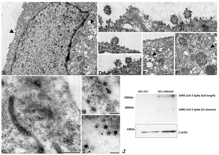
Figure 4.
SARS-CoV-2 virions can be detected in GCs infected cells. (A–F) Representative electron microscopy images of GCs infected with SARS-CoV2 for 24 h. Viral particles are arranged around the cell membrane (A–D) and sometimes grouped in aggregates (B–D). Small viral infiltration clusters can be observed within the cytoplasmic compartment (E–F), and the ultrastructural organization is not altered (A bar = 1 µm; B–F, bar 500 nm). (G–I) Post-embedding immunogold electron microscopy localization of the spike protein. Localization of spike protein was shown in the cytoplasm of GCs (G, bar = 200 nm); in some cases, gold immunolocalization was observed close to viral particles (H,I, bar = 50 nm). (J) Western blot of SARS-CoV-2 spike in human GCs uninfected (GCs Ctrl) or infected with SARS-CoV-2 (GCs infected) for 24 h. The image shows a band of about 170 kD supporting the proteolytic activation of spike associated with viral entry. Equal protein loading of the two preparations was verified using the housekeeping beta-actin. Western blot was repeated twice on a pool of 3 patients.

