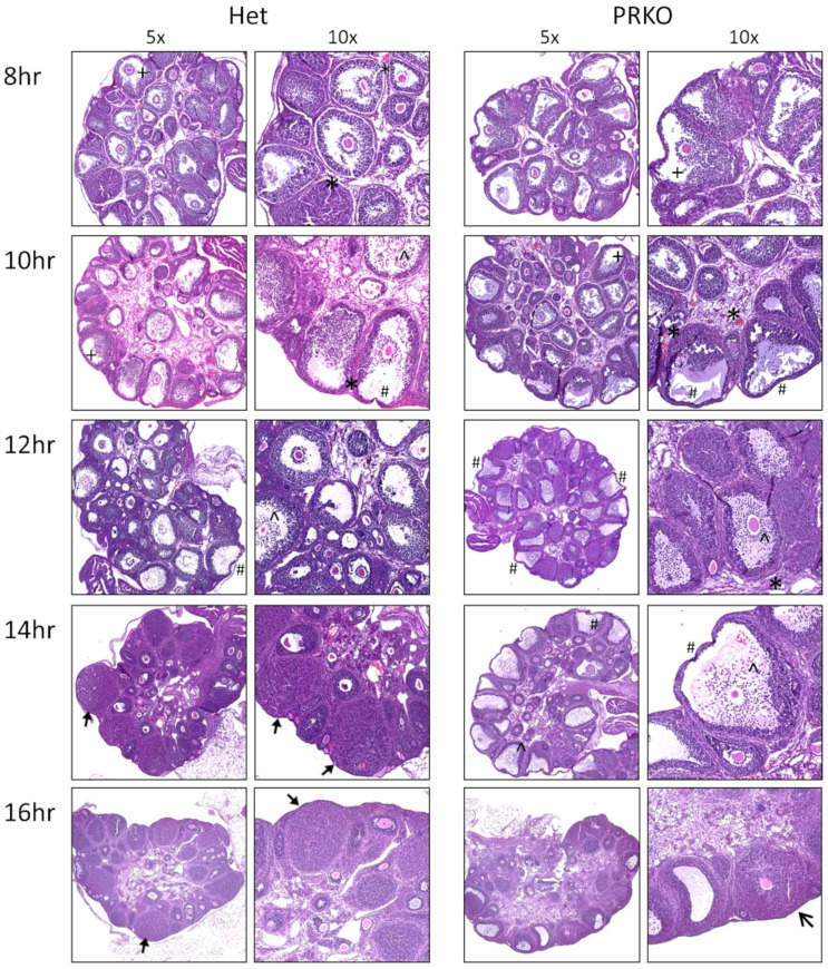
Figure 2.
Histological analysis of PRKO ovaries demonstrates specific defect in follicular rupture. Ovaries were collected from PRKO and heterozygous (Het) littermates at 8, 10, 12 h timepoints post-hCG and 14 h, and 16 h timepoints, after ovulation has normally occurred. Ovary sections were stained with H&E to analyze histological phenotypes including ^ COC expansion, + granulosa cell accumulation at the follicle base, # thinning of the apical wall, * vascularization, luteinization, unruptured luteinized follicles. Representative examples from histology performed on ovaries from 3 mice per timepoint.

