Abstract
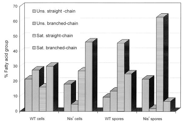
The membrane fatty acids, thermal resistance, and germination of a nisin-resistant (Nisr) mutant of Clostridium botulinum 169B were compared with those of the wild-type (WT) strain. In the membranes of WT cells, almost 50% of the total fatty acids were unsaturated, but in those of Nisr cells, only 23% of the fatty acids were unsaturated. WT and Nisr spores contained similar amounts (approximately 23%) of unsaturated fatty acids, but the saturated straight-chain/branched-chain ratio was significantly higher in Nisr spores than in WT spores. These fatty acid differences suggest that Nisr cell and spore membranes may be more rigid, a characteristic which would interfere with the pore-forming ability of nisin. Nisr C. botulinum did not produce an extracellular nisin-degrading enzyme, nor were there any differences in the sodium dodecyl sulfate-polyacrylamide gel electrophoresis patterns of coat proteins extracted from WT and Nisr spores, eliminating these as possible reasons for nisin resistance. Nisr spores had thermal resistance parameters similar to those of WT spores. In WT spores, but not in Nisr spores, nisin caused a 40% reduction in thermal resistance and a twofold increase in the germination rate. Because the nisin-induced increase in the germination rate of WT spores occurred only in the presence of a germinant (a molecule that triggers germination), nisin can be classified as a progerminant (a molecule that stimulates germination only in the presence of a germinant).
The action of nisin, a bacteriocin with “Generally Recognized as Safe” status, against vegetative cells has been extensively studied. The addition of nisin to cells causes the efflux of cations and amino acids (39), the efflux of ATP (51), and the dissipation of the proton motive force (5, 36). Nisin also induces the release of entrapped small molecules from artificial lipid vesicles in a time- and concentration-dependent fashion (12, 13, 52). The cytoplasmic membrane is widely regarded as the primary target of nisin, and a model for the action of nisin on cells suggests that nisin acts through a multistep process, which includes the binding of nisin to the cell, insertion into the membrane, and pore formation (13, 41, 51). The formation of pores by nisin in cell membranes, membrane vesicles, and liposomes is widely accepted (2, 10, 13, 30, 42, 51). However, much less is known about the action of nisin against spores.
Nisin can prevent Clostridium botulinum spore outgrowth and toxin production under certain conditions (32, 35, 43, 44, 46–48). Nisin decreases the D value (defined as the time required at a given temperature to decrease the number of viable organisms 10-fold) of spores from spoilage bacteria (6, 7, 23). Heated spores are very sensitive to nisin (16), and nisin-treated spores become more heat sensitive (49). Spore sensitivity to nisin increases with heating independently of the intrinsic heat resistance of the spores. When Heinemann and coworkers studied four sporeformers (16), the most heat-resistant organism, Bacillus stearothermophilus, was the most sensitive to the action of nisin, and the least heat-resistant organism, B. coagulans, had intermediate nisin sensitivity. Ramseier (37) suggested that spores are insensitive to nisin when dormant but become sensitive upon germination. Spores exposed to nisin turn phase dark, lose their heat resistance, and halt outgrowth when they begin to swell (14, 24). Nisin interacts with sulfhydryl groups in membranes of freshly germinated spores (25, 33), but the effect of this interaction is unclear. The action of nisin on germination per se is examined for the first time in this paper.
The emergence of nisin-resistant (Nisr) pathogens would threaten the safety of nisin-preserved products and compromise a key component in many multiple-level preservation systems. Knowledge of the physiological characteristics of nisin resistant strains and the conditions that prevent the emergence of nisin resistance can establish usage conditions to minimize this problem. In Listeria monocytogenes, resistance to nisin correlates with changes in membrane fatty acid composition, phospholipid composition, membrane fluidity, and the cell wall and requires divalent cations (8, 9, 26, 29, 50). It is thus clearly established that nisin resistance is a complex phenotype and may be the cumulative result of multiple phenotypic changes (8, 50). Although nisin-resistant strains of L. monocytogenes are stable after growth for many generations in the absence of nisin (15, 26), there is no genetic evidence on whether this change corresponds to a single mutation or multiple mutations.
In contrast, studies on nisin resistance in sporeformers and particularly in C. botulinum are limited. Type A and type B C. botulinum strains have higher frequencies of nisin resistance than strains of L. monocytogenes and other spore-forming bacteria, and higher nisin concentrations are required to minimize the emergence of resistant strains (27). Stable, toxigenic Nisr C. botulinum strains were previously isolated in our laboratory. Spores produced from nisin-resistant vegetative cells are also nisin resistant (27). Nisin resistance in spores is especially difficult to study because the spore germination process and the mechanism of action of nisin on spores are not well understood. A few studies on bacillus spores have reported that resistance to nisin is associated with an enzyme that is produced during germination (18) and that acts on the C-terminal lanthionine ring of nisin (19).
In this study, we characterize the phenotypic changes in Nisr C. botulinum 169B compared to the parental wild-type (WT) strain with respect to fatty acid composition, germination, and heat resistance in the presence and absence of nisin. The involvement of nisin-degrading enzymes and gross changes in spore coat proteins were excluded as factors in the nisin resistance of this strain.
MATERIALS AND METHODS
Bacterial strains and culture conditions.
Proteolytic C. botulinum 169B was obtained from the U.S. Department of Agriculture Eastern Regional Research Center (Wyndmoor, Pa.). Nisr C. botulinum 169B was previously isolated in our laboratory (27). Vegetative cells were maintained at 4°C in cooked-meat medium (Difco Laboratories, Detroit, Mich.). Spores were prepared by the biphasic method (3) and maintained at −80°C as spore suspensions (108 to 109 spores/ml) in distilled water. Spores were purified from vegetative cells and debris by suspension in an aqueous-polymer two-phase system (40). Pure spore suspensions were obtained by washing the polyethylene glycol phase with distilled water several times. Spores were heat shocked for 10 min at 80°C immediately prior to use. Lactobacillus sake ATCC 15521, the indicator strain in nisin assays (described below), was grown in lactobacillus MRS broth (Difco) at 30°C and maintained as stab cultures by adding 1.5% Bacto Agar (Difco) to the broth. Stab cultures were kept at 4°C. Stock cultures were maintained at −80°C with 20% glycerol added to the broth. In all of the experiments, C. botulinum manipulation and incubation were done with two anaerobic chambers containing an atmosphere of 5% H2, 10% CO2, and 85% N2 and connected by an air lock (Coy Laboratory Products, Inc., Grass Lake, Mich.). One chamber was maintained at 30°C for incubation, and the other was maintained at room temperature. Pure nisin (a gift from Aplin and Barrett, Trowbridge, England) solutions were prepared with 0.02 M HCl and 0.75% NaCl (nisin diluent). All media and reagents were prereduced in the room-temperature chamber for at least 24 h prior to use.
Fatty acid composition.
To determine if the Nisr strain had an altered lipid composition, the fatty acid compositions of WT and Nisr C. botulinum 169B were determined by gas-liquid chromatography of their methyl esters. The WT strain was grown in 200 ml of brain heart infusion (BHI) broth, and the Nisr strain was grown in the same broth containing 103 IU of nisin per ml. After overnight incubation in the anaerobic chamber, the cultures were centrifuged at 10,000 × g for 10 min, washed with 0.1% peptone water, and centrifuged again. A loop from each pellet was transferred to a glass tube with a Teflon-lined cap. Fatty acid methyl esters were prepared and extracted into hexane by a protocol supplied by MIDI, Inc. (Newark, Del.) (42a). Lipids resuspended in hexane–tert-butyl ether (1:1) were maintained at −20°C for no more than 2 weeks. The fatty acids were analyzed by Analytical Services, Inc. (Essex, Vt.). Values given are the means for three independent preparations, and variances were analyzed and compared by a general linear model procedure at the 95% confidence interval with SAS statistical software (SAS Institute Inc., Cary, N.C.). To examine the fatty acids of spores, spores were disrupted with five times their weight of glass beads (0.11 to 1.12 μm) in chloroform to form a paste (21). Chloroform was evaporated first under a stream of nitrogen and later in a vacuum centrifuge. The fatty acids were then extracted and analyzed by the same procedure as that used for vegetative cells.
Assay for a putative extracellular nisin-inactivating enzyme.
The possible involvement of a nisin-degrading enzyme was investigated with cultures and germinating spores of Nisr C. botulinum 169B by quantification of residual nisin after incubation of known nisin concentrations with culture supernatants and germinating spore exudates. Overnight cultures of WT and Nisr C. botulinum 169B grown in BHI broth without nisin were centrifuged, and the supernatants were sterilized by passage through 0.45-μm-pore-size syringe filters (Fisher) and adjusted to pH 7 with 0.1 M HCl. Spores were inoculated into fresh BHI broth to a final concentration of 108 spores/ml and incubated anaerobically at 30°C for 2 h. Spores were then centrifuged, and the germination exudate was filter sterilized and adjusted to pH 7. A solution containing 104 IU of nisin per ml was serially diluted in the cell supernatants, in the spore exudates, and in uninoculated broth to obtain final concentrations of 102, 103, and 104 IU of nisin per ml. Tubes were incubated for 2 h at 30°C. Cell supernatants and spore exudates were also incubated without nisin. The remaining nisin activity was then quantified from the diameter of the inhibition zone on a lawn of L. sake ATCC 15521, the indicator organism (38).
Extraction of spore coat proteins.
Spore coat proteins might also play a role in nisin resistance by binding nisin nonspecifically, as potential germinant receptors, or by other mechanisms. The coat protein patterns of WT and Nisr C. botulinum 169B spores were compared by sodium dodecyl sulfate (SDS)-polyacrylamide gel electrophoresis (PAGE) as described below. The exosporium layer was removed by the technique of Du and Nickerson (11) with some modifications. Briefly, pure spore suspensions were centrifuged at 10,000 × g. The wet weight of the spore pellet was recorded; glass beads (0.1 mm in diameter) suspended in distilled water were added in a 1:1 ratio (wt/wt). The mixture was cooled in an ice-water bath and homogenized twice on a Beadbeater (model 3110BX; Biospec Products, Bartlesville, Okla.) for 2 min each time with cooling in ice in between. The glass beads were removed by suspending the spores in an aqueous-polymer two-phase system as described above. The spores and exosporia (which were retained in the polyethylene glycol phase) were washed three times with distilled water to remove the polyethylene glycol. The exposure and debris were then separated by slow centrifugation (10 min, 1,200 × g) and washing of the spore pellet with distilled water. The spores were analyzed for purity under phase-contrast microscopy. The spore coat proteins were extracted with a detergent by the technique of Nicholson and Setlow (34). The protein concentration in the samples was determined by modified (20) Bradford assay (4) that makes use of the Coomassie Plus assay (Pierce, Rockford, Ill.) and bovine serum albumin as a standard. Coat proteins were resolved for visual analysis by SDS-PAGE (22) on 15% slab gels. Proteins on polyacrylamide gels were visualized by a silver stain procedure (28).
Determination of heat resistance of nisin-treated spores.
To determine if nisin resistance affected the heat resistance of spores, the heat resistance of WT and Nisr C. botulinum 169B was assayed at 80, 100, and 110°C. Pyrex tubing (Fisher) was cut into 6-in. lengths, sealed on one end, and autoclaved. Under anaerobic conditions, WT or Nisr C. botulinum 169B spores were suspended in germination buffer (see below) containing 103 IU of nisin per ml or an equal volume of nisin diluent as a control. Aliquots of 1.5 ml were then dispensed into separate series of tubes and immediately sealed by use of a torch outside the anaerobic chamber. The tubes were completely submerged in a water bath at 80°C or in an oil bath (model K6; MGW Lauda, Königshofen, Germany) at 100 and 110°C for different times. After the heat treatment, the tubes were cooled in a room temperature water bath for 5 s before being transferred to an ice-water bath for 2 min. Inside the anaerobic chamber, the tubes were cut open, and aliquots were spread plated from the appropriate decimal dilutions in sterile distilled water onto BHI agar containing 0.02% resazurin (Sigma Chemical Company, St. Louis, Mo.). Plates were incubated anaerobically at 30°C. After 48 h, the colonies formed were counted, and the log CFU per milliliter was plotted against time to calculate the D values by linear regression. Values given are the means for three independent experiments, and variances were analyzed and compared by a general linear model procedure at the 95% confidence interval with SAS statistical software.
Measurement of spore germination.
Spore germination was studied because its stimulation or inhibition could play an important role in nisin action and resistance. To quantify germination by the phase darkening of spores, WT and Nisr C. botulinum 169B spores were suspended under anaerobic conditions in germination buffer containing 103 IU of nisin per ml or an equal volume of nisin diluent as a control. Germination buffer contained 100 mM 2-(N-morpholino)-ethanesulfonate buffer at pH 6 and 100 mM l-alanine and 50 mM l-cysteine as germinants. Tubes were sealed with rubber stoppers in an anaerobic chamber to avoid the inclusion of oxygen during the germination experiment, which was conducted outside the chamber with a water bath at 40°C. At specific times during the incubation, 20-μl aliquots were removed from each tube through the rubber stopper by use of a sterile syringe. Germination was determined microscopically as a change in spores from phase bright to phase dark (31). Germination rates were calculated from the change in the percentage of germinated spores with respect to time. Extent of germination was the maximum percent of spores which germinated at the end of the experiment. The experiment was done in triplicate.
As an independent assay of germination, the efflux of dipicolinic acid (DPA) (which accompanies germination) was also measured. Spores of WT and Nisr C. botulinum 169B were centrifuged, the supernatant was discarded inside the anaerobic chamber, the spores were resuspended in germination buffer at 4 × 10⁹ spores/ml, and 3-ml quantities were dispensed into tubes containing 30 μl of a solution containing 104 IU of nisin per ml or 30 μl of nisin diluent. The tubes were sealed with rubber stoppers and incubated outside the anaerobic chamber with a water bath at 40°C. Samples of 0.5 ml were taken at different times and centrifuged in a microcentrifuge, and the release of DPA was measured by the colorimetric method of Janssen et al. (17). A calibration curve for pure DPA (Sigma) was constructed to determine the DPA released from the spores (34). The experiment was done in triplicate.
Safety considerations.
All experiments were done in compliance with Centers for Disease Control and Prevention Biosafety Level II guidelines in a university-approved facility by personnel immunized against botulinum toxin.
RESULTS
The major fatty acids of WT and Nisr C. botulinum 169B vegetative cells and spores are listed in Table 1 in the order of their retention times. Vegetative cells contained mainly 18-carbon fatty acids, whereas spores contained mainly 16-carbon fatty acids. The branched (cyclo) 19-carbon fatty acid found in vegetative cells of both strains was present at significantly lower levels in spores. Fatty acid 15:1, present as a low percentage in vegetative cells, was absent in spores. Spores contained the long straight-chain fatty acid 22:0, accounting for 8.58 and 15.75% of the total fatty acids in WT and Nisr spores, respectively. This fatty acid was not present in vegetative cells. The Nisr strain lacked the fatty acid 18:1 iso, which accounted for a high percentage of the total fatty acids in the WT strain. Other fatty acids varied in proportions from cells to spores and from WT to Nisr strains (Table 1).
TABLE 1.
Fatty acid compositions of WT and Nisr C. botulinum 169B vegetative cells and spores
| Sample | % (mean ± SD) (relative to total fatty acid) of:
|
|||||||||
|---|---|---|---|---|---|---|---|---|---|---|
| 13:1 | 14:0 | 3-OH 13:0 | 14:1 cis 7 | 15:1 | 15:0 | 16:1 | 16:0 | 2-OH 16:0 | 17:1 | |
| WT cells | 0.79 ± 0.12 | 2.78 ± 3.80 | 1.89 ± 0.73 | 2.48 ± 1.10 | 1.63 ± 0.93 | 2.91 ± 1.30 | 8.36 ± 0.21 | 8.51 ± 3.12 | 5.16 ± 5.1 | 2.64 ± 1.70 |
| Nisr cells | 3.58 ± 2.31 | 8.96 ± 0.51 | 17.25 ± 3.28 | 1.48 ± 0.73 | 0.33 ± 0.20 | 3.98 ± 2.13 | 4.87 ± 0.27 | 7.13 ± 2.98 | 6.72 ± 0.04 | 1.05 ± 0.98 |
| WT spores | 0.00 ± 0.00 | 31.2 ± 1.24 | 0.51± 0.08 | 0.00 ± 0.00 | 0.00 ± 0.00 | 6.20 ± 1.99 | 3.81 ± 1.74 | 20.73 ± 0.38 | 7.34 ± 0.52 | 0.00 ± 0.00 |
| Nisr spores | 0.24 ± 0.15 | 9.40 ± 1.81 | 0.56 ± 0.35 | 1.92 ± 1.49 | 0.00 ± 0.00 | 7.11 ± 0.79 | 11.45 ± 0.13 | 14.49 ± 1.92 | 5.41 ± 1.34 | 2.03 ± 0.55 |
| % (mean ± SD) (relative to total fatty acid) of:
| |||||||||
|---|---|---|---|---|---|---|---|---|---|
| 17:1 anteis | 17:0 cyc | 18:2 | 18:1 iso | 18:0 iso | 18:0 | 18:1 cis 9 | 19:0 iso | 19:0 cyc | 22:0 |
| 5.46 ± 1.50 | 4.73 ± 1.32 | 4.23 ± 4.32 | 19.64 ± 3.86 | 5.72 ± 1.21 | 1.90 ± 1.42 | 3.90 ± 2.14 | 3.95 ± 2.41 | 8.64 ± 0.25 | 0.00 ± 0.00 |
| 3.27 ± 3.01 | 3.60 ± 0.10 | 6.91 ± 2.59 | 0.00 ± 0.00 | 3.33 ± 2.14 | 6.90 ± 4.54 | 1.43 ± 0.57 | 2.24 ± 1.34 | 7.22 ± 2.27 | 0.00 ± 0.00 |
| 2.21 ± 0.86 | 2.37 ± 1.25 | 5.66 ± 4.42 | 11.32 ± 2.41 | 11.11 ± 4.98 | 7.07 ± 3.99 | 0.00 ± 0.00 | 2.08 ± 0.94 | 1.63 ± 0.14 | 8.58 ± 2.35 |
| 0.00 ± 0.00 | 0.00 ± 0.00 | 5.52 ± 5.86 | 0.00 ± 0.00 | 0.00 ± 0.00 | 16.16 ± 1.22 | 2.30 ± 1.03 | 0.43 ± 0.17 | 0.31 ± 0.41 | 15.75 ± 2.36 |
The data were grouped in four main classes based on whether the fatty acids were saturated or unsaturated and straight or branched (Fig. 1). Unsaturated fatty acids accounted for almost 50% of the total fatty acids in WT vegetative cells and, among them, the percentages of branched- and straight-chain fatty acids were similar. Unsaturated fatty acids comprised only ∼23% of the total fatty acids in spores and in Nisr vegetative cells. The ratio of unsaturated straight-chain to unsaturated branched-chain fatty acids was higher in the Nisr strain than in the WT strain. Nisr spores contained a higher percentage of straight-chain, saturated fatty acids than did WT spores.
FIG. 1.
Distribution of fatty acids into main groups (unsaturated [Uns.] straight-chain and branched-chain fatty acids and saturated [Sat.] straight-chain and branched-chain fatty acids) for WT and Nisr C. botulinum 169B vegetative cells and spores.
The pattern of silver-stained coat proteins from the Nisr strain separated by SDS-PAGE was identical to that for the WT strain (data not shown). The possible production of an extracellular nisin-degrading enzyme was investigated. The remaining nisin activity after incubation in the Nisr strain cell supernatant or germination exudates was not significantly different from the activity of an equivalent nisin concentration that remained after incubation in the WT controls (data not shown). C. botulinum did not produce any substance that inhibited the indicator organism, L. sake, and that could have interfered with this assay, because nisin-free supernatant from WT or Nisr cells or spores did not produce inhibition zones around the wells.
Table 2 shows the decimal reduction times (D values) for WT and Nisr C. botulinum 169B spores in the presence or absence of nisin at three different temperatures. In the absence of nisin, spores from both strains had similar heat resistance parameters. In the presence of nisin, the thermal resistance of WT spores was reduced by about 40% at all temperatures. Nisin did not reduce the thermal resistance of Nisr spores. The z values (defined as the number of degrees required to change the D value by a factor of 10) for both strains in the absence and presence of nisin were not significantly different. The z value for the WT strain in the absence of nisin was 10.9°C, and that in the presence of nisin was 10.7°C. For the Nisr strain, the z value was 10.9°C in the absence and in the presence of nisin.
TABLE 2.
Decimal reduction times (D values) for WT and Nisr C. botulinum 169B spores at three different temperatures in the presence or absence of 103 IU of nisin per mla
| Strain and condition |
D value (min) at temp (°C) of:
|
||
|---|---|---|---|
| 80 | 100 | 110 | |
| WT control | 883.7 ± 43.9 | 27.2 ± 7.9 | 1.4 ± 0.01 |
| WT + nisin | 577.0 ± 95.7b | 16.5 ± 3.6b | 0.8 ± 0.04b |
| Nisr control | 909.1 ± 0.1 | 23.5 ± 2.5 | 1.4 ± 0.06 |
| Nisr + nisin | 871.2 ± 53.6 | 22.9 ± 3.5 | 1.4 ± 0.06 |
Results represent the mean ± SD for three independent experiments.
Significantly different from values obtained with the other strains and conditions.
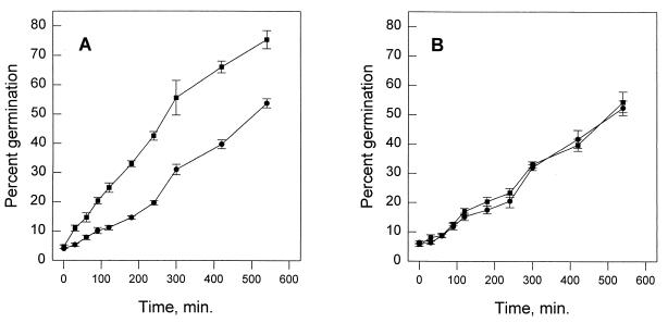
Results from three independent experiments in which germination was determined by phase-contrast microscopy are shown in Fig. 2. The germination rate was calculated from the slope of the percent germination-versus-time plot, which was linear to 300 min. The rate at which WT spores turned phase dark in germination buffer was 0.08% ± 0.002% min−1; in the presence of 103 IU of nisin per ml, the rate was 0.16% ± 0.016% min−1. The germination rate for Nisr spores in the absence of nisin was 0.080% ± 0.002% min−1; that in the presence of nisin was 0.087% ± 0.004% min−1. An analysis of variance (α, 0.05) showed that the only significant difference among the germination rates was when nisin was added to germination buffer for the WT strain. For this strain, the extent of germination also increased when nisin was incorporated in germination buffer. After incubation for 24 h, WT spores in the presence of nisin germinated to an extent of 81% ± 4%, while in the absence of nisin the extent of germination was only 60% ± 2%.
FIG. 2.
Percentage of germinated C. botulinum 169B spores at different times in the presence (■) or absence (•) of 103 IU of nisin per ml. Germinated (phase-dark) and nongerminated (phase-bright) spores were counted by phase-contrast microscopy. (A) Average of three independent experiments for WT spores. (B) Average of three independent experiments for Nisr spores. Bars show standard deviations.
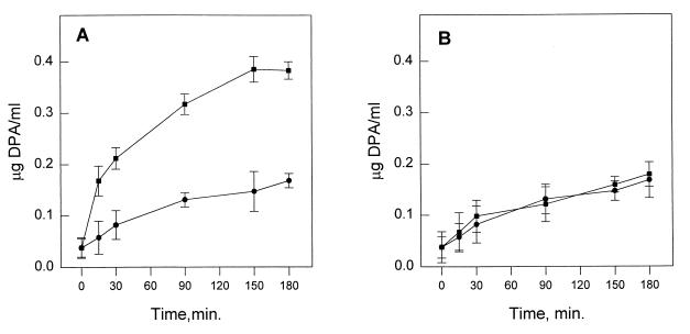
Nisin also increased the release of DPA from germinating WT spores. First, a release of DPA corresponding to 3.7 × 10⁻⁹ μg/spore occurred after the heat shock period. During the first 30 min of incubation after the addition of nisin to WT C. botulinum 169B, the release of DPA took place at a rate 5-fold higher than in the absence of nisin and continued at a rate 2.8-fold higher (Fig. 3A). Nisr spores released DPA at a slightly lower rate than WT spores and were not influenced by the addition of nisin (Fig. 2B). Neither germination nor efflux of DPA occurred when WT or Nisr spores were exposed to nisin in buffer that did not contain l-alanine and l-cysteine as germinants (data not shown).
FIG. 3.
DPA released into the supernatant in the presence (■) or absence (•) of 103 IU of nisin per ml from WT (A) and Nisr (B) C. botulinum 169B spores. Bars show standard deviations.
DISCUSSION
The phenotypic characterization of the Nisr strain in comparison with the WT strain was a useful tool for identifying components correlated with nisin resistance. The correlation of altered membrane lipid composition with nisin resistance was first established with L. monocytogenes nisin-resistant strains (8, 26, 29). The altered fatty acid composition of the nisin-resistant strain derived from C. botulinum 169B extends this correlation to another genera and to spores. In Nisr C. botulinum cells, the decrease in total and branched-chain unsaturated fatty acids and the corresponding increase in saturated fatty acids (relative to the levels in the nisin-sensitive WT strain) are consistent with an ordering of the fatty acids that would result in a more rigid membrane. The changes found in the fatty acid composition of Nisr spores were also consistent with increased membrane rigidity.
Nisr C. botulinum did not produce a nisin-degrading enzyme, since there was no loss of nisin activity after incubation of nisin in the Nisr cell supernatant and spore exudate. The Bacillus nisinase reported by Jarvis and Farr (19) acts as a dehydroalanine reductase in the C-terminal region of nisin. The enzyme was purified from a culture at the onset of sporulation. Since the spore coat proteins contain unusual amino acids (45), it is possible that the primary action of this enzyme is the packing of the spore coat during sporulation or the opening of the spore coat during germination. Nisin may be a secondary coincidental substrate. Moreover, a specific enzyme directed toward nisin dihydroamino acid residues cannot explain the cross-resistance of the Nisr strain to class IIa bacteriocins, which do not contain dehydroamino acids (27). No nisinases have been detected in nisin-resistant isolates from other species (26, 29).
The passage of nisin through the spore coat may be affected by changes in the coat protein composition. The characteristic proteins of the C. botulinum spore coat have not been studied, but the proteins of C. botulinum 169B spores were similar for the Nisr and the WT strains, as determined by SDS-PAGE.
Nisr C. botulinum 169B spores were not more heat resistant than WT spores. Thus, the generation of Nisr C. botulinum would not pose a public health threat for thermally processed foods. Nisin did sensitize WT C. botulinum 169B spores to heat, reducing the D values by 40%, but the heat sensitivity of Nisr spores was not increased by nisin. While nisin decreased the D values of the WT spores, it did not affect the z values. Different z values would suggest different mechanisms of heat inactivation, such as are found when spores are heated in oils (1). The similar z values obtained for WT spores in the presence or absence of nisin suggest that nisin does not change the mechanism of thermal inactivation.
When the germination responses of Nisr spores were compared to the germination responses of WT spores in buffer containing l-alanine and l-cysteine, the germination responses were similar, and nisin did not affect the rate nor the extent of germination in Nisr spores. However, the results for germination in WT spores brought a new insight into the action of nisin. Nisin doubled their germination rate and increased the extent of germination. Since these actions required an exogenous germinant (l-alanine or l-cysteine), nisin can be characterized as a potent progerminant in WT spores.
The progerminant action of nisin may explain the increased heat sensitivity of the nisin-treated spores. Changes that make Nisr spores resistant to nisin appear to prevent this progerminant action, so that Nisr spores are not sensitized by nisin during heat treatment. In the presence of nisin, the increased darkening of the WT spores correlates with the faster release of dipicolinic acid, and the loss of the dormant properties may be the underlying cause of the increased heat sensitivity of the nisin-treated spores. Since nisin also sensitizes Bacillus spores to heat (7), it is possible that this progerminant action is a general mechanism by which nisin sensitizes spores to heat. Nisin may then arrest the outgrowth of the spores by forming pores in the membrane of the emerging cell. Nisr spores did not germinate faster in the presence of nisin and were not heat sensitized by the antimicrobial agent. In practical applications, this finding can be related to a “botulism risk.” Processing could achieve the same lethality at lower time-temperature combinations if nisin were added to the product. However, if nisin-resistant strains emerged, the reduced process would be inadequate. Nevertheless, the emergence of nisin-resistant spores can be minimized by manipulating different environmental conditions (27).
ACKNOWLEDGMENTS
Research in our laboratory and preparation of the manuscript were supported by state appropriations, U.S. Hatch Act Funds, and the U.S. Department of Agriculture CSRS NRI Food Safety Program (grant 94-37201-0994).
We thank Peter Setlow for advice during the course of this research.
Footnotes
Manuscript D-10974-1-98 of the New Jersey Agricultural Experiment Station.
REFERENCES
- 1.Ababouch L H, Grimit L, Eddafry R, Busta F F. Thermal inactivation kinetics of Bacillus subtilis spores suspended in buffer and in oils. J Appl Bacteriol. 1995;78:669–676. doi: 10.1111/j.1365-2672.1995.tb03114.x. [DOI] [PubMed] [Google Scholar]
- 2.Abee T. Pore-forming bacteriocins of Gram-positive bacteria and self-protection mechanisms of producer organisms. FEMS Microbiol Lett. 1995;129:1–10. doi: 10.1016/0378-1097(95)00137-T. [DOI] [PubMed] [Google Scholar]
- 3.Anellis A, Berkowitz D, Kemper D, Rowley D B. Production of Clostridium botulinum type A and B spores by the biphasic methods: effects on spore population, radiation resistance, and toxigenicity. Appl Microbiol. 1972;23:734–739. doi: 10.1128/am.23.4.734-739.1972. [DOI] [PMC free article] [PubMed] [Google Scholar]
- 4.Bradford M M. A rapid and sensitive method for the quantitation of microgram quantities of protein utilizing the principle of protein-dye binding. Anal Biochem. 1976;72:248–254. doi: 10.1016/0003-2697(76)90527-3. [DOI] [PubMed] [Google Scholar]
- 5.Bruno M E, Kaiser A, Montville T J. Depletion of proton motive force by nisin in Listeria monocytogenes cells. Appl Environ Microbiol. 1992;58:2255–2259. doi: 10.1128/aem.58.7.2255-2259.1992. [DOI] [PMC free article] [PubMed] [Google Scholar]
- 6.Campbell L L, O’Brien R T. Antibiotics in food preservation. Food Technol. 1955;9:461–465. [Google Scholar]
- 7.Campbell L L, Sniff E E, O’Brien R T. Subtilin and nisin as additives that lower the heat-process requirements of canned foods. Food Technol. 1959;13:462–464. [Google Scholar]
- 8.Crandall A D, Montville T J. Nisin resistance in Listeria monocytogenes ATCC 700302 is a complex phenotype. Appl Environ Microbiol. 1998;64:231–237. doi: 10.1128/aem.64.1.231-237.1998. [DOI] [PMC free article] [PubMed] [Google Scholar]
- 9.Davies E A, Falahee M B, Adams M R. Involvement of the cell envelope of Listeria monocytogenes in the acquisition of nisin resistance. J Appl Bacteriol. 1996;81:139–146. doi: 10.1111/j.1365-2672.1996.tb04491.x. [DOI] [PubMed] [Google Scholar]
- 10.Driessen A J, Van Den Hooven H W, Kuiper W, Van De Kamp M, Sahl H G, Konings R N, Konings W N. Mechanistic studies of lantibiotic-induced permeabilization of phospholipid vesicles. Biochemistry. 1995;34:1606–1614. doi: 10.1021/bi00005a017. [DOI] [PubMed] [Google Scholar]
- 11.Du C, Nickerson K W. Bacillus thuringiensis HD-73 spores have surface-localized Cry1Ac toxin: physiological and pathogenic consequences. Appl Environ Microbiol. 1996;62:3722–3726. doi: 10.1128/aem.62.10.3722-3726.1996. [DOI] [PMC free article] [PubMed] [Google Scholar]
- 12.Gao F J, Abee T, Konings W N. Mechanism of action of the peptide antibiotic nisin in liposomes and cytochrome c oxidase-containing proteoliposomes. Appl Environ Microbiol. 1991;57:2164–2170. doi: 10.1128/aem.57.8.2164-2170.1991. [DOI] [PMC free article] [PubMed] [Google Scholar]
- 13.Garcia Garcera M J, Elferink M G L, Driessen A J M, Konings W N. In vitro pore-forming activity of the lantibiotic nisin. Role of protonmotive force and lipid composition. Euro J Biochem. 1993;212:417–422. doi: 10.1111/j.1432-1033.1993.tb17677.x. [DOI] [PubMed] [Google Scholar]
- 14.Gould G W. Effect of food preservatives on the growth of bacteria from spores. In: Molin M, editor. Microbial inhibitors in food. IVth International Symposium on Food Microbiology. Stockholm, Sweden: Almquist and Wiksel; 1964. pp. 17–24. [Google Scholar]
- 15.Harris L J, Fleming H P, Klaenhammer T R. Sensitivity and resistance of Listeria monocytogenes ATCC 19115, Scott A and UAL 500 to nisin. J Food Prot. 1991;54:836–840. doi: 10.4315/0362-028X-54.11.836. [DOI] [PubMed] [Google Scholar]
- 16.Heinemann B, Voris L, Stumbo C R. Use of nisin in processing food products. Food Technol. 1965;19:592–596. [Google Scholar]
- 17.Janssen F W, Lund A J, Andreson L E. Colorimetric assay for dipicolinic acid in bacterial spores. Science. 1958;127:26–27. doi: 10.1126/science.127.3288.26. [DOI] [PubMed] [Google Scholar]
- 18.Jarvis B. Resistance to nisin and production of nisin-inactivating enzymes by several Bacillus species. J Gen Microbiol. 1967;47:33–48. doi: 10.1099/00221287-47-1-33. [DOI] [PubMed] [Google Scholar]
- 19.Jarvis B, Farr J. Partial purification, specificity and mechanism of action of the nisin-inactivating enzyme from Bacillus cereus. Biochim Biophys Acta. 1971;227:232–240. doi: 10.1016/0005-2744(71)90056-8. [DOI] [PubMed] [Google Scholar]
- 20.Kaiser A, Montville T J. Purification of the bacteriocin bavaricin MN and characterization of its mode of action against Listeria monocytogenes Scott A cells and lipid vesicles. Appl Environ Microbiol. 1996;62:4529–4535. doi: 10.1128/aem.62.12.4529-4535.1996. [DOI] [PMC free article] [PubMed] [Google Scholar]
- 21.Kimble C E, McCollough M L, Paterno V A, Anderson A W. Comparison of the fatty acids of proteolytic type B and nonproteolytic types E and F of Clostridium botulinum. Appl Microbiol. 1969;18:883–888. doi: 10.1128/am.18.5.883-888.1969. [DOI] [PMC free article] [PubMed] [Google Scholar]
- 22.Laemmli U K. Cleavage of structural proteins during the assembly of the head of bacteriophage T4. Nature (London) 1970;227:680–685. doi: 10.1038/227680a0. [DOI] [PubMed] [Google Scholar]
- 23.Lewis J C, Michener H D, Stumbo C R, Titus D S. Additives accelerating death of spores by moist heat. J Agric Food Chem. 1954;2:298–301. [Google Scholar]
- 24.Lipinska E. Nisin and its applications. In: Woodbine M, editor. Antibiotics and antibiosis in agriculture. London, England: Butterworths; 1977. pp. 103–130. [Google Scholar]
- 25.Liu W, Hansen J N. Enhancement of the chemical and antimicrobial properties of subtilin by site directed mutagenesis. J Biol Chem. 1992;267:25078–25085. [PubMed] [Google Scholar]
- 26.Mazzotta A S, Montville T J. Nisin induces changes in membrane fatty acid composition of Listeria monocytogenes nisin-resistant strains at 10°C and 30°C. J Appl Microbiol. 1997;82:32–38. doi: 10.1111/j.1365-2672.1997.tb03294.x. [DOI] [PubMed] [Google Scholar]
- 27.Mazzotta A S, Crandall A D, Montville T J. Nisin resistance in Clostridium botulinum spores and vegetative cells. Appl Environ Microbiol. 1997;63:2654–2659. doi: 10.1128/aem.63.7.2654-2659.1997. [DOI] [PMC free article] [PubMed] [Google Scholar]
- 28.Merril C R, Dunau M L, Goldman D. A rapid sensitive silver stain for polypeptides in polyacrylamide gels. Anal Biochem. 1981;110:201–207. doi: 10.1016/0003-2697(81)90136-6. [DOI] [PubMed] [Google Scholar]
- 29.Ming X, Daeschel M A. Nisin resistance of foodborne bacteria and the specific resistance responses of Listeria monocytogenes Scott A. J Food Prot. 1993;56:944–948. doi: 10.4315/0362-028X-56.11.944. [DOI] [PubMed] [Google Scholar]
- 30.Moll G N, Roberts G C, Konings W N, Driessen A J. Mechanism of lantibiotic-induced pore-formation. Antonie Leeuwenhoek. 1996;69:185–191. doi: 10.1007/BF00399423. [DOI] [PubMed] [Google Scholar]
- 31.Montville T J. Characterization of a halo-acid-tolerant variant of Clostridium botulinum B-aphis. Appl Environ. 1984;48:311–316. doi: 10.1128/aem.48.2.311-316.1984. [DOI] [PMC free article] [PubMed] [Google Scholar]
- 32.Montville T J, Rogers A M, Okereke A. Differential sensitivity of Clostridium botulinum strains to nisin is not biotype-associated. J Food Prot. 1992;55:444–448. doi: 10.4315/0362-028X-55.6.444. [DOI] [PubMed] [Google Scholar]
- 33.Morris S L, Walsh R C, Hansen J N. Identification and characterization of some bacterial membrane sulfhydryl groups which are targets of bacteriostatic and antibiotic action. J Biol Chem. 1984;259:13590–13594. [PubMed] [Google Scholar]
- 34.Nicholson W L, Setlow P. Sporulation, germination and outgrowth. In: Harwood C R, Cutting S M, editors. Molecular biological methods for Bacillus. New York, N.Y: John Wiley & Sons, Inc.; 1990. pp. 391–450. [Google Scholar]
- 35.Okereke A, Montville T J. Bacteriocin-mediated inhibition of Clostridium botulinum spores by lactic acid bacteria at refrigeration and abuse temperatures. Appl Environ Microbiol. 1991;57:3423–3428. doi: 10.1128/aem.57.12.3423-3428.1991. [DOI] [PMC free article] [PubMed] [Google Scholar]
- 36.Okereke A, Montville T J. Nisin dissipates the proton motive force of the obligate anaerobe Clostridium sporogenes PA 3679. Appl Environ Microbiol. 1992;58:2463–2467. doi: 10.1128/aem.58.8.2463-2467.1992. [DOI] [PMC free article] [PubMed] [Google Scholar]
- 37.Ramseier H R. The action of nisin on Clostridium butyricum. Arch Mikrobiol. 1960;37:57. doi: 10.1007/BF00414627. [DOI] [PubMed] [Google Scholar]
- 38.Rogers A M, Montville T J. Improved agar diffusion assay for nisin quantification. Food Biotechnol. 1991;5:161–168. [Google Scholar]
- 39.Ruhr E, Sahl H G. Mode of action of the peptide antibiotic nisin and influence on the membrane potential of whole cells and cytoplasmic and artificial membrane vesicles. Antimicrob Agents Chemother. 1985;27:841–845. doi: 10.1128/aac.27.5.841. [DOI] [PMC free article] [PubMed] [Google Scholar]
- 40.Sacks L E, Alderton G. Behavior of bacterial spores in aqueous polymer two-phase systems. J Bacteriol. 1961;82:331–341. doi: 10.1128/jb.82.3.331-341.1961. [DOI] [PMC free article] [PubMed] [Google Scholar]
- 41.Sahl H G. Pore formation in bacterial membranes by cationic lantibiotics. In: Jung G, Sahl H G, editors. Nisin and novel lantibiotics. Leiden, The Netherlands: Escom Publishers; 1991. pp. 347–358. [Google Scholar]
- 42.Sahl H G, Kordel M, Benz R. Voltage-dependent depolarization of bacterial membranes and artificial lipid bilayers by the peptide antibiotic nisin. Arch Microbiol. 1987;149:120–124. doi: 10.1007/BF00425076. [DOI] [PubMed] [Google Scholar]
- 42a.Sasser M. MIDI Technical Note 101: identification of bacteria by gas chromatography of cellular fatty acids. Newark, Del: MIDI, Inc.; 1990. [Google Scholar]
- 43.Scott V N, Taylor S L. Effect of nisin on the outgrowth of Clostridium botulinum spores. J Food Sci. 1981;46:117–120. [Google Scholar]
- 44.Scott V N, Taylor S L. Temperature, pH and spore load effects on the ability of nisin to prevent the outgrowth of Clostridium botulinum spores. J Food Sci. 1981;46:121–126. [Google Scholar]
- 45.Setlow P. Spore structural proteins. In: Hoch J A, Losick R, Sonenshein A L, editors. Bacillus subtilis and other gram-positive bacteria: biochemistry, physiology, and molecular genetics. Washington, D.C: American Society for Microbiology; 1993. pp. 801–809. [Google Scholar]
- 46.Somers E B, Taylor S L. Further studies on the antibotulinal effectiveness of nisin in acidic media. J Food Sci. 1981;46:1972–1973. [Google Scholar]
- 47.Taylor L Y, Cann D D, Welch B J. Antibotulinal properties of nisin in fresh fish packaged in an atmosphere of carbon dioxide. J Food Prot. 1990;53:953–957. doi: 10.4315/0362-028X-53.11.953. [DOI] [PubMed] [Google Scholar]
- 48.Taylor S L, Somers E B, Krueger L A. Antibotulinal effectiveness of nisin-nitrite combinations in culture medium and chicken frankfurter emulsions. J Food Prot. 1985;48:234–239. doi: 10.4315/0362-028X-48.3.234. [DOI] [PubMed] [Google Scholar]
- 49.Thorpe R H. The action of nisin on spoilage bacteria. I. The effect of nisin on the heat resistance of Bacillus stearothermophilus. J Appl Bacteriol. 1960;23:136–139. [Google Scholar]
- 50.Verheul A, Russell N J, Van’t Hof R, Rombouts F M, Abee T. Modifications of membrane phospholipid composition in nisin-resistant Listeria monocytogenes Scott A. Appl Environ Microbiol. 1997;63:3451–3454. doi: 10.1128/aem.63.9.3451-3457.1997. [DOI] [PMC free article] [PubMed] [Google Scholar]
- 51.Winkowski K, Bruno M E C, Montville T J. Correlation of bioenergetic parameters with cell death in Listeria monocytogenes cells exposed to nisin. Appl Environ Microbiol. 1994;60:4186–4188. doi: 10.1128/aem.60.11.4186-4188.1994. [DOI] [PMC free article] [PubMed] [Google Scholar]
- 52.Winkowski K, Ludescher R D, Montville T J. Physicochemical characterization of the nisin-membrane interaction with liposomes derived from Listeria monocytogenes. Appl Environ Microbiol. 1996;62:323–327. doi: 10.1128/aem.62.2.323-327.1996. [DOI] [PMC free article] [PubMed] [Google Scholar]


