

Abstract
3D cell cultures are becoming the new standard for cell-based in vitro research, due to their higher transferrability toward in vivo biology. The lack of established techniques for the non-destructive quantification of relevant variables, however, constitutes a major barrier to the adoption of these technologies, as it increases the resources needed for the experimentation and reduces its accuracy. In this review, we aim at addressing this limitation by providing an overview of different non-destructive approaches for the evaluation of biological features commonly quantified in a number of studies and applications. In this regard, we will cover cell viability, gene expression, population distribution, cell morphology and interactions between the cells and the environment. This analysis is expected to promote the use of the showcased technologies, together with the further development of these and other monitoring methods for 3D cell cultures. Overall, an extensive technology shift is required, in order for monolayer cultures to be superseded, but the potential benefit derived from an increased accuracy of in vitro studies, justifies the effort and the investment.
Keywords: 3D cell culture, Non-destructive technology, in-vitro quantitative analysis
Introduction
Culturing cells in vitro is a cornerstone of biomedical research, which has been instrumental for furthering our scientific knowledge and has enabled the more ethical development of safe and effective drugs. Indeed, the possibility of maintaining cells in the laboratory has provided researchers with a platform for the study of physiological and pathological phenomena which was effective and inexpensive, and afforded them a higher level of control with respect to entire organisms.
In recent years, however, relevant differences between in vitro and in vivo behavior have emerged (Breslin & ODriscoll, 2013; Waring et al., 2015). These are generally attributed to the over simplification of 2D cell monolayers, which are unable to capture relevant features of in vivo biology and consequently results in poor translation of laboratory data in clinical studies (Honek, 2017; Liston & Davis, 2017; Menshykau, 2017; Jo et al., 2018).
The main limitation of 2D cell cultures is that the microenvironment where the cells are maintained completely disregards the contribution of the physical cues associated with the physiological extracellular matrix (ECM) on cell behaviour. This limits cell-to-cell signalling and impacts over their functions. For example Edmondson et al. (2014) showed that 2D cell culture can affect the expression of cell surface receptors which, in turn, could alter the response to drugs targeting these receptors (Attwood et al., 2020; Abbas et al., 2021). Furthermore, a comparison of 2D vs 3D cell cultures led to the identification of important differences in proliferation and treatment response (Li et al., 2021b) .
A partial solution to these issues involves substituting the flat polystyrene plates with micro or nano-patterned surfaces. While different techniques and materials can be used to produce the grooved pattern, and specific motifs tend to be better suited for different applications, this strategy has been shown to be effective in (i) recapitulating cell response to multi-cue environments, (ii) enhancing adhesion, proliferation, migration and stem-cell differentiation, and (iii) enable large scale cell alignment (Table 1).
Table 1. Micro and nano patterning effect on cell cultures.
| Application | References |
|---|---|
| Response to multi-cue environments | Van Der Putten et al. (2021), Chang & Taniguchi (2021) |
| Adhesion, proliferation, migration and | Han et al. (2021), Kang et al. (2021), Wang et al. (2021b) |
| Differentiation enhancement | Blin (2021), Nagayama & Hanzawa (2022) |
| Large-scale cell alignment | Yu et al. (2021) |
3D cell culture models represent a more comprehensive solution. These setups integrate additional features of biological tissues (e.g., tridimensional cell disposition, interaction among cells and with the surrogate extracellular matrix, non-uniform nutrients availability) and as such have been shown to reduce the gap between in vitro and in vivo behavior (Duval et al., 2017; Kapałczyńska et al., 2018; Nunes et al., 2019; Costard et al., 2021; Belfiore et al., 2021).
The widespread use of these setups and their integration in the drug development pipeline are however hampered by the techniques used for their analysis. Indeed, methods that require the fixation or disgregation of the culture can be generally considered a standard de facto. This inability to monitor 3D cell cultures over time affects the accuracy of the results, as small differences in initial cell number or structure size cannot be accounted for. Additionally, a larger number of constructs will be needed, hence increasing the resources necessary to conduct the experiments.
In an effort to overcome this adoption barrier, in this review paper, we showcase non-destructive techniques for the analysis and characterization of relevant features in 3D cell cultures. Each section analyzes a specific kind of assay and a comparative analysis of non-destructive and destructive techniques will be presented, together with its perspective evolution in the near future. Additionally, an introductory paragraph, aimed at describing the main 3D cell culture approaches currently available, and a concluding section summarizing the status of non-destructive analysis of 3D cell cultures have been included.
3D Cell Culture Models
While a comprehensive analysis of 3D cell culture methods is beyond the scope this review, in this section we provide an overview of the most commonly available methods, to lay the foundation for the understanding of this work. For a more comprehensive description of 3D cell culture technologies, the reader is referred to other recently published review papers such as (Lv et al., 2017; Badekila, Kini & Jaiswal, 2021; Duval et al., 2017).
3D cell cultures are classically divided between scaffold-based (SB), that is relying on an external support material, and scaffold-free (SF), where cells self-assemble in clusters or spheroids (Fig. 1). Within these groups, multiple practical protocols are available.
Figure 1. Summary of 3D cell culture models.

In (A) the scaffold-based (SB) setups are presented, while (B) and (C) illustrate scaffold free (SF) and hybrid methods, respectively.
For SB systems, the scaffolds can either be produced independently from cell seeding (Liverani et al., 2019; Dinescu et al., 2019) or aggregated with cells already included (Dollinger et al., 2017; Pasini et al., 2021). The former generally grants more flexibility in scaffold production, allowing for a wider range of materials and technologies to be used. Non-vital techniques, such as freeze-drying, two- photons polymerization, and electrospinning enable the fine-tuning of the scaffold’s mechanical properties and superficial features (Remuzzi et al., 2020; Dinescu et al., 2019; Wang et al., 2021a), which results in structures that are stable over time and well suited to withstand mechanical stimulation (Lovecchio et al., 2019).
Including cells in the scaffold’s liquid phase tends to be associated with their more uniform distribution throughout the structure, as they can be dispersed within the solution, rather than having to migrate from the surface. This feature limits the possible scaffold materials to polymers that become solid through temperature changes or gentle chemical processes that do not compromise cell viability. The resulting structure, also called hydrogel, is often less structurally robust and more prone to macroscopic changes in size due to cell activity (James-Bhasin, Siegel & Nazhat, 2018).
A fundamental advancement in scaffold production is represented by bioplotting, which relies on fused deposition modeling (FDM) technology to print 3D structures with high precision and fidelity (Buenzli et al., 2020). In most cases, cells can be directly dispersed within the ink, thus combining high accuracy in scaffold production techniques with uniform cell distribution (Ahlfeld et al., 2020).
SF set-ups, on the other hand, rely on cells’ self-aggregation/organization properties to produce approximately spherical 3D cultures. These are generally smaller than the ones obtained with SB approaches, with starting populations of about 10 k cells (Shoval et al., 2017; Gamerith et al., 2017; Ahmad et al., 2017; Skeberdyté et al., 2018) as opposed to 100 k cells (Pasini et al., 2021; Picone et al., 2020; Rivero et al., 2020), but tend to be simple and, in some cases, well suited for high throughput analyses (Benien & Swami, 2014). The reduced size of SF cultures has however been shown to be an important drawback of these systems, as the number of cells that they can support is very low, when compared with those that compose human tissues (De Pieri, Rochev & Zeugolis, 2021). Furthermore, the lack of independent support structure results in densely packed cells which can be associated with the presence of hypoxic cores and macromolecular crowding (De Pieri, Rochev & Zeugolis, 2021), that further reduce the accuracy of these systems.
The ease of production of SF 3D cell cultures, which relies on various methods to prevent or disrupt cell adhesion to the culturing surface, however, makes them an attractive setup. Low attachment plates are widely used in this regard (Takagi et al., 2007), as the hydrophilic polymer coating on their surface prevents cells from attaching and thus enables spheroid formation. This approach is simple, and generally leads to spheroid formation within a few hours, but provides little control on the size of each culture and thus tends to be associated with higher variability. To address this limitation, culture surfaces micro-patterned with microscopic valleys of defined shape and size can be used. In this case, cells are generally allowed to adhere to a micro-patterned thermosensitive substrate, to ensure an approximately uniform cell density within each valley. The temperature of the structure is then changed, to induce a shape change in the substrate which, in turn, leads to cell detachment and spheroid formation. An example of this approach is reported in Kim et al. (2018a), where decreasing the temperature to 4 °C led to a significant increase in micropattern area which resulted in spheroid formation within minutes. Hanging drops is another widely used method in which cells are cultured within a suspended drop of medium and aggregate, due to gravity, to its bottom (Wang et al., 2017; Cho et al., 2020; Zibaei et al., 2021). This technique was developed at the beginning of the twentieth century to visualize small live organisms and study their motility (Jain, Jain & Jain, 2020) and then adapted to the production of spheroids.
The dichotomy between SB and SF methods, however, is not perfect and numerous approaches combine elements typical of both methods. Indeed, polymeric mixtures such as Matrigel can be used when producing spheroids to allow for the aggregation of more cells and increase repeatability (Badea et al., 2019). At the same time, integrating external support structures and cells’ self-aggregating properties results in organoids, 3D cultures integrating multiple cell types that aim at replicating the structure and functionality of organised tissues (Lu et al., 2019; Asai et al., 2017). Microspheres are another notable example, as they serve the double purpose of cell culture support and precision delivery system (Dong et al., 2021). Many different techniques are available for their production (Zhang et al., 2021), and some of them allow for the inclusion of living cells which have been shown to be able to survive, proliferate and differentiate within these setups (Zhang et al., 2018).
This wealth of options for 3D cultures effectively allows to choose the best set-up for each application, as different models are best suited to replicate specific aspects of the corresponding in vivo system (Kapałczyńska et al., 2018). At the same time, the lack of a single established substitute for 2D cultures has contributed to delaying the introduction of 3D cultures in the drug development pipeline (Kelm et al., 2019).
This work, and the experimental techniques it showcases, are expected to contribute to the translational value of 3D cell cultures and their general applicability in biomedical research.
Cell Viability and Proliferation
The evaluation of cell viability is key to a number of in vitro studies. Indeed, it is used to test the feasibility of new cell culture methods (Yu et al., 2020; Paez et al., 2020), assess the efficacy of potential therapeutic treatments (Marrella et al., 2021; Yang et al., 2020), and generally monitor the health and status of a population of cells (Vitale et al., 2020; Rivero et al., 2020).
The most common approaches for the evaluation of cell viability are metabolic membrane structural competence and DNA-content based assays which repeated over time allow for the quantification of proliferation (Table 2).
Table 2. Cell viability and proliferation assays.
Bromodeoxyuridine (BrdU) assay is another established way of quantifying cell proliferation, through the visualisation of newly synthetised DNA in proliferating cells (Table 2).
Metabolic methods are a class of assays in which an optical (absorbance or fluorescence) signal, proportional to the number of living cells, is generated through a chemical modification of a soluble reagent added to the culture. These methods were initially developed for 2D cultures and, depending on the reagent’s composition, can preserve cell viability. One of the main drawbacks of translating this approach to 3D cell cultures is that it requires the reagent to diffuse through the whole structure. This condition might not be verified, especially for larger SB cultures, thus the results might be partial or inaccurate. For this reason, live-dead staining tends to be preferred. This method consists in staining a thin slice of the culture with both calcein-AM and ethidium homodimer-1. These compounds produce strong fluorescent signals, at different wavelengths, when reacting with alive or dead cells respectively and can thus be used to quantify their density.
This technique has the advantage of maintaining cell spatial distribution, and thus allows to study how different positions within the structure affect cell behaviour. On the other hand, it does not preserve cell viability and requires the sectioning of the culture, thus making this method poorly suited for the long term monitoring or high throughput experiments.
Another class of non-viable methods for viability quantification relies on the evaluation of DNA content using fluorescent probes. These assays are well established, accurate, and can be used for 3D cell cultures with minimal adjustment with respect to 2D monolayers, but they require the DNA to be extracted, thus resulting in the impossibility of monitoring the same culture over time. Additionally, this is an indirect measure, that determines the number of cells as the ratio of total amount of DNA in the sample and the assumed quantity of DNA present within each cell. As the latter can vary widely between different cells (Gillooly, Hein & Damiani, 2015) and with the different phases of the cell cycle (Cooper, 2000), this technique could result in non-negligible errors, especially when comparing samples where cells have different sizes or where the length of the cell cycle might be affected.
BrdU assay directly quantifies proliferation through a thymidine analog that, when added to the culture, is incorporated in the DNA of proliferating cells. The quantification of the BrdU, however, is based on immunostaining, which requires cells to be fixed and permeabilized, in order for antibodies to effectively bind their target. While DNA probes capable of penetrating live cells have been developed (Veetil et al., 2020; Bucevičius, Gilat & Lukinavicius, 2020), their use is still fairly limited and generally confined within 2D monolayers.
The vital approaches for the quantification of cell viability and proliferation can be divided in three main classes: (i) chemical, (ii) optical and (iii) electrical.
Chemical assays (Fig. 2A) rely on the quantification of Lactate Dehydrogenase (LDH) in the culture’s supernatant (Lam et al., 2020; Vormann et al., 2018; Liaudanskaya et al., 2019; Liaudanskaya et al., 2020). This enzyme, present in all cells, is released in the media upon membrane damage and is thus an effective indicator of cell death (Kumar, Nagarajan & Uchil, 2018). For this reason, LDH quantification is particularly suited for the monitoring of cytotoxicity when testing new pharmacological treatments. Additionally, being a measurement conducted on the culture’s media, it eliminates any possible interference of the acquisition on cellular function and easily adapts to high throughput screenings and long term monitoring. On the flip side, it is an indirect method and it requires a non-negligible amount of cell death. As such, its scope of application is rather narrow and mainly focused on drug testing. The estimate of LDH concentration, furthermore, is an average of the whole culture that cannot capture behaviourally different microenvironments within the culture.
Figure 2. Non-destructive methods for viability quantification.

(A) Working principle of the LDH assay. (B) Representative image of a 3D cell culture obtained with light-sheet microscopy. Image reproduced from Alladin et al. (2020). (C) Example of 2D conductivity map reconstructed from EIT measurements. Image extracted from Wu et al. (2018).
Optical methods that preserve the culture’s structural integrity address this limitation (Li et al., 2016; Pan, Onda & Hirano, 2019; Christoffersson et al., 2019; Alladin et al., 2020). (Fig. 2B). They rely on advanced microscopy technologies (e.g., laser-based confocal, light-sheet) and fluorescent probes that preserve cell viability (Li et al., 2021c; Cheng et al., 2017) to produce a stack of images corresponding to different depths within the culture. This approach is characterized by high spatial resolution (∼100–500 nm Fouquet et al., 2015) and by the possibility of monitoring more than one variable at the same time. As an example, Liaudanskaya et al. (2020) combined the study of neural network destruction due to impact injury with the quantification of changes in collagen fibrillary structure, while Aguet et al. (2016) analysed how membrane remodeling changes throughout the cell cycle. The versatility and potential of these methods is, however, associated with lengthy and complex experimental procedures and with the need of acquiring highly specific, expensive instrumentation. In addition, the constraints on the maximum axial scan range of the microscope (i.e., the maximum depth that can be effectively imaged) make these techniques better suited for smaller SF cultures.
Electrical measurements represent an effective compromise. Indeed, while their resolution is not as high as that of the optical methods, they pose fewer limitations on culture size, and the instruments required for the measurement are not so expensive. This is partly due to the fact that impedance spectroscopy has been used for a number of years to monitor viability and growth in cell monolayers (Pérez et al., 2018; Voiculescu, Li & Nordin, 2020). The working principle behind this methods relies on the measurement of changes over time in electrical current, upon stimulation of the culture with the same sinusoidal voltage signal (Benson, Cramer & Galla, 2013; Elbrecht, Long & Hickman, 2016; Srinivasan et al., 2015). Cells are generally grown on conductive electrodes and their well defined electrical model, that combines resistant and capacitive elements, is used to infer the status of the culture. As such, the major issue when translating this approach in 3D is the lack of direct contact between the electrode and the cells. This is commonly addressed by including the whole culture (e.g., the scaffold or other extracellular components) in the measurement with the assumption of negligible changes in the electrical properties of the support structure (Pan et al., 2019; De León, Pupovac & McArthur, 2020; Pan et al., 2020). An alternative solution has been proposed by Inal et al. (2017), where a SB culture featuring a conductive polymer scaffold is presented.
Beside impedance spectroscopy, which provides a population-wise quantification of cell viability, electrical impedance tomography (EIT) has recently been proposed (Wu et al., 2018; Wu et al., 2019; Yang et al., 2019). This technique has been extensively used for the non-invasive visualization of internal structures of the human body (Shiraz et al., 2019; Murphy et al., 2017; Samoré et al., 2017), as it provides a map of the conductivity of one or more sections of the object of interest (Fig. 2C). While this resolution is not yet available for 3D cell cultures, where a uniform change in electrical properties is generally recovered, EIT holds great potential for the non-destructive monitoring of viability because changes in the setup (e.g., increasing the number of electrodes) and/or a more advanced reconstruction algorithm are expected to resolve this issue.
Several non-destructive approaches are already available for the monitoring of cell viability and proliferation. However, no method has emerged as overall better than the others and a case by case evaluation taking into account culture type and study aim is generally recommended.
Gene Expression
Gene expression profiles are an established approach for the quantitative evaluation of cell behaviour and how it changes upon external stimulation (Picone et al., 2020; Ciardulli et al., 2020) or when environmental conditions are modified (Gamerith et al., 2017; Brady et al., 2020; Lewis, Green & Shah, 2018).
As for the evaluation of cell viability, the most common approaches for the quantification of specific proteins or RNA sequences is associated with the destruction of the culture. Indeed a prerequisite of techniques such as micro-arrays, RT-PCR or RNA-sequencing is the lysis of the cells, while flow cytometry requires culture disgregation and immunohistochemistry/immunofluorescence, its fixation and slicing (Table 3). High throughput and transcriptome-wide technologies, are also widely used, but have similar requirements (Waylen et al., 2020).
Table 3. Gene expression assays.
| Assay | References |
|---|---|
| Microarray | Gamerith et al. (2017), Brady et al. (2020) |
| RT-PCR | Henriksson, Gatenholm & Hägg (2017), Zhou et al. (2017), Lewis, Green & Shah (2018) |
| He et al. (2020), Henrionnet et al. (2020), Brady et al. (2020) | |
| Devall et al. (2020), Schneider et al. (2020), Ciardulli et al. (2020) | |
| RNA sequencing | Devall et al. (2020) |
| Flow cytometry | Zhou et al. (2017) |
| Immunohistochemistry/ immunofluorescence | Zhou et al. (2017), Henrionnet et al. (2020), Brady et al. (2020) |
| Ciardulli et al. (2020), Schneider et al. (2020) |
The scarcity of RNA and proteins is often responsible for the need to have direct access to the cells or their content. Fluorescent probes able to enter the cells and bind to specific RNA sequences have been shown to provide sufficiently strong signals which can also be measured in entire organisms (Okamoto, 2019; Braselmann et al., 2018; Suseela et al., 2018; He et al., 2020). This feature suggests the potential transferability of this approach to 3D cell cultures even though specificity, accuracy and uptake efficiency can still be challenging and depend on the specific target.
An alternative strategy, that allows for the quantification of specific proteins, relies on genome editing techniques to couple the production of a fluorescent reporter to the expression level of a gene of interest (Koch et al., 2018; Di Blasi et al., 2021; Ceroni & Ellis, 2018). This approach is commonly employed in the synthetic biology field (Cortesi et al., 2017; Bandiera et al., 2016; Xu & Qi, 2019) and is characterized by high precision and accuracy. Modifying the genome of eukaryote/human cells is however an additional procedural step, which might not be feasible, or result in side interactions and unexpected behavioural changes.
Both vital fluorescent probes and gene editing allow for the possibility of monitoring gene expression changes over time and, preserving the distribution of cells within the culture, they allow to study how different microenvironments within the culture affect cell behaviour. They are however affected by the same limitations discussed in the previous section for optical methods. Indeed, advanced microscopy set-ups (e.g., confocal microscope) are needed to properly visualize the internal culture’s regions, and limitations on the maximum axial scan range might preclude the use of this technique for larger SB systems.
In addition, the number of genes that can be monitored at the same time is fairly limited, both due to the maximum number of fluorophores that can be effectively distinguished (about five but depends on instrument and application; Kleeman et al., 2018) and the need to modify each gene independently when a gene editing procedure is required.
As such, additional improvements are needed to open to the non-destructive large-scale evaluation of gene expression. A possible strategy to increase the throughput of these assays with technologies already available, relies on the combination of different readout methods. This approach has already been proposed for the visualization of complex structures within 3D plant tissues (Ursache et al., 2018) (Fig. 3), but it could be adapted, using vital fluorescence- and colorimetric-based methods, to the study of gene expression in 3D cell cultures. While potentially useful to increase the number of markers that can be monitored at one time, this approach has limited scalability and is substantially affected by the same limitations detailed above. As such transcriptome/proteome-wide vital analyses require significant advances with respect to current technology.
Figure 3. Image exemplifying the combination of fluorescent and colorimetric readout methods.
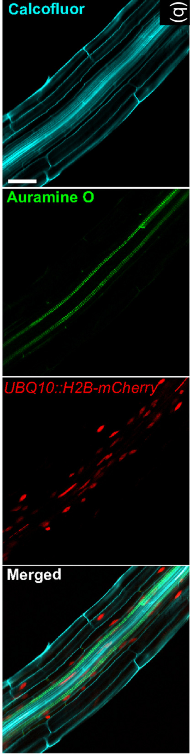
Image extracted from Ursache et al. (2018). © 2017 The Authors The Plant Journal © 2017 John Wiley and Sons Ltd.
Disposition and Morphology
A key feature of 3D cell cultures, that sets them apart from 2D approaches is the presence of gradients within the structure (e.g., nutrients, oxygen, waste products, drug levels) which effectively create multiple microenvironments capable of influencing cell behaviour. Hence the study of cell disposition within the culture, and how it changes over time acquires great relevance.
Optical methods are naturally suited for this analysis as they provide direct visualization of the culture. Common approaches involve staining of different cellular components (e.g., nucleus, cytoplasm, specific proteins) and their imaging through different microscopy techniques (Table 4). These methods were initially developed for tissue slices and as such require the sectioning of the culture. They are, however, fairly straightforward and a wealth of resources are available for their optimization and execution. More advanced techniques, like light-sheet and two-photon microscopy have also been used to study cell disposition within the culture (Table 4). These methods afford more comprehensive results, as the whole structure can be taken into account, but are associated with higher costs and the need for specific instrumentation.
Table 4. Cell disposition assays.
| Assay | References |
|---|---|
| Staining | Griffith & Swartz (2006), Trappmann et al. (2017), Liverani et al. (2019) |
| Shichi et al. (2019), Zoetemelk et al. (2019), Bassi et al. (2020) | |
| Lamparelli et al. (2021), Pasini et al. (2021) | |
| Light-sheet/two-photon microscopy | Accardo et al. (2018), Cong et al. (2019), Pan, Onda & Hirano (2019) |
| Christoffersson et al. (2019) | |
| Computational models | Celià-Terrassa et al. (2018), Kuzmic et al. (2019) |
| Cortesi et al. (2020a), Kim et al. (2020) |
As described in the cell viability and proliferation section, viable fluorophores are available and can thus be used to monitor cell status and position within the culture. As an example, Wang et al. (2018) were able to evaluate cell migration over a period of 3 h. The use of this approach is however limited by the need for a microscope environmental chamber, to maintain standard culturing conditions, and by the detrimental effect that long term exposure to the excitation light can have on the fluorophore and the cells.
Computational modeling has been shown to be an alternative approach for the study of cell distribution within the culture (Table 4). Indeed, multiple set-ups are available for the simulation of cell migration and the study of how different variables (e.g., cell–cell, cell–matrix interactions, phenotype) influence it. Among these tools, the computational scaffold simulator (SALSA) that we recently developed (Cortesi et al., 2020b; Cortesi et al., 2021a) is particularly relevant, as it is programmable and hence adaptable to different setups.
While computational models are not measurements techniques, validated mathematical models have been shown to be able to provide relevant insights on complex biological phenomena (Cortesi et al., 2019; Jin et al., 2017b; Imle et al., 2019), effectively optimize experimental conditions (Hyndman et al., 2020; Tajsoleiman et al., 2018; Cortesi et al., 2020b) and predict experimental results (Tripathi et al., 2020; Trac et al., 2019; Celià-Terrassa et al., 2018). As such, a more extensive integration of these techniques within the experimental studies is warranted and expected to improve the analysis of 3D cell cultures.
Cell morphology is another key aspect of culturing cells in 3D, as different environments have been shown to be associated with alternative cell shapes (Randles et al., 2020; Zhang et al., 2020; Miller, Hu & Barker, 2020) which, in turn, result in radical changes in behaviour (Leggett et al., 2021; Venturini et al., 2020; Esfahani & Knöll, 2020).
Imaging techniques are again widely used, as they provide qualitative information on cell shape and dimension while allowing for the quantification of indices like eccentricity or aspect ratio (Pasini et al., 2021; Cortesi et al., 2018; Costa-Almeida et al., 2019). Furthermore, high resolution techniques such as transmission electron microscopy, allow for the visualization of subcellular features and of how they change when cells are cultured in 3D (Remuzzi et al., 2020).
Raman imaging has also been shown to be able to retrieve cell shape both in isolated cells (Jin et al., 2017a) and from within a 3D culture (Baldock et al., 2019) (Fig. 4). This technique relies on the acquisition of Raman spectra at different points within the sample, according to a specific grid pattern. Integrating this information allows to effectively map the whole culture with a micrometric resolution. The need for specific instrumentation reduces the applicability of this method that however remains very promising, as it has been shown to be proposed also as a non-destructive technique (Kallepitis et al., 2017).
Figure 4. 3D Raman map of a cell adhered to a scaffold.

Image extracted from Baldock et al. (2019).
Computational modeling has also been shown to be useful to study how cells move and change shape (Ruan & Murphy, 2019; Peng, Vermolen & Weihs, 2021; Van Liedekerke et al., 2020; Ziebert & Aranson, 2016). These tools provide an accessible alternative to investigate cell morphology that could be fundamental for the study of this aspect in the short term.
Comprehensively, the measurement of cell disposition and morphology with non-destructive techniques is possible but still affected by many limitations. While technology developments are expected to bridge this gap, computational models offer a low cost versatile alternative which could be used both to replicate experimental behaviours and to evaluate the effect of changing parameters and environmental conditions.
Matrix Interaction
Another aspect tightly connected with cells disposition within the culture and their migration abilities is their interaction between the cells and among the cells and the extracellular matrix (Yamada & Sixt, 2019). This is a key feature of in vivo biology, connected with changes in gene expression (Tajik et al., 2016) and behaviour (Liverani et al., 2019), whose study in the lab has been enabled by 3D cell cultures. As an example, in vivo-like cell alignment, induced by the surrogate ECM features, was shown to result in improved tissue regeneration and repair (Lu et al., 2021; Li et al., 2021a). While a comprehensive analysis of cell–cell and cell-environment interactions is beyond the scope of this review, the interested reader is referred to Delle Cave et al. (2021) and Bechtel et al. (2021) for more information.
Multiple techniques are available for the analysis of the interaction between the cells and their environment. Advanced microscopy setups (transmission/scanning electron microscopes) grant a resolution high enough to resolve specific features of the material and how they change due to cell activity (Ahmad et al., 2017; Hermenean et al., 2017; Dinescu et al., 2019; Lee et al., 2018). These assays are generally coupled with the analysis of the expression of key genetic markers (Ahmad et al., 2017; Jang et al., 2017; Ma et al., 2017; Dinescu et al., 2019; Lee et al., 2018), the evaluation of the activity of specific enzymes (e.g., Alkaline phosphatase) (Ahmad et al., 2017; Hermenean et al., 2017) or the staining of relevant compounds (Dinescu et al., 2019; Lee et al., 2018).
As detailed in the previous sections, these methods are largely connected with cell destructive processing. While the alternative approaches already described maintain their general validity, other methods specific for the evaluation of changes in matrix properties have been proposed for the study of artificial bone substitutes (Cortesi et al., 2021b; Lovecchio et al., 2022; Arunngam et al., 2018). They are based on either EIT or spectroscopic techniques and aim at quantifying different calcium-based components that are typical of cell-induced mineralization.
Raman spectroscopy was shown in Arunngam et al. (2018) to be able to measure hydroxyapatite in microspheres of gelatin hydrogel containing pre-osteoblasts. This data, compared with the corresponding cell density, allowed for the analysis of intra-culture variability.
Our works on the topic (Cortesi et al., 2021b; Lovecchio et al., 2022) rely on a different experimental model that grants better reproducibility and a higher level of control. Indeed, the amount of minerals produced by the cells is highly dependent on the type of cells and the experimental protocol employed. To overcome this issue, we used alginate-based scaffolds that can be polymerised in presence of defined amounts of calcium carbonate to produce phantoms with highly reproducible mineral content. This difference could be quantified with either a custom-made spectrometer (Lovecchio et al., 2022) or an EIT system (Cortesi et al., 2021b) (Fig. 5). The former is a miniaturized system that yields scaffold-wide measurements and could be potentially integrated within a bioreactor to enable the automatic monitoring of mineralization. The latter, on the other hand, is procedurally more complex and time consuming, but it retrieves a bidimensional conductivity map of a section of the scaffold, which might potentially enable sub-scaffold resolution.
Figure 5. Schematic representation of the phantoms and results presented in Cortesi et al. (2021b), Lovecchio et al. (2022).

Measuring mineralization in artificial bone substitutes is at the forefront of the study of the interaction between the cells and their environment, due to the prominent changes in matrix composition and properties caused by the cells, but this is not the only situation in which cells have been shown to remodel their environment (Liverani et al., 2017). As such further analyses, and other technologies, are warranted to enable the monitoring of matrix properties in different setups and applications.
Conclusion
3D cell cultures hold great potential for the improvement of in vitro experimentation. Indeed they enable the study of the interaction among the cells and between the cells and the environment in a controlled yet realistic setting. As such, these methods could be used to study complex biological phenomena and further our understanding of cell behaviours normally observed only in vivo, like 3D cell migration or matrix remodelling.
The establishment of these advanced cell culture methods as a standard could also contribute to the reduction and replacement of animal studies, as the improved accuracy of in vitro results could decrease the need for in vivo experimentation without compromising the validity of the analysis (Ingber, 2020). This would result in both a reduction of the drug development costs, as laboratory studies require less resources than animal ones, and also in an improvement of the efficacy of the whole drug development pipeline. Indeed, most of the therapeutic treatments that enter the testing process don’t get approved due to lack of effectiveness (Takebe, Imai & Ono, 2018; Hay et al., 2014) and thus more accurate in vitro models, could enable the early identification of ultimately unsuccessful compounds.
Beside de novo drug development, 3D cell culture models could also aid the definition of new therapeutic indications for drugs already approved. This approach, often called drug repurposing, is more cost effective, as the safety of the compound has already been proven, and its large scale production is already available. However, it is affected by the same issues discussed previously (i.e., high withdrawal rate due to ineffectiveness). As such, the use of 3D cell culture models in the initial phases of screening could be fundamental to further improve the effectiveness of this approach.
Another key advantage of 3D in vitro cultures is the possibility of creating patient-specific models that, yielding information on each subject’s response to different treatments/stimuli, represents an important resource for personalized medicine. This novel treatment paradigm promises increased response and better prognosis, by taking subject-specific features into account when defining the therapeutic protocol (Mathur & Sutton, 2017; Goetz & Schork, 2018).
Accurate and realistic experimental models are, however, only part of the equation. Indeed the techniques used to quantify relevant features are also key, as they determine the usability of each system and its scope of application. The methods presented in this review are an important step in this regard, showing how multiple key features can be evaluated non-destructively. However, not all the proposed techniques have the same relevance, as some allow to measure more than one variable, potentially at the same time (Fig. 6). Optical and spectroscopic techniques are the most convenient, spanning three variables each, while chemical measurements quantifies only one. In Fig. 6 computational modelling is also associate to the evaluation of a single variable. This is not entirely true, as specialized models can be used to simulate all the quantities considered within this work (Cortesi et al., 2020b; Cortesi et al., 2021a; Cortesi et al., 2020a; Kim et al., 2018b; Sun & Hu, 2018; M Cortesi & E Giorda, 2022, unpublished data), but this analysis hasn’t been included, as measurement techniques were preferred whenever available.
Figure 6. Schematic representation of the techniques presented within this work (coloured ovals) and of their use to quantify different biological variables (text fields).

Comprehensively, the proposed methods open to the non-destructive monitoring of 3D cell cultures dynamic behaviour and thus offer the unprecedented opportunity of characterizing complex biological phenomena in controlled yet realistic conditions.
Acknowledgments
The authors are grateful to Joseph Lovecchio, PhD. for helpful feedbacks and discussion.
Funding Statement
This article is the result of the research project funded by POR-FESR (2014-2020) Emilia Romagna (PG/2018/632022 DINAMICA). This work was supported by the Italian Ministry for Education, University and Research (MIUR) under the program “Dipartimenti di Eccellenza (2018-2022)”. This project received funding from the European Union’s Horizon 2020 research and innovation programme under the Marie Sklodowska-Curie grant agreement No 883172”. There was no additional external funding received for this study. The funders had no role in study design, data collection and analysis, decision to publish, or preparation of the manuscript.
Additional Information and Declarations
Competing Interests
The authors declare there are no competing interests.
Author Contributions
Marilisa Cortesi conceived and designed the study, conducted the literature research, prepared figures and/or tables, authored or reviewed drafts of the paper and approved the final draft.
Emanuele Giordano conceived and designed the study, authored and reviewed drafts of the paper and approved the final draft.
Data Availability
The following information was supplied regarding data availability:
This is a literature review, no raw data/code were produced.
References
- Abbas et al. (2021).Abbas M, Moradi F, Hu W, Regudo KL, Osborne M, Pettipas J, Atallah DS, Hachem R, Ott-Peron N, Stuart JA. Vertebrate cell culture as an experimental approach–limitations and solutions. Comparative Biochemistry and Physiology Part B: Biochemistry and Molecular Biology. 2021;254:110570. doi: 10.1016/j.cbpb.2021.110570. [DOI] [PubMed] [Google Scholar]
- Accardo et al. (2018).Accardo A, Blatché M-C, Courson R, Loubinoux I, Vieu C, Malaquin L. Two-photon lithography and microscopy of 3D hydrogel scaffolds for neuronal cell growth. Biomedical Physics & Engineering Express. 2018;4(2):027009. doi: 10.1088/2057-1976/aaab93. [DOI] [Google Scholar]
- Aguet et al. (2016).Aguet F, Upadhyayula S, Gaudin R, Chou Y-y, Cocucci E, He K, Chen B-C, Mosaliganti K, Pasham M, Skillern W. Membrane dynamics of dividing cells imaged by lattice light-sheet microscopy. Molecular Biology of the Cell. 2016;27(22):3418–3435. doi: 10.1091/mbc.e16-03-0164. [DOI] [PMC free article] [PubMed] [Google Scholar]
- Ahlfeld et al. (2017).Ahlfeld T, Cidonio G, Kilian D, Duin S, Akkineni A, Dawson J, Yang S, Lode A, Oreffo R, Gelinsky M. Development of a clay based bioink for 3D cell printing for skeletal application. Biofabrication. 2017;9(3):034103. doi: 10.1088/1758-5090/aa7e96. [DOI] [PubMed] [Google Scholar]
- Ahlfeld et al. (2020).Ahlfeld T, Cubo-Mateo N, Cometta S, Guduric V, Vater C, Bernhardt A, Akkineni AR, Lode A, Gelinsky M. A novel plasma-based bioink stimulates cell proliferation and differentiation in bioprinted, mineralized constructs. ACS Applied Materials & Interfaces. 2020;12(11):12557–12572. doi: 10.1021/acsami.0c00710. [DOI] [PubMed] [Google Scholar]
- Ahmad et al. (2017).Ahmad T, Lee J, Shin YM, Shin HJ, Perikamana S. KM, Park SH, Kim SW, Shin H. Hybrid-spheroids incorporating ECM like engineered fragmented fibers potentiate stem cell function by improved cell/cell and cell/ECM interactions. Acta Biomaterialia. 2017;64:161–175. doi: 10.1016/j.actbio.2017.10.022. [DOI] [PubMed] [Google Scholar]
- Alkildani, Jung & Barbeck (2021).Alkildani S, Jung O, Barbeck M. In Vitro investigation of jellyfish collagen as a tool in cell culture and (bone) tissue engineering. Anticancer Research. 2021;41(2):707–717. doi: 10.21873/anticanres.14822. [DOI] [PubMed] [Google Scholar]
- Alladin et al. (2020).Alladin A, Chaible L, Del Valle LG, Sabine R, Loeschinger M, Wachsmuth M, Hériché J-K, Tischer C, Jechlinger M. Tracking cells in epithelial acini by light sheet microscopy reveals proximity effects in breast cancer initiation. Elife. 2020;9:e54066. doi: 10.7554/eLife.54066. [DOI] [PMC free article] [PubMed] [Google Scholar]
- Arunngam et al. (2018).Arunngam P, Mahardika A, Hiroko M, Andriana BB, Tabata Y, Sato H. Study of the cell activity in three-dimensional cell culture by using Raman spectroscopy. Imaging, manipulation, and analysis of biomolecules, cells, and tissues XVI; International Society for Optics and Photonics; 2018. p. 1049726. [Google Scholar]
- Asai et al. (2017).Asai A, Aihara E, Watson C, Mourya R, Mizuochi T, Shivakumar P, Phelan K, Mayhew C, Helmrath M, Takebe T. Paracrine signals regulate human liver organoid maturation from induced pluripotent stem cells. Development. 2017;144(6):1056–1064. doi: 10.1242/dev.142794. [DOI] [PMC free article] [PubMed] [Google Scholar]
- Attwood et al. (2020).Attwood MM, Jonsson J, Rask-Andersen M, Schiöth HB. Soluble ligands as drug targets. Nature Reviews Drug Discovery. 2020;19(10):695–710. doi: 10.1038/s41573-020-0078-4. [DOI] [PubMed] [Google Scholar]
- Badea et al. (2019).Badea MA, Balas M, Hermenean A, Ciceu A, Herman H, Ionita D, Dinischiotu A. Influence of Matrigel on single-and multiple-spheroid cultures in breast cancer research. Slas Discovery: Advancing Life Sciences R&D. 2019;24(5):563–578. doi: 10.1177/2472555219834698. [DOI] [PubMed] [Google Scholar]
- Badekila, Kini & Jaiswal (2021).Badekila AK, Kini S, Jaiswal AK. Fabrication techniques of biomimetic scaffolds in three-dimensional cell culture: a review. Journal of Cellular Physiology. 2021;236(2):741–762. doi: 10.1002/jcp.29935. [DOI] [PubMed] [Google Scholar]
- Baldock et al. (2019).Baldock SJ, Talari AC, Smith R, Wright KL, Ashton L. Single-cell Raman microscopy of microengineered cell scaffolds. Journal of Raman Spectroscopy. 2019;50(3):371–379. doi: 10.1002/jrs.5525. [DOI] [Google Scholar]
- Bandiera et al. (2016).Bandiera L, Pasini A, Pasotti L, Zucca S, Mazzini G, Magni P, Giordano E, Furini S. Experimental measurements and mathematical modeling of biological noise arising from transcriptional and translational regulation of basic synthetic gene circuits. Journal of Theoretical Biology. 2016;395:153–160. doi: 10.1016/j.jtbi.2016.02.004. [DOI] [PubMed] [Google Scholar]
- Bassi et al. (2020).Bassi G, Panseri S, Dozio SM, Sandri M, Campodoni E, Dapporto M, Sprio S, Tampieri A, Montesi M. Scaffold-based 3D cellular models mimicking the heterogeneity of osteosarcoma stem cell niche. Scientific Reports. 2020;10(1):1–12. doi: 10.1038/s41598-019-56847-4. [DOI] [PMC free article] [PubMed] [Google Scholar]
- Bechtel et al. (2021).Bechtel TJ, Reyes-Robles T, Fadeyi OO, Oslund RC. Strategies for monitoring cell–cell interactions. Nature Chemical Biology. 2021;17(6):641–652. doi: 10.1038/s41589-021-00790-x. [DOI] [PubMed] [Google Scholar]
- Belfiore et al. (2021).Belfiore L, Aghaei B, Law AM, Dobrowolski JC, Raftery LJ, Tjandra AD, Yee C, Piloni A, Volkerling A, Ferris CJ. Generation and analysis of 3D cell culture models for drug discovery. European Journal of Pharmaceutical Sciences. 2021;163:105876. doi: 10.1016/j.ejps.2021.105876. [DOI] [PubMed] [Google Scholar]
- Benien & Swami (2014).Benien P, Swami A. 3D tumor models: history, advances and future perspectives. Future oncology. 2014;10(7):1311–1327. doi: 10.2217/fon.13.274. [DOI] [PubMed] [Google Scholar]
- Benson, Cramer & Galla (2013).Benson K, Cramer S, Galla H-J. Impedance-based cell monitoring: barrier properties and beyond. Fluids and Barriers of the CNS. 2013;10(1):1–11. doi: 10.1186/2045-8118-10-1. [DOI] [PMC free article] [PubMed] [Google Scholar]
- Blin (2021).Blin G. Quantitative developmental biology in vitro using micropatterning. Development. 2021;148(15):dev186387. doi: 10.1242/dev.186387. [DOI] [PMC free article] [PubMed] [Google Scholar]
- Brady et al. (2020).Brady L, Gil da Costa RM, Coleman IM, Matson CK, Risk MC, Coleman RT, Nelson PS. A comparison of prostate cancer cell transcriptomes in 2D monoculture vs 3D xenografts identify consistent gene expression alterations associated with tumor microenvironments. The Prostate. 2020;80(6):491–499. doi: 10.1002/pros.23963. [DOI] [PMC free article] [PubMed] [Google Scholar]
- Braselmann et al. (2018).Braselmann E, Wierzba AJ, Polaski JT, Chromiński M, Holmes ZE, Hung S-T, Batan D, Wheeler JR, Parker R, Jimenez R. A multicolor riboswitch-based platform for imaging of RNA in live mammalian cells. Nature Chemical Biology. 2018;14(10):964–971. doi: 10.1038/s41589-018-0103-7. [DOI] [PMC free article] [PubMed] [Google Scholar]
- Breslin & ODriscoll (2013).Breslin S, ODriscoll L. Three-dimensional cell culture: the missing link in drug discovery. Drug Discovery Today. 2013;18(5–6):240–249. doi: 10.1016/j.drudis.2012.10.003. [DOI] [PubMed] [Google Scholar]
- Bucevičius, Gilat & Lukinavicius (2020).Bucevičius J, Gilat T, Lukinavičius G. Far-red switching DNA probes for live cell nanoscopy. Chemical Communications. 2020;56(94):14797–14800. doi: 10.1039/D0CC06759H. [DOI] [PubMed] [Google Scholar]
- Buenzli et al. (2020).Buenzli PR, Lanaro M, Wong CS, McLaughlin MP, Allenby MC, Woodruff MA, Simpson MJ. Cell proliferation and migration explain pore bridging dynamics in 3D printed scaffolds of different pore size. Acta Biomaterialia. 2020;114:285–295. doi: 10.1016/j.actbio.2020.07.010. [DOI] [PubMed] [Google Scholar]
- Celià-Terrassa et al. (2018).Celià-Terrassa T, Bastian C, Liu DD, Ell B, Aiello NM, Wei Y, Zamalloa J, Blanco AM, Hang X, Kunisky D. Hysteresis control of epithelial-mesenchymal transition dynamics conveys a distinct program with enhanced metastatic ability. Nature Communications. 2018;9(1):1–12. doi: 10.1038/s41467-017-02088-w. [DOI] [PMC free article] [PubMed] [Google Scholar]
- Ceroni & Ellis (2018).Ceroni F, Ellis T. The challenges facing synthetic biology in eukaryotes. Nature Reviews Molecular Cell Biology. 2018;19(8):481–482. doi: 10.1038/s41580-018-0013-2. [DOI] [PubMed] [Google Scholar]
- Chang & Taniguchi (2021).Chang C-J, Taniguchi A. Establishment of a nanopatterned renal disease model by mimicking the physical and chemical cues of a diseased mesangial cell microenvironment. ACS Applied Bio Materials. 2021;4(2):1573–1583. doi: 10.1021/acsabm.0c01406. [DOI] [PubMed] [Google Scholar]
- Cheng et al. (2017).Cheng D, Xu W, Yuan L, Zhang X. Investigation of drug-induced hepatotoxicity and its remediation pathway with reaction-based fluorescent probes. Analytical Chemistry. 2017;89(14):7693–7700. doi: 10.1021/acs.analchem.7b01671. [DOI] [PubMed] [Google Scholar]
- Cho et al. (2020).Cho C-Y, Chiang T-H, Hsieh L-H, Yang W-Y, Hsu H-H, Yeh C-K, Huang C-C, Huang J-H. Development of a novel hanging drop platform for engineering controllable 3D microenvironments. Frontiers in Cell and Developmental Biology. 2020;8:327. doi: 10.3389/fcell.2020.00327. [DOI] [PMC free article] [PubMed] [Google Scholar]
- Christoffersson et al. (2019).Christoffersson J, Meier F, Kempf H, Schwanke K, Coffee M, Beilmann M, Zweigerdt R, Mandenius C-F. Cell-based assays using iPSCs for drug development and testing. Springer; New York: 2019. Evaluating the effect of drug compounds on cardiac spheroids using the cardiac cell outgrowth assay; pp. 185–193. [DOI] [PubMed] [Google Scholar]
- Ciardulli et al. (2020).Ciardulli MC, Marino L, Lovecchio J, Giordano E, Forsyth NR, Selleri C, Maffulli N, Porta GD. Tendon and cytokine marker expression by human bone marrow mesenchymal stem cells in a hyaluronate/poly-lactic-co-glycolic acid (PLGA)/fibrin three-dimensional (3D) scaffold. Cells. 2020;9(5):1268. doi: 10.3390/cells9051268. [DOI] [PMC free article] [PubMed] [Google Scholar]
- Cong et al. (2019).Cong A, Pimenta RM, Lee HB, Mereddy V, Holy J, Heikal AA. Two-photon fluorescence lifetime imaging of intrinsic NADH in three-dimensional tumor models. Cytometry Part A. 2019;95(1):80–92. doi: 10.1002/cyto.a.23632. [DOI] [PubMed] [Google Scholar]
- Cooper (2000).Cooper G. The cell: a molecular approach. Sinauer Associates; Southerland: 2000. [Google Scholar]
- Cortesi et al. (2017).Cortesi M, Bandiera L, Pasini A, Bevilacqua A, Gherardi A, Furini S, Giordano E. Reliable measurement of E. coli single cell fluorescence distribution using a standard microscope set-up. Journal of Biological Engineering. 2017;11(1):1–9. doi: 10.1186/s13036-016-0043-2. [DOI] [PMC free article] [PubMed] [Google Scholar]
- Cortesi et al. (2020a).Cortesi M, Liverani C, Mercatali L, Ibrahim T, Giordano E. Computational models to explore the complexity of the epithelial to mesenchymal transition in cancer. Wiley Interdisciplinary Reviews: Systems Biology and Medicine. 2020a;12(6):e1488. doi: 10.1002/wsbm.1488. [DOI] [PubMed] [Google Scholar]
- Cortesi et al. (2020b).Cortesi M, Liverani C, Mercatali L, Ibrahim T, Giordano E. An in-silico study of cancer cell survival and spatial distribution within a 3D microenvironment. Scientific Reports. 2020b;10(1):1–14. doi: 10.1038/s41598-019-56847-4. [DOI] [PMC free article] [PubMed] [Google Scholar]
- Cortesi et al. (2021a).Cortesi M, Liverani C, Mercatali L, Ibrahim T, Giordano E. Development and validation of an in-silico tool for the study of therapeutic agents in 3D cell cultures. Computers in Biology and Medicine. 2021a;130:104211. doi: 10.1016/j.compbiomed.2021.104211. [DOI] [PubMed] [Google Scholar]
- Cortesi et al. (2018).Cortesi M, Llamosas E, Henry CE, Kumaran R-YA, Ng B, Youkhana J, Ford CE. I-AbACUS: a reliable software tool for the semi-automatic analysis of invasion and migration transwell assays. Scientific Reports. 2018;8(1):1–9. doi: 10.1038/s41598-018-22091-5. [DOI] [PMC free article] [PubMed] [Google Scholar]
- Cortesi et al. (2019).Cortesi M, Pasini A, Furini S, Giordano E. Identification via numerical computation of transcriptional determinants of a cell phenotype decision making. Frontiers in Genetics. 2019;10:575. doi: 10.3389/fgene.2019.00575. [DOI] [PMC free article] [PubMed] [Google Scholar]
- Cortesi et al. (2021b).Cortesi M, Samoré A, Lovecchio J, Ramilli R, Tartagni M, Giordano E, Crescentini M. Development of an electrical impedance tomography set-up for the quantification of mineralization in biopolymer scaffolds. Physiological Measurements. 2021b;42:064001. doi: 10.1088/1361-6579/ac023b. [DOI] [PubMed] [Google Scholar]
- Costa-Almeida et al. (2019).Costa-Almeida R, Calejo I, Altieri R, Domingues RM, Giordano E, Reis RL, Gomes ME. Exploring platelet lysate hydrogel-coated suture threads as biofunctional composite living fibers for cell delivery in tissue repair. Biomedical Materials. 2019;14(3):034104. doi: 10.1088/1748-605X/ab0de6. [DOI] [PubMed] [Google Scholar]
- Costard et al. (2021).Costard LS, Hosn RR, Ramanayake H, O’Brien FJ, Curtin CM. Influences of the 3D microenvironment on cancer cell behaviour and treatment responsiveness: a recent update on lung, breast and prostate cancer models. Acta Biomaterialia. 2021;132:360–378. doi: 10.1016/j.actbio.2021.01.023. [DOI] [PubMed] [Google Scholar]
- De León, Pupovac & McArthur (2020).De León SE, Pupovac A, McArthur SL. Three-Dimensional (3D) cell culture monitoring: Opportunities and challenges for impedance spectroscopy. Biotechnology and Bioengineering. 2020;117(4):1230–1240. doi: 10.1002/bit.27270. [DOI] [PubMed] [Google Scholar]
- De Pieri, Rochev & Zeugolis (2021).De Pieri A, Rochev Y, Zeugolis DI. Scaffold-free cell-based tissue engineering therapies: advances, shortfalls and forecast. NPJ Regenerative Medicine. 2021;6(1):1–15. doi: 10.1038/s41536-020-00111-1. [DOI] [PMC free article] [PubMed] [Google Scholar]
- Delle Cave et al. (2021).Delle Cave D, Rizzo R, Sainz B, Gigli G, Del Mercato LL, Lonardo E. The revolutionary roads to study cell–cell interactions in 3d in vitro pancreatic cancer models. Cancers. 2021;13(4):930. doi: 10.3390/cancers13040930. [DOI] [PMC free article] [PubMed] [Google Scholar]
- Devall et al. (2020).Devall M, Jennelle LT, Bryant J, Bien S, Peters U, Powell S, Casey G. Modeling the effect of prolonged ethanol exposure on global gene expression and chromatin accessibility in normal 3D colon organoids. PLOS ONE. 2020;15(1):e0227116. doi: 10.1371/journal.pone.0227116. [DOI] [PMC free article] [PubMed] [Google Scholar]
- Di Blasi et al. (2021).Di Blasi R, Zouein A, Ellis T, Ceroni F. Genetic toolkits to design and build mammalian synthetic systems. Trends in Biotechnology. 2021;39:P1004–P1018. doi: 10.1016/j.tibtech.2020.12.007. [DOI] [PubMed] [Google Scholar]
- Dinescu et al. (2019).Dinescu S, Ionita M, Ignat S-R, Costache M, Hermenean A. Graphene oxide enhances chitosan-based 3D scaffold properties for bone tissue engineering. International Journal of Molecular Sciences. 2019;20(20):5077. doi: 10.3390/ijms20205077. [DOI] [PMC free article] [PubMed] [Google Scholar]
- Dollinger et al. (2017).Dollinger BR, Gupta MK, Martin JR, Duvall CL. Reactive oxygen species shielding hydrogel for the delivery of adherent and nonadherent therapeutic cell types. Tissue Engineering Part A. 2017;23(19–20):1120–1131. doi: 10.1089/ten.tea.2016.0495. [DOI] [PMC free article] [PubMed] [Google Scholar]
- Dong et al. (2021).Dong Z, Meng X, Yang W, Zhang J, Sun P, Zhang H, Fang X, Wang D-A, Fan C. Progress of gelatin-based microspheres (GMSs) as delivery vehicles of drug and cell. Materials Science and Engineering: C. 2021;122:111949. doi: 10.1016/j.msec.2021.111949. [DOI] [PubMed] [Google Scholar]
- Duval et al. (2017).Duval K, Grover H, Han L-H, Mou Y, Pegoraro AF, Fredberg J, Chen Z. Modeling physiological events in 2D vs. 3D cell culture. Physiology. 2017;32(4):266–277. doi: 10.1152/physiol.00036.2016. [DOI] [PMC free article] [PubMed] [Google Scholar] [Research Misconduct Found]
- Edmondson et al. (2014).Edmondson R, Broglie JJ, Adcock AF, Yang L. Three-dimensional cell culture systems and their applications in drug discovery and cell-based biosensors. Assay and Drug Development Technologies. 2014;12(4):207–218. doi: 10.1089/adt.2014.573. [DOI] [PMC free article] [PubMed] [Google Scholar]
- Elbrecht, Long & Hickman (2016).Elbrecht DH, Long CJ, Hickman JJ. Transepithelial/endothelial Electrical Resistance (TEER) theory and applications for microfluidic body-on-a-chip devices. Journal of Rare Diseases Research & Treatment. 2016;1:46–52. [Google Scholar]
- Ergene et al. (2020).Ergene E, Bilecen DS, Kaya B, Huri PY, Hasirci V. 3D cellular alignment and biomimetic mechanical stimulation enhance human adipose-derived stem cell myogenesis. Biomedical Materials. 2020;15(5):055017. doi: 10.1088/1748-605X/ab95e2. [DOI] [PubMed] [Google Scholar]
- Esfahani & Knöll (2020).Esfahani PH, Knöll R. Cell shape: effects on gene expression and signaling. Biophysical Reviews. 2020;12:1–7. doi: 10.1007/s12551-020-00722-4. [DOI] [PMC free article] [PubMed] [Google Scholar]
- Eswaramoorthy et al. (2021).Eswaramoorthy SD, Dhiman N, Joshi A, Rath SN. 3D bioprinting of mesenchymal stem cells and endothelial cells in an alginate-gelatin-based bioink. Journal of 3D Printing in Medicine. 2021;5(1):23–36. doi: 10.2217/3dp-2020-0026. [DOI] [Google Scholar]
- Fontoura et al. (2020).Fontoura JC, Viezzer C, Dos Santos FG, Ligabue RA, Weinlich R, Puga RD, Antonow D, Severino P, Bonorino C. Comparison of 2D and 3D cell culture models for cell growth, gene expression and drug resistance. Materials Science and Engineering: C. 2020;107:110264. doi: 10.1016/j.msec.2019.110264. [DOI] [PubMed] [Google Scholar]
- Fouquet et al. (2015).Fouquet C, Gilles J-F, Heck N, Dos Santos M, Schwartzmann R, Cannaya V, Morel M-P, Davidson RS, Trembleau A, Bolte S. Improving axial resolution in confocal microscopy with new high refractive index mounting media. PLOS ONE. 2015;10(3):e0121096. doi: 10.1371/journal.pone.0121096. [DOI] [PMC free article] [PubMed] [Google Scholar]
- Gamerith et al. (2017).Gamerith G, Rainer J, Huber JM, Hackl H, Trajanoski Z, Koeck S, Lorenz E, Kern J, Kofler R, Kelm JM. 3D-cultivation of NSCLC cell lines induce gene expression alterations of key cancer-associated pathways and mimic in-vivo conditions. Oncotarget. 2017;8(68):112647. doi: 10.18632/oncotarget.22636. [DOI] [PMC free article] [PubMed] [Google Scholar]
- Gillooly, Hein & Damiani (2015).Gillooly JF, Hein A, Damiani R. Nuclear DNA content varies with cell size across human cell types. Cold Spring Harbor Perspectives in Biology. 2015;7(7):a019091. doi: 10.1101/cshperspect.a019091. [DOI] [PMC free article] [PubMed] [Google Scholar]
- Goetz & Schork (2018).Goetz LH, Schork NJ. Personalized medicine: motivation, challenges, and progress. Fertility and Sterility. 2018;109(6):952–963. doi: 10.1016/j.fertnstert.2018.05.006. [DOI] [PMC free article] [PubMed] [Google Scholar]
- Griffith & Swartz (2006).Griffith LG, Swartz MA. Capturing complex 3D tissue physiology in vitro. Nature Reviews Molecular Cell Biology. 2006;7(3):211–224. doi: 10.1038/nrm1858. [DOI] [PubMed] [Google Scholar]
- Han et al. (2021).Han S-S, Cho M-O, Huh KM, Kang S-W. Effects of nanopatterned-surface dishes on chondrocyte growth and cell cycle progression. RSC Advances. 2021;11(1):39–47. doi: 10.1039/D0RA08256B. [DOI] [PMC free article] [PubMed] [Google Scholar]
- Hay et al. (2014).Hay M, Thomas DW, Craighead JL, Economides C, Rosenthal J. Clinical development success rates for investigational drugs. Nature Biotechnology. 2014;32(1):40–51. doi: 10.1038/nbt.2786. [DOI] [PubMed] [Google Scholar]
- He et al. (2020).He X, Chen H, Xu C, Fan J, Xu W, Li Y, Deng H, Shen J. Ratiometric and colorimetric fluorescent probe for hypochlorite monitor and application for bioimaging in living cells, bacteria and zebrafish. Journal of Hazardous Materials. 2020;388:122029. doi: 10.1016/j.jhazmat.2020.122029. [DOI] [PubMed] [Google Scholar]
- Henriksson, Gatenholm & Hägg (2017).Henriksson I, Gatenholm P, Hägg D. Increased lipid accumulation and adipogenic gene expression of adipocytes in 3D bioprinted nanocellulose scaffolds. Biofabrication. 2017;9(1):015022. doi: 10.1088/1758-5090/aa5c1c. [DOI] [PubMed] [Google Scholar]
- Henrionnet et al. (2020).Henrionnet C, Pourchet L, Neybecker P, Messaoudi O, Gillet P, Loeuille D, Mainard D, Marquette C, Pinzano A. Combining innovative bioink and low cell density for the production of 3D-Bioprinted cartilage substitutes: a pilot study. Stem Cells International. 2020;2020:1–16. doi: 10.1155/2020/2487072. [DOI] [PMC free article] [PubMed] [Google Scholar]
- Hercog et al. (2020).Hercog K, Štampar M, Štern A, Filipič M, Žegura B. Application of advanced HepG2 3D cell model for studying genotoxic activity of cyanobacterial toxin cylindrospermopsin. Environmental Pollution. 2020;265:114965. doi: 10.1016/j.envpol.2020.114965. [DOI] [PubMed] [Google Scholar]
- Hermenean et al. (2017).Hermenean A, Codreanu A, Herman H, Balta C, Rosu M, Mihali CV, Ivan A, Dinescu S, Ionita M, Costache M. Chitosan-graphene oxide 3D scaffolds as promising tools for bone regeneration in critical-size mouse calvarial defects. Scientific Reports. 2017;7(1):1–12. doi: 10.1038/s41598-016-0028-x. [DOI] [PMC free article] [PubMed] [Google Scholar]
- Hilderbrand et al. (2020).Hilderbrand AM, Ford EM, Guo C, Sloppy JD, Kloxin AM. Hierarchically structured hydrogels utilizing multifunctional assembling peptides for 3D cell culture. Biomaterials Science. 2020;8(5):1256–1269. doi: 10.1039/C9BM01894H. [DOI] [PMC free article] [PubMed] [Google Scholar]
- Honek (2017).Honek J. Preclinical research in drug development. Medical Writing. 2017;26:5–8. [Google Scholar]
- Huang, Yu & Tang (2020).Huang Z, Yu P, Tang J. Characterization of triple-negative breast cancer MDA-MB-231 cell spheroid model. OncoTargets and Therapy. 2020;13:5395. doi: 10.2147/OTT.S249756. [DOI] [PMC free article] [PubMed] [Google Scholar]
- Hyndman et al. (2020).Hyndman L, McKee S, Mottram NJ, Singh B, Webb SD, McGinty S. Mathematical modelling of fluid flow and solute transport to define operating parameters for in vitro perfusion cell culture systems. Interface Focus. 2020;10(2):20190045. doi: 10.1098/rsfs.2019.0045. [DOI] [PMC free article] [PubMed] [Google Scholar]
- Imle et al. (2019).Imle A, Kumberger P, Schnellbächer ND, Fehr J, Carrillo-Bustamante P, Ales J, Schmidt P, Ritter C, Godinez WJ, Müller B. Experimental and computational analyses reveal that environmental restrictions shape HIV-1 spread in 3D cultures. Nature Communications. 2019;10(1):1–18. doi: 10.1038/s41467-018-07882-8. [DOI] [PMC free article] [PubMed] [Google Scholar]
- Inal et al. (2017).Inal S, Hama A, Ferro M, Pitsalidis C, Oziat J, Iandolo D, Pappa A-M, Hadida M, Huerta M, Marchat D. Conducting polymer scaffolds for hosting and monitoring 3D cell culture. Advanced Biosystems. 2017;1(6):1700052. doi: 10.1002/adbi.201700052. [DOI] [Google Scholar]
- Ingber (2020).Ingber DE. Is it time for reviewer 3 to request human organ chip experiments instead of animal validation studies? Advanced Science. 2020;7(22):2002030. doi: 10.1002/advs.202002030. [DOI] [PMC free article] [PubMed] [Google Scholar]
- Jain, Jain & Jain (2020).Jain A, Jain R, Jain S. Basic techniques in biochemistry, microbiology and molecular biology. Springer; New York: 2020. Motility testing–hanging drop method and stab; pp. 121–122. [Google Scholar]
- James-Bhasin, Siegel & Nazhat (2018).James-Bhasin M, Siegel PM, Nazhat SN. A three-dimensional dense collagen hydrogel to model cancer cell/osteoblast interactions. Journal of Functional Biomaterials. 2018;9(4):72. doi: 10.3390/jfb9040072. [DOI] [PMC free article] [PubMed] [Google Scholar]
- Jang et al. (2017).Jang M, Koh I, Lee SJ, Cheong J-H, Kim P. Droplet-based microtumor model to assess cell-ECM interactions and drug resistance of gastric cancer cells. Scientific Reports. 2017;7(1):1–10. doi: 10.1038/s41598-016-0028-x. [DOI] [PMC free article] [PubMed] [Google Scholar]
- Jin et al. (2017a).Jin Q, Li M, Polat B, Paidi SK, Dai A, Zhang A, Pagaduan JV, Barman I, Gracias DH. Mechanical trap surface-enhanced Raman spectroscopy for three-dimensional surface molecular imaging of single live cells. Angewandte Chemie. 2017a;129(14):3880–3884. doi: 10.1002/ange.201700695. [DOI] [PubMed] [Google Scholar]
- Jin et al. (2017b).Jin W, Penington CJ, McCue SW, Simpson MJ. A computational modelling framework to quantify the effects of passaging cell lines. PLOS ONE. 2017b;12(7):e0181941. doi: 10.1371/journal.pone.0181941. [DOI] [PMC free article] [PubMed] [Google Scholar]
- Jo et al. (2018).Jo Y, Choi N, Kim K, Koo H-J, Choi J, Kim HN. Chemoresistance of cancer cells: requirements of tumor microenvironment-mimicking in vitro models in anti-cancer drug development. Theranostics. 2018;8(19):5259. doi: 10.7150/thno.29098. [DOI] [PMC free article] [PubMed] [Google Scholar]
- Kallepitis et al. (2017).Kallepitis C, Bergholt MS, Mazo MM, Leonardo V, Skaalure SC, Maynard SA, Stevens MM. Quantitative volumetric Raman imaging of three dimensional cell cultures. Nature Communications. 2017;8(1):1–9. doi: 10.1038/s41467-016-0009-6. [DOI] [PMC free article] [PubMed] [Google Scholar]
- Kang et al. (2021).Kang E-S, Kim H, Han Y, Cho Y-W, Son H, Luo Z, Kim T-H. Enhancing osteogenesis of adipose-derived mesenchymal stem cells using gold nanostructure/peptide-nanopatterned graphene oxide. Colloids and Surfaces B: Biointerfaces. 2021;204:111807. doi: 10.1016/j.colsurfb.2021.111807. [DOI] [PubMed] [Google Scholar]
- Kapałczyńska et al. (2018).Kapałczyńska M, Kolenda T, Przybyła W, Zajaczkowska M, Teresiak A, Filas V, Ibbs M, Bliźniak R, Łuczewski Ł, Lamperska K. 2D and 3D cell cultures–a comparison of different types of cancer cell cultures. Archives of Medical Science: AMS. 2018;14(4):910. doi: 10.5114/aoms.2016.63743. [DOI] [PMC free article] [PubMed] [Google Scholar]
- Kelm et al. (2019).Kelm JM, Lal-Nag M, Sittampalam GS, Ferrer M. Translational in vitro research: integrating 3D drug discovery and development processes into the drug development pipeline. Drug Discovery Today. 2019;24(1):26–30. doi: 10.1016/j.drudis.2018.07.007. [DOI] [PubMed] [Google Scholar]
- Khan et al. (2020).Khan AH, Cook JK, Wortmann III WJ, Kersker ND, Rao A, Pojman JA, Melvin AT. Synthesis and characterization of thiol-acrylate hydrogels using a base-catalyzed Michael addition for 3D cell culture applications. Journal of Biomedical Materials Research Part B: Applied Biomaterials. 2020;108(5):2294–2307. doi: 10.1002/jbm.b.34565. [DOI] [PubMed] [Google Scholar]
- Kim et al. (2018a).Kim EM, Lee YB, Byun H, Chang H-K, Park J, Shin H. Fabrication of spheroids with uniform size by self-assembly of a micro-scaled cell sheet (μCS): the effect of cell contraction on spheroid formation. ACS Applied Materials & Interfaces. 2018a;11(3):2802–2813. doi: 10.1021/acsami.8b18048. [DOI] [PubMed] [Google Scholar]
- Kim et al. (2020).Kim J, Zheng Y, Alobaidi AA, Nan H, Tian J, Jiao Y, Sun B. Geometric dependence of 3D collective cancer invasion. Biophysical Journal. 2020;118(5):1177–1182. doi: 10.1016/j.bpj.2020.01.008. [DOI] [PMC free article] [PubMed] [Google Scholar]
- Kim et al. (2018b).Kim M-C, Silberberg YR, Abeyaratne R, Kamm RD, Asada HH. Computational modeling of three-dimensional ECM-rigidity sensing to guide directed cell migration. Proceedings of the National Academy of Sciences of the United States of America. 2018b;115(3):E390–E399. doi: 10.1073/pnas.1717230115. [DOI] [PMC free article] [PubMed] [Google Scholar]
- Kleeman et al. (2018).Kleeman B, Olsson A, Newkold T, Kofron M, DeLay M, Hildeman D, Grimes HL. A guide to choosing fluorescent protein combinations for flow cytometric analysis based on spectral overlap. Cytometry Part A. 2018;93(5):556–562. doi: 10.1002/cyto.a.23360. [DOI] [PMC free article] [PubMed] [Google Scholar]
- Koch et al. (2018).Koch B, Nijmeijer B, Kueblbeck M, Cai Y, Walther N, Ellenberg J. Generation and validation of homozygous fluorescent knock-in cells using CRISPR–Cas9 genome editing. Nature Protocols. 2018;13(6):1465. doi: 10.1038/nprot.2018.042. [DOI] [PMC free article] [PubMed] [Google Scholar]
- Kumar, Nagarajan & Uchil (2018).Kumar P, Nagarajan A, Uchil PD. Analysis of cell viability by the lactate dehydrogenase assay. Cold Spring Harbor Protocols. 2018;2018(6):pdb–prot095497. doi: 10.1101/pdb.prot095497. [DOI] [PubMed] [Google Scholar]
- Kuzmic et al. (2019).Kuzmic N, Moore T, Devadas D, Young EW. Modelling of endothelial cell migration and angiogenesis in microfluidic cell culture systems. Biomechanics and Modeling in Mechanobiology. 2019;18(3):717–731. doi: 10.1007/s10237-018-01111-3. [DOI] [PubMed] [Google Scholar]
- Lam et al. (2020).Lam D, Enright HA, Peters SK, Moya ML, Soscia DA, Cadena J, Alvarado JA, Kulp KS, Wheeler EK, Fischer NO. Optimizing cell encapsulation condition in ECM-Collagen I hydrogels to support 3D neuronal cultures. Journal of Neuroscience Methods. 2020;329:108460. doi: 10.1016/j.jneumeth.2019.108460. [DOI] [PubMed] [Google Scholar]
- Lamparelli et al. (2021).Lamparelli EP, Lovecchio J, Ciardulli MC, Giudice V, Dale TP, Selleri C, Forsyth N, Giordano E, Maffulli N, Della Porta G. Chondrogenic commitment of human bone marrow mesenchymal stem cells in a perfused collagen hydrogel seeded with hTGF- β1-releasing PLGA microcarrier. Pharmaceutics. 2021;13(3):399. doi: 10.3390/pharmaceutics13030399. [DOI] [PMC free article] [PubMed] [Google Scholar]
- Lee et al. (2018).Lee YB, Kim EM, Byun H, Chang H-k, Jeong K, Aman ZM, Choi YS, Park J, Shin H. Engineering spheroids potentiating cell-cell and cell-ECM interactions by self-assembly of stem cell microlayer. Biomaterials. 2018;165:105–120. doi: 10.1016/j.biomaterials.2018.02.049. [DOI] [PubMed] [Google Scholar]
- Leggett et al. (2021).Leggett SE, Hruska AM, Guo M, Wong IY. The epithelial-mesenchymal transition and the cytoskeleton in bioengineered systems. Cell Communication and Signaling. 2021;19(1):1–24. doi: 10.1186/s12964-020-00683-x. [DOI] [PMC free article] [PubMed] [Google Scholar]
- Lewis, Green & Shah (2018).Lewis PL, Green RM, Shah RN. 3D-printed gelatin scaffolds of differing pore geometry modulate hepatocyte function and gene expression. Acta Biomaterialia. 2018;69:63–70. doi: 10.1016/j.actbio.2017.12.042. [DOI] [PMC free article] [PubMed] [Google Scholar]
- Li et al. (2021c).Li X, Liang X, Yin J, Lin W. Organic fluorescent probes for monitoring autophagy in living cells. Chemical Society Reviews. 2021c;50(1):102–119. doi: 10.1039/d0cs00896f. [DOI] [PubMed] [Google Scholar]
- Li et al. (2021b).Li S, Yang K, Chen X, Zhu X, Zhou H, Li P, Chen Y, Jiang Y, Li T, Qin X. Simultaneous 2D and 3D cell culture array for multicellular geometry, drug discovery and tumor microenvironment reconstruction. Biofabrication. 2021b;13(4):045013. doi: 10.1088/1758-5090/ac1ea8. [DOI] [PubMed] [Google Scholar]
- Li et al. (2021a).Li G, Zheng T, Wu L, Han Q, Lei Y, Xue L, Zhang L, Gu X, Yang Y. Bionic microenvironment-inspired synergistic effect of anisotropic micro-nanocomposite topology and biology cues on peripheral nerve regeneration. Science Advances. 2021a;7(28):eabi5812. doi: 10.1126/sciadv.abi5812. [DOI] [PMC free article] [PubMed] [Google Scholar]
- Li et al. (2016).Li L, Zhou Q, Voss TC, Quick KL, LaBarbera DV. High-throughput imaging: focusing in on drug discovery in 3D. Methods. 2016;96:97–102. doi: 10.1016/j.ymeth.2015.11.013. [DOI] [PMC free article] [PubMed] [Google Scholar]
- Liaudanskaya et al. (2020).Liaudanskaya V, Chung JY, Mizzoni C, Rouleau N, Berk AN, Wu L, Turner JA, Georgakoudi I, Whalen MJ, Nieland TJ. Modeling controlled cortical impact injury in 3D brain-like tissue cultures. Advanced Healthcare Materials. 2020;9(12):2000122. doi: 10.1002/adhm.202000122. [DOI] [PMC free article] [PubMed] [Google Scholar]
- Liaudanskaya et al. (2019).Liaudanskaya V, Jgamadze D, Berk AN, Bischoff DJ, Gu BJ, Hawks-Mayer H, Whalen MJ, Chen HI, Kaplan DL. Engineering advanced neural tissue constructs to mitigate acute cerebral inflammation after brain transplantation in rats. Biomaterials. 2019;192:510–522. doi: 10.1016/j.biomaterials.2018.11.031. [DOI] [PMC free article] [PubMed] [Google Scholar]
- Liston & Davis (2017).Liston DR, Davis M. Clinically relevant concentrations of anticancer drugs: a guide for nonclinical studies. Clinical Cancer Research. 2017;23(14):3489–3498. doi: 10.1158/1078-0432.CCR-16-3083. [DOI] [PMC free article] [PubMed] [Google Scholar]
- Liverani et al. (2019).Liverani C, De Vita A, Minardi S, Kang Y, Mercatali L, Amadori D, Bongiovanni A, La Manna F, Ibrahim T, Tasciotti E. A biomimetic 3D model of hypoxia-driven cancer progression. Scientific Reports. 2019;9(1):1–13. doi: 10.1038/s41598-019-48701-4. [DOI] [PMC free article] [PubMed] [Google Scholar]
- Liverani et al. (2017).Liverani C, Mercatali L, Cristofolini L, Giordano E, Minardi S, Della Porta G, De Vita A, Miserocchi G, Spadazzi C, Tasciotti E. Investigating the mechanobiology of cancer cell–ECM interaction through collagen-based 3D scaffolds. Cellular and Molecular Bioengineering. 2017;10(3):223–234. doi: 10.1007/s12195-017-0483-x. [DOI] [PMC free article] [PubMed] [Google Scholar]
- Lovecchio et al. (2022).Lovecchio J, Betti V, Cortesi M, Ravagli E, Severi S, Giordano E. Design of a custom-made device for real-time optical measurement of differential mineral concentrations in 3D scaffolds for bone tissue engineering. Royal Society Open Sciences. 2022;9(1):210791. doi: 10.1098/rsos.210791. [DOI] [PMC free article] [PubMed] [Google Scholar]
- Lovecchio et al. (2019).Lovecchio J, Gargiulo P, Luna J. LV, Giordano E, Sigurjónsson ÓE. A standalone bioreactor system to deliver compressive load under perfusion flow to hBMSC-seeded 3D chitosan-graphene templates. Scientific Reports. 2019;9(1):1–11. doi: 10.1038/s41598-019-53319-7. [DOI] [PMC free article] [PubMed] [Google Scholar]
- Lu et al. (2019).Lu M, Henry CE, Lai H, Khine YY, Ford CE, Stenzel MH. A new 3D organotypic model of ovarian cancer to help evaluate the antimetastatic activity of RAPTA-C conjugated micelles. Biomaterials Science. 2019;7(4):1652–1660. doi: 10.1039/C8BM01326H. [DOI] [PubMed] [Google Scholar]
- Lu et al. (2021).Lu K, Qian Y, Gong J, Zhu Z, Yin J, Ma L, Yu M, Wang H. Biofabrication of aligned structures that guide cell orientation and applications in tissue engineering. Bio-Design and Manufacturing. 2021;4(2):258–277. doi: 10.1007/s42242-020-00104-5. [DOI] [Google Scholar]
- Lv et al. (2017).Lv D, Hu Z, Lu L, Lu H, Xu X. Three-dimensional cell culture: a powerful tool in tumor research and drug discovery. Oncology Letters. 2017;14(6):6999–7010. doi: 10.3892/ol.2017.7134. [DOI] [PMC free article] [PubMed] [Google Scholar]
- Ma et al. (2017).Ma Y, Ji Y, Zhong T, Wan W, Yang Q, Li A, Zhang X, Lin M. Bioprinting-based PDLSC-ECM screening for in vivo repair of alveolar bone defect using cell-laden, injectable and photocrosslinkable hydrogels. ACS Biomaterials Science & Engineering. 2017;3(12):3534–3545. doi: 10.1021/acsbiomaterials.7b00601. [DOI] [PubMed] [Google Scholar]
- Marrella et al. (2019).Marrella A, Dondero A, Aiello M, Casu B, Olive D, Regis S, Bottino C, Pende D, Meazza R, Caluori G. Cell-laden hydrogel as a clinical-relevant 3D model for analyzing neuroblastoma growth, immunophenotype, and susceptibility to therapies. Frontiers in Immunology. 2019;10:1876. doi: 10.3389/fimmu.2019.01876. [DOI] [PMC free article] [PubMed] [Google Scholar]
- Marrella et al. (2021).Marrella A, Varani G, Aiello M, Vaccari I, Vitale C, Mojzisek M, Degrassi C, Scaglione S. 3D fluid-dynamic ovarian cancer model resembling systemic drug administration for efficacy assay. ALTEX-Alternatives to Animal Experimentation. 2021;38(1):82–94. doi: 10.14573/altex.2003131. [DOI] [PubMed] [Google Scholar]
- Mathur & Sutton (2017).Mathur S, Sutton J. Personalized medicine could transform healthcare. Biomedical Reports. 2017;7(1):3–5. doi: 10.3892/br.2017.922. [DOI] [PMC free article] [PubMed] [Google Scholar]
- Menshykau (2017).Menshykau D. Emerging technologies for prediction of drug candidate efficacy in the preclinical pipeline. Drug Discovery Today. 2017;22(11):1598–1603. doi: 10.1016/j.drudis.2017.04.019. [DOI] [PubMed] [Google Scholar]
- Miller, Hu & Barker (2020).Miller AE, Hu P, Barker TH. Feeling things out: bidirectional signaling of the cell–ECM Interface, implications in the mechanobiology of cell spreading, migration, proliferation, and differentiation. Advanced Healthcare Materials. 2020;9(8):1901445. doi: 10.1002/adhm.201901445. [DOI] [PMC free article] [PubMed] [Google Scholar]
- Murphy et al. (2017).Murphy E, Mahara A, Khan S, Hyams E, Schned A, Pettus J, Halter R. Comparative study of separation between ex vivo prostatic malignant and benign tissue using electrical impedance spectroscopy and electrical impedance tomography. Physiological Measurements. 2017;38(6):1242–1261. doi: 10.1088/1361-6579/aa660e. [DOI] [PMC free article] [PubMed] [Google Scholar]
- Nagayama & Hanzawa (2022).Nagayama K, Hanzawa T. Cell type-specific orientation and migration responses for a microgrooved surface with shallow grooves. Bio-Medical Materials and Engineering. 2022 doi: 10.3233/BME-211356. Preprint. [DOI] [PubMed] [Google Scholar]
- Nunes et al. (2019).Nunes AS, Barros AS, Costa EC, Moreira AF, Correia IJ. 3D tumor spheroids as in vitro models to mimic in vivo human solid tumors resistance to therapeutic drugs. Biotechnology and Bioengineering. 2019;116(1):206–226. doi: 10.1002/bit.26845. [DOI] [PubMed] [Google Scholar]
- Okamoto (2019).Okamoto A. Next-generation fluorescent nucleic acids probes for microscopic analysis of intracellular nucleic acids. Applied Microscopy. 2019;49(1):1–7. doi: 10.1007/s42649-019-0003-7. [DOI] [PMC free article] [PubMed] [Google Scholar]
- Paez et al. (2020).Paez JI, Farrukh A, Valbuena-Mendoza R, Włodarczyk-Biegun MK, Del Campo A. Thiol-methylsulfone-based hydrogels for 3D cell encapsulation. ACS Applied Materials & Interfaces. 2020;12(7):8062–8072. doi: 10.1021/acsami.0c00709. [DOI] [PubMed] [Google Scholar]
- Pan et al. (2019).Pan Y, Hu N, Wei X, Gong L, Zhang B, Wan H, Wang P. 3D cell-based biosensor for cell viability and drug assessment by 3D electric cell/matrigel-substrate impedance sensing. Biosensors and Bioelectronics. 2019;130:344–351. doi: 10.1016/j.bios.2018.09.046. [DOI] [PubMed] [Google Scholar]
- Pan et al. (2020).Pan Y, Jiang D, Gu C, Qiu Y, Wan H, Wang P. 3D microgroove electrical impedance sensing to examine 3D cell cultures for antineoplastic drug assessment. Microsystems & Nanoengineering. 2020;6(1):1–10. doi: 10.1038/s41378-019-0121-y. [DOI] [PMC free article] [PubMed] [Google Scholar]
- Pan, Onda & Hirano (2019).Pan C, Onda K, Hirano T. Bevacizumab versus anti-preeclamptic drugs: evaluation with three-dimensionally co-cultured human mini tumors. Anticancer Research. 2019;39(7):3543–3551. doi: 10.21873/anticanres.13500. [DOI] [PubMed] [Google Scholar]
- Pasini et al. (2021).Pasini A, Lovecchio J, Cortesi M, Liverani C, Spadazzi C, Mercatali L, Ibrahim T, Giordano E. Perfusion flow enhances viability and migratory phenotype in 3D-cultured breast cancer cells. Annals of Biomedical Engineering. 2021;49:1–11. doi: 10.1007/s10439-021-02727-w. [DOI] [PMC free article] [PubMed] [Google Scholar]
- Peng, Vermolen & Weihs (2021).Peng Q, Vermolen F, Weihs D. A formalism for modelling traction forces and cell shape evolution during cell migration in various biomedical processes. Biomechanics and Modeling in Mechanobiology. 2021;20:1–17. doi: 10.1007/s10237-021-01456-2. [DOI] [PMC free article] [PubMed] [Google Scholar]
- Pepelanova et al. (2018).Pepelanova I, Kruppa K, Scheper T, Lavrentieva A. Gelatin-methacryloyl (GelMA) hydrogels with defined degree of functionalization as a versatile toolkit for 3D cell culture and extrusion bioprinting. Bioengineering. 2018;5(3):55. doi: 10.3390/bioengineering5030055. [DOI] [PMC free article] [PubMed] [Google Scholar]
- Pérez et al. (2018).Pérez P, Huertas G, Maldonado-Jacobi A, Martín M, Serrano JA, Olmo A, Daza P, Yúfera A. Sensing cell-culture assays with low-cost circuitry. Scientific Reports. 2018;8(1):1–11. doi: 10.1038/s41598-018-27295-3. [DOI] [PMC free article] [PubMed] [Google Scholar]
- Picone et al. (2020).Picone G, Cappadone C, Pasini A, Lovecchio J, Cortesi M, Farruggia G, Lombardo M, Gianoncelli A, Mancini L, Ralf H M. Analysis of intracellular magnesium and mineral depositions during osteogenic commitment of 3d cultured saos2 cells. International Journal of Molecular Sciences. 2020;21(7):2368. doi: 10.3390/ijms21072368. [DOI] [PMC free article] [PubMed] [Google Scholar]
- Racané et al. (2021).Racané L, Cindrić M, Zlatar I, Kezele T, Milić A, Brajša K, Hranjec M. Preclinical in vitro screening of newly synthesised amidino substituted benzimidazoles and benzothiazoles. Journal of Enzyme Inhibition and Medicinal Chemistry. 2021;36(1):163–174. doi: 10.1080/14756366.2020.1850711. [DOI] [PMC free article] [PubMed] [Google Scholar]
- Randles et al. (2020).Randles MJ, Lausecker F, Humphries JD, Byron A, Clark SJ, Miner JH, Zent R, Humphries MJ, Lennon R. Basement membrane ligands initiate distinct signalling networks to direct cell shape. Matrix Biology. 2020;90:61–78. doi: 10.1016/j.matbio.2020.02.005. [DOI] [PMC free article] [PubMed] [Google Scholar]
- Raphael et al. (2017).Raphael B, Khalil T, Workman VL, Smith A, Brown CP, Streuli C, Saiani A, Domingos M. 3D cell bioprinting of self-assembling peptide-based hydrogels. Materials Letters. 2017;190:103–106. doi: 10.1016/j.matlet.2016.12.127. [DOI] [Google Scholar]
- Remuzzi et al. (2020).Remuzzi A, Bonandrini B, Tironi M, Longaretti L, Figliuzzi M, Conti S, Zandrini T, Osellame R, Cerullo G, Raimondi MT. Effect of the 3D artificial nichoid on the morphology and mechanobiological response of mesenchymal stem cells cultured in vitro. Cells. 2020;9(8):1873. doi: 10.3390/cells9081873. [DOI] [PMC free article] [PubMed] [Google Scholar]
- Rivero et al. (2020).Rivero RE, Capella V, Liaudat AC, Bosch P, Barbero CA, Rodríguez N, Rivarola CR. Mechanical and physicochemical behavior of a 3D hydrogel scaffold during cell growth and proliferation. RSC Advances. 2020;10(10):5827–5837. doi: 10.1039/C9RA08162C. [DOI] [PMC free article] [PubMed] [Google Scholar]
- Ruan & Murphy (2019).Ruan X, Murphy RF. Evaluation of methods for generative modeling of cell and nuclear shape. Bioinformatics. 2019;35(14):2475–2485. doi: 10.1093/bioinformatics/bty983. [DOI] [PMC free article] [PubMed] [Google Scholar]
- Samoré et al. (2017).Samoré A, Guermandi M, Placati S, Guerrieri R. Parametric detection and classification of compact conductivity contrasts with electrical impedance tomography. IEEE Transactions on Instrumentation and Measurement. 2017;66:2666–2679. doi: 10.1109/TIM.2017.2711818. [DOI] [Google Scholar]
- Santos, Custódio & Mano (2022).Santos SC, Custódio CA, Mano JF. Human protein-based porous scaffolds as platforms for xeno-free 3D cell culture. Advanced Healthcare Materials. 2022 doi: 10.1002/adhm.202102383. Epub ahead of print Feb 19 2022. [DOI] [PubMed] [Google Scholar]
- Schneider et al. (2020).Schneider I, Baumgartner W, Gröninger O, Stark WJ, Märsmann S, Calcagni M, Cinelli P, Wolint P, Buschmann J. 3D microtissue–derived human stem cells seeded on electrospun nanocomposites under shear stress: modulation of gene expression. Journal of the Mechanical Behavior of Biomedical Materials. 2020;102:103481. doi: 10.1016/j.jmbbm.2019.103481. [DOI] [PubMed] [Google Scholar]
- Sevimli et al. (2022).Sevimli M, Bayram D, Özgöçmen M, Armağan I, Sevimli TS. Boric acid suppresses cell proliferation by TNF signaling pathway mediated apoptosis in SW-480 human colon cancer line. Journal of Trace Elements in Medicine and Biology. 2022;71:126958. doi: 10.1016/j.jtemb.2022.126958. [DOI] [PubMed] [Google Scholar]
- Shichi et al. (2019).Shichi Y, Sasaki N, Michishita M, Hasegawa F, Matsuda Y, Arai T, Gomi F, Aida J, Takubo K, Toyoda M. Enhanced morphological and functional differences of pancreatic cancer with epithelial or mesenchymal characteristics in 3D culture. Scientific Reports. 2019;9(1):1–10. doi: 10.1038/s41598-019-47416-w. [DOI] [PMC free article] [PubMed] [Google Scholar]
- Shiraz et al. (2019).Shiraz A, Khodadad D, Nordebo S, Yerworth R, Frerichs I, van Kaam A, Kallio M, Papadouri T, Bayford R, Demosthenous A. Compressive sensing in electrical impedance tomography for breathing monitoring. Physiological Measurements. 2019;40:034010. doi: 10.1088/1361-6579/ab0daa. [DOI] [PubMed] [Google Scholar]
- Shoval et al. (2017).Shoval H, Karsch-Bluman A, Brill-Karniely Y, Stern T, Zamir G, Hubert A, Benny O. Tumor cells and their crosstalk with endothelial cells in 3D spheroids. Scientific Reports. 2017;7(1):1–11. doi: 10.1038/s41598-016-0028-x. [DOI] [PMC free article] [PubMed] [Google Scholar]
- Skeberdyté et al. (2018).Skeberdyté A, Sarapinienė I, Aleksander-Krasko J, Stankevičius V, Sužiedėlis K, Jarmalaitė S. Dichloroacetate and salinomycin exert a synergistic cytotoxic effect in colorectal cancer cell lines. Scientific Reports. 2018;8(1):1–13. doi: 10.1038/s41598-018-35815-4. [DOI] [PMC free article] [PubMed] [Google Scholar]
- Skeberdyte et al. (2020).Skeberdyte A, Sarapinienė I, Krasko JA, Barakauskienė A, Žilionytė K, Prokarenkaitė R, Sužiedėlis K, Bukelskienė V, Jarmalaitė S. Salinomycin and dichloroacetate synergistically inhibit Lewis lung carcinoma cell proliferation, tumor growth and metastasis. Biochemical and Biophysical Research Communications. 2020;523(4):874–879. doi: 10.1016/j.bbrc.2019.12.107. [DOI] [PubMed] [Google Scholar]
- Srinivasan et al. (2015).Srinivasan B, Kolli AR, Esch MB, Abaci HE, Shuler ML, Hickman JJ. TEER measurement techniques for in vitro barrier model systems. Journal of Laboratory Automation. 2015;20(2):107–126. doi: 10.1177/2211068214561025. [DOI] [PMC free article] [PubMed] [Google Scholar]
- Sun & Hu (2018).Sun X, Hu B. Mathematical modeling and computational prediction of cancer drug resistance. Briefings in Bioinformatics. 2018;19(6):1382–1399. doi: 10.1093/bib/bbx065. [DOI] [PMC free article] [PubMed] [Google Scholar]
- Suseela et al. (2018).Suseela Y, Narayanaswamy N, Pratihar S, Govindaraju T. Far-red fluorescent probes for canonical and non-canonical nucleic acid structures: current progress and future implications. Chemical Society Reviews. 2018;47(3):1098–1131. doi: 10.1039/C7CS00774D. [DOI] [PubMed] [Google Scholar]
- Tajik et al. (2016).Tajik A, Zhang Y, Wei F, Sun J, Jia Q, Zhou W, Singh R, Khanna N, Belmont AS, Wang N. Transcription upregulation via force-induced direct stretching of chromatin. Nature Materials. 2016;15(12):1287–1296. doi: 10.1038/nmat4729. [DOI] [PMC free article] [PubMed] [Google Scholar]
- Tajsoleiman et al. (2018).Tajsoleiman T, Abdekhodaie MJ, Gernaey KV, Krühne U. Efficient computational design of a scaffold for cartilage cell regeneration. Bioengineering. 2018;5(2):33. doi: 10.3390/bioengineering5020033. [DOI] [PMC free article] [PubMed] [Google Scholar]
- Takagi et al. (2007).Takagi A, Watanabe M, Ishii Y, Morita J, Hirokawa Y, Matsuzaki T, Shiraishi T. Three-dimensional cellular spheroid formation provides human prostate tumor cells with tissue-like features. Anticancer Research. 2007;27(1A):45–53. [PubMed] [Google Scholar]
- Takebe, Imai & Ono (2018).Takebe T, Imai R, Ono S. The current status of drug discovery and development as originated in United States academia: the influence of industrial and academic collaboration on drug discovery and development. Clinical and Translational Science. 2018;11(6):597–606. doi: 10.1111/cts.12577. [DOI] [PMC free article] [PubMed] [Google Scholar]
- Trac et al. (2019).Trac D, Hoffman JR, Bheri S, Maxwell JT, Platt MO, Davis ME. Predicting functional responses of progenitor cell exosome potential with computational modeling. Stem Cells Translational Medicine. 2019;8(11):1212–1221. doi: 10.1002/sctm.19-0059. [DOI] [PMC free article] [PubMed] [Google Scholar]
- Trappmann et al. (2017).Trappmann B, Baker BM, Polacheck WJ, Choi CK, Burdick JA, Chen CS. Matrix degradability controls multicellularity of 3D cell migration. Nature Communications. 2017;8(1):1–8. doi: 10.1038/s41467-016-0009-6. [DOI] [PMC free article] [PubMed] [Google Scholar]
- Tripathi et al. (2020).Tripathi S, Chakraborty P, Levine H, Jolly MK. A mechanism for epithelial-mesenchymal heterogeneity in a population of cancer cells. PLOS Computational Biology. 2020;16(2):e1007619. doi: 10.1371/journal.pcbi.1007619. [DOI] [PMC free article] [PubMed] [Google Scholar]
- Ursache et al. (2018).Ursache R, Andersen TG, Marhavỳ P, Geldner N. A protocol for combining fluorescent proteins with histological stains for diverse cell wall components. The Plant Journal. 2018;93(2):399–412. doi: 10.1111/tpj.13784. [DOI] [PubMed] [Google Scholar]
- Van Der Putten et al. (2021).Van Der Putten C, Buskermolen AB, Werner M, Brouwer HF, Bartels PA, Dankers PY, Bouten CV, Kurniawan NA. Protein micropatterning in 2.5 D: an approach to investigate cellular responses in multi-cue environments. ACS Applied Materials & Interfaces. 2021;13(22):25589–25598. doi: 10.1021/acsami.1c01984. [DOI] [PMC free article] [PubMed] [Google Scholar]
- Van Liedekerke et al. (2020).Van Liedekerke P, Neitsch J, Johann T, Warmt E, Gonzàlez-Valverde I, Hoehme S, Grosser S, Kaes J, Drasdo D. A quantitative high-resolution computational mechanics cell model for growing and regenerating tissues. Biomechanics and Modeling in Mechanobiology. 2020;19(1):189–220. doi: 10.1007/s10237-019-01204-7. [DOI] [PMC free article] [PubMed] [Google Scholar]
- Veetil et al. (2020).Veetil AT, Zou J, Henderson KW, Jani MS, Shaik SM, Sisodia SS, Hale ME, Krishnan Y. DNA-based fluorescent probes of NOS2 activity in live brains. Proceedings of the National Academy of Sciences of the United States of America. 2020;117(26):14694–14702. doi: 10.1073/pnas.2003034117. [DOI] [PMC free article] [PubMed] [Google Scholar]
- Venturini et al. (2020).Venturini V, Pezzano F, Castro FC, Häkkinen H-M, Jiménez-Delgado S, Colomer-Rosell M, Marro M, Tolosa-Ramon Q, Paz-López S, Valverde MA. The nucleus measures shape changes for cellular proprioception to control dynamic cell behavior. Science. 2020;370(6514):eaba2644. doi: 10.1126/science.aba2644. [DOI] [PubMed] [Google Scholar]
- Vitale et al. (2020).Vitale C, Fedi A, Marrella A, Varani G, Fato M, Scaglione S. 3D perfusable hydrogel recapitulating the cancer dynamic environment to in vitro investigate metastatic colonization. Polymers. 2020;12(11):2467. doi: 10.3390/polym12112467. [DOI] [PMC free article] [PubMed] [Google Scholar]
- Voiculescu, Li & Nordin (2020).Voiculescu I, Li F, Nordin AN. Impedance spectroscopy of adherent mammalian cell culture for biochemical applications: a review. IEEE Sensors Journal 2020 [Google Scholar]
- Vormann et al. (2018).Vormann MK, Gijzen L, Hutter S, Boot L, Nicolas A, van den Heuvel A, Vriend J, Ng CP, Nieskens TT, van Duinen V. Nephrotoxicity and kidney transport assessment on 3D perfused proximal tubules. The AAPS Journal. 2018;20(5):1–11. doi: 10.1208/s12248-017-0160-y. [DOI] [PubMed] [Google Scholar]
- Wang et al. (2018).Wang P, Dreger M, Madrazo E, Williams CJ, Samaniego R, Hodson NW, Monroy F, Baena E, Sánchez-Mateos P, Hurlstone A. WDR5 modulates cell motility and morphology and controls nuclear changes induced by a 3D environment. Proceedings of the National Academy of Sciences of the United States of America. 2018;115(34):8581–8586. doi: 10.1073/pnas.1719405115. [DOI] [PMC free article] [PubMed] [Google Scholar]
- Wang et al. (2021a).Wang G, Qian G, Zan J, Qi F, Zhao Z, Yang W, Peng S, Shuai C. A co-dispersion nanosystem of graphene oxide@ silicon-doped hydroxyapatite to improve scaffold properties. Materials & Design. 2021a;199:109399. doi: 10.1016/j.matdes.2020.109399. [DOI] [Google Scholar]
- Wang et al. (2017).Wang S, Wang X, Boone J, Wie J, Yip K-P, Zhang J, Wang L, Liu R. Application of hanging drop technique for kidney tissue culture. Kidney and Blood Pressure Research. 2017;42(2):220–231. doi: 10.1159/000476018. [DOI] [PMC free article] [PubMed] [Google Scholar]
- Wang et al. (2021b).Wang Y, Yang Y, Wang X, Kawazoe N, Yang Y, Chen G. The varied influences of cell adhesion and spreading on gene transfection of mesenchymal stem cells on a micropatterned substrate. Acta Biomaterialia. 2021b;125:100–111. doi: 10.1016/j.actbio.2021.01.042. [DOI] [PubMed] [Google Scholar]
- Waring et al. (2015).Waring MJ, Arrowsmith J, Leach AR, Leeson PD, Mandrell S, Owen RM, Pairaudeau G, Pennie WD, Pickett SD, Wang J. An analysis of the attrition of drug candidates from four major pharmaceutical companies. Nature Reviews Drug discovery. 2015;14(7):475–486. doi: 10.1038/nrd4609. [DOI] [PubMed] [Google Scholar]
- Waylen et al. (2020).Waylen LN, Nim HT, Martelotto LG, Ramialison M. From whole-mount to single-cell spatial assessment of gene expression in 3D. Communications Biology. 2020;3(1):1–11. doi: 10.1038/s42003-019-0734-6. [DOI] [PMC free article] [PubMed] [Google Scholar]
- Wu et al. (2019).Wu H, Yang Y, Bagnaninchi P, Jia J. Calibrated frequency-difference electrical impedance tomography for 3D tissue culture monitoring. IEEE Sensors Journal. 2019;21(5) doi: 10.1109/JSEN.2019.2919182. [DOI] [Google Scholar]
- Wu et al. (2018).Wu H, Zhou W, Yang Y, Jia J, Bagnaninchi P. Exploring the potential of electrical impedance tomography for tissue engineering applications. Materials. 2018 doi: 10.3390/ma11060930. [DOI] [PMC free article] [PubMed] [Google Scholar]
- Xu & Qi (2019).Xu X, Qi LS. A CRISPR–dCas toolbox for genetic engineering and synthetic biology. Journal of Molecular Biology. 2019;431(1):34–47. doi: 10.1016/j.jmb.2018.06.037. [DOI] [PubMed] [Google Scholar]
- Yamada & Sixt (2019).Yamada KM, Sixt M. Mechanisms of 3D cell migration. Nature Reviews Molecular Cell Biology. 2019;20(12):738–752. doi: 10.1038/s41580-019-0172-9. [DOI] [PubMed] [Google Scholar]
- Yang et al. (2020).Yang K, Lee M, Jones PA, Liu SS, Zhou A, Xu J, Sreekanth V, Wu JL, Vo L, Lee EA. A 3D culture platform enables development of zinc-binding prodrugs for targeted proliferation of β cells. Science Advances. 2020;6(47):eabc3207. doi: 10.1126/sciadv.abc3207. [DOI] [PMC free article] [PubMed] [Google Scholar]
- Yang et al. (2018).Yang X, Lu Z, Wu H, Li W, Zheng L, Zhao J. Collagen-alginate as bioink for three-dimensional (3D) cell printing based cartilage tissue engineering. Materials Science and Engineering: C. 2018;83:195–201. doi: 10.1016/j.msec.2017.09.002. [DOI] [PubMed] [Google Scholar]
- Yang et al. (2019).Yang Y, Wu H, Jia J, Bagnaninchi P. Scaffold-based 3-D cell culture imaging using a miniature electrical impedance tomography sensor. IEEE Sensors Journal. 2019;19:9071–9080. doi: 10.1109/JSEN.2019.2924154. [DOI] [Google Scholar]
- Yu et al. (2021).Yu S, Liu D, Wang T, Lee YZ, Wong J. CN, Song X. Micropatterning of polymer substrates for cell culture. Journal of Biomedical Materials Research Part B: Applied Biomaterials. 2021;109(10):1525–1533. doi: 10.1002/jbm.b.34811. [DOI] [PubMed] [Google Scholar]
- Yu et al. (2020).Yu C-T, Wang F-M, Liu Y-T, Ng HY, Jhong Y-R, Hung C-H, Chen Y-W. Effect of bone morphogenic protein-2-loaded mesoporous strontium substitution calcium silicate/recycled fish gelatin 3D cell-laden scaffold for bone tissue engineering. Processes. 2020;8(4):493. doi: 10.3390/pr8040493. [DOI] [Google Scholar]
- Zargarzadeh et al. (2022).Zargarzadeh M, Silva AS, Nunes C, Coimbra MA, Custódio CA, Mano JF. Self-glucose feeding hydrogels by enzyme empowered degradation for 3D cell culture. Materials Horizons. 2022;9:694–707. doi: 10.1039/d0mh01982h. [DOI] [PubMed] [Google Scholar]
- Zhang et al. (2018).Zhang S, Ma B, Wang S, Duan J, Qiu J, Li D, Sang Y, Ge S, Liu H. Mass-production of fluorescent chitosan/graphene oxide hybrid microspheres for in vitro 3D expansion of human umbilical cord mesenchymal stem cells. Chemical Engineering Journal. 2018;331:675–684. doi: 10.1016/j.cej.2017.09.014. [DOI] [Google Scholar]
- Zhang et al. (2020).Zhang G, Varkey M, Wang Z, Xie B, Hou R, Atala A. ECM concentration and cell-mediated traction forces play a role in vascular network assembly in 3D bioprinted tissue. Biotechnology and Bioengineering. 2020;117(4):1148–1158. doi: 10.1002/bit.27250. [DOI] [PubMed] [Google Scholar]
- Zhang et al. (2021).Zhang J, Wang Y, Qu Q, Lu T, Li F, Wang J, Yang A, Zou Y, Huang C. Preparation of single, heteromorphic microspheres, and their progress for medical applications. Macromolecular Materials and Engineering. 2021;306(2):2000593. doi: 10.1002/mame.202000593. [DOI] [Google Scholar]
- Zhou et al. (2017).Zhou Y, Chen H, Li H, Wu Y. 3D culture increases pluripotent gene expression in mesenchymal stem cells through relaxation of cytoskeleton tension. Journal of Cellular and Molecular Medicine. 2017;21(6):1073–1084. doi: 10.1111/jcmm.12946. [DOI] [PMC free article] [PubMed] [Google Scholar]
- Zibaei et al. (2021).Zibaei Z, Babaei E, Rezaie Nezhad Zamani A, Rahbarghazi R, Azeez HJ. Curcumin-enriched Gemini surfactant nanoparticles exhibited tumoricidal effects on human 3D spheroid HT-29 cells in vitro. Cancer Nanotechnology. 2021;12(1):1–15. doi: 10.1186/s12645-020-00071-7. [DOI] [PMC free article] [PubMed] [Google Scholar]
- Ziebert & Aranson (2016).Ziebert F, Aranson IS. Computational approaches to substrate-based cell motility. NPJ Computational Materials. 2016;2(1):1–16. doi: 10.1038/s41524-016-0001-z. [DOI] [Google Scholar]
- Zoetemelk et al. (2019).Zoetemelk M, Rausch M, Colin DJ, Dormond O, Nowak-Sliwinska P. Short-term 3D culture systems of various complexity for treatment optimization of colorectal carcinoma. Scientific Reports. 2019;9(1):1–14. doi: 10.1038/s41598-019-42836-0. [DOI] [PMC free article] [PubMed] [Google Scholar]
Associated Data
This section collects any data citations, data availability statements, or supplementary materials included in this article.
Data Availability Statement
The following information was supplied regarding data availability:
This is a literature review, no raw data/code were produced.